DOI: 10.5935/2359-4802.20170035
Abstract
Background: Hypoxia is a physiological condition that may affect the cardiac autonomic modulation, which can be assessed by spontaneous fluctuations in heart rate, know as heart rate variability (HRV). Studies have reported reductions or maintenance of HRV in hypoxic situation presenting controversial effects. There is a knowledge gap in relation to changes in HRV during hypoxia.
Objective: The aim of this study was to systematically review the effects of hypoxia on HRV in unacclimatized healthy adults at rest.
Methods: This systematic review was performed according to PRISMA guidelines. Search terms used in MEDLINE, SCOPUS, LILACS and EUROPE PMC database were: “heart rate variability” OR “cardiac autonomic modulation” OR “cardiac autonomic regulation” AND NOT intermittent NOT sleep (hypoxia OR altitude). Records were filtered by species, age group and language. Results: At the end of the screening and eligibility, 13 manuscripts remained for qualitative synthesis.
Discussion: The studies used different experimental protocols involving difference in barometric pressure, oxygen level, time of exposure to hypoxia and control of respiratory rate. Possibly the influence of these factors and also the interindividual variation to hypoxia may justify different responses in HRV.
Conclusion: Based on the investigated studies, hypoxia has been capable of generating a decrease in HRV, either by reduction or maintenance of vagal modulation, or by sympathetic predominance or even the combination of these responses in healthy adults unacclimatized to hypoxia. This effect appears to be dependent on altitude level and barometric pressure. (Int J Cardiovasc Sci. 2017;30(3):251-261)
Keywords: Heart Rate, Hipóxia, Altitude, Atmospheric Pressure, Autonomic Nervous System, Review.
Introduction
Several physiological conditions, such as hypoxia, can exert influence on the cardiac autonomic modulation.1 This
condition consists of a lower availability of oxygen in the tissues and can be caused during the ascent to altitude. At high altitudes hypoxia occurs due to a high altitude pressure of oxygen in the atmosphere (PO2) compared to sea level. The reduction in the fraction of inspired oxygen (FiO2), made with specific equipment, also leads to hypoxia,
and has been used to simulate the altitude at normobaric condition. Both types of hypoxia reduces the partial pressure of arterial oxygen (PaO2) leading to decreased arterial oxygen saturation (SaO2). These effects stimulate peripheral chemoreceptors to adjust lung ventilation (LV) in order to try to restore arterial oxygen levels.2
Breathing3 and the activity of the chemoreceptors
influence the modulation of heart rate (HR) during exposure to hypoxia.4 At rest in normoxia, HR and
blood pressure (BP) are modulated beat to beat mainly
REVIEW ARTICLE
Mailing Address: Pedro Paulo da Silva Soares
R u a P r o f . H e r n a n i M e l o , 1 0 1 , s a l a 3 0 4 Y . P o s t a l C o d e : 2 4 . 2 1 0 - 1 3 0 , S ã o D o m i n g o s , N i t e r ó i , R J – B r a z i l . E m a i l : p p s s o a r e s @ i d . u f f . b r
Effects of Hypoxia on Heart Rate Variability in Healthy Individuals: A Systematic Review
A n d r é L u i z M u s m a n n o B r a n c o O l i v e i r a 1 , 2, P h i l i p p e d e A z e r e d o R o h a n 1 , 3, T h i a g o R o d r i g u e s G o n ç a l v e s 1 , 2 , P e d r o
P a u l o d a S i l v a S o a r e s 1 , 2 , 3
Laboratório de Fisiologia do Exercício Experimental e Aplicada (LAFE-EA) – Universidade Federal Fluminense (UFF)1; Programa de Pós-graduação em Ciências Cardiovasculares (PPGCCV) – Faculdade de Medicina (UFF)2; Programa de Pós-graduação em Ciências Biomédicas - Departamento de Fisiologia e Farmacologia (UFF)3, Niterói, Rio de Janeiro – Brazil
by arterial baroreceptors.5 However, during exposure
to hypoxia, peripheral chemoreceptors also act as regulators of autonomic activity and resets baroreflex control of heart rate and sympathetic activity, allowing higher levels of HR, BP and sympathetic drive, but without changing baroreflex sensitivity.4
The autonomic nervous system controls the heart beats through its sympathetic and parasympathetic discharging on the sinus node. Each branch performs different functions, where increased sympathetic activation reflects in an increase in HR, while increased parasympathetic or vagal activation leads to decreased heart beats.5
The combination of these humoral and neural mechanisms modulate HR. A widely used non-invasive tool to assess cardiac autonomic modulation by fluctuations in R-R intervals is HR variability (HRV).6,7
HRV may be assessed by the dynamics of RR intervals time series in the time-domain and in the frequency-domain by the spectral method,7 the indexes: the square root of the
mean of the squares of the successive differences between adjacent RR intervals (rMSSD) and percentage of adjacent R-R intervals that varied by more than 50 ms (pNN50) represent parasympathetic modulation, while the standard deviation of all normal RR intervals (SDNN) and the standard deviation of the means of the normal RR intervals (SDANN) represent all cyclical components related to variability during the recording period. In the frequency-domain,6,7
obtained using spectral analysis, the three major indexes used are: Very low frequency (VLF: 0 - 0.04 Hz) which although its physiological application is not well defined seems to correspond to the influence of the thermoregulatory and renin-angiotensin-aldosterone systems; the spectral component of low frequency (LF: 0.04 – 0.15 Hz) at rest refers to sympathetic and parasympathetic modulation, but mostly sympathetic, being related to the tonic baroreflex activity, and high frequency spectral component (HF: 0.15 – 0.40 Hz) corresponding to vagal and respiratory modulation. The components obtained using spectral analysis may be described in absolute values (ms²), normalized units, or normalized by neperian logarithm based on absolute values.6,7
Although many studies have used HRV to assess cardiac autonomic modulation during hypoxia, the results have been controversial, particularly due to differences present in the experimental designs,3,8,9 such
as the altitude level,10 hypoxia in hypobaric (HH) or
normobaric (NH) environment11, length of exposure,12
natives or non-natives in altitude13 and acclimation
condition.14 Furthermore, other factors may interfere
with the HRV even in normoxia, such as respiratory frequency (RF), change in corporal position,15,16 physical
capacity,17 age,18 temperature,19 which if not controlled
may lead to conflicts in the results and undermine the interpretation of the impact of hypoxia on HR modulation. In this sense, the objective of this study is to systematically review this issue to clarify the effects of hypoxia on cardiac autonomic modulation in unacclimatized healthy young adults at rest.
Methods
In order to structure the methods applied to this manuscript, PRISMA (Preferred Reporting Items for Systematic Reviews and Meta-Analyses)was used.20
Types of participants
Healthy, non-altitude resident adults aged 19-44 years.
Types of interventions.
Acute exposure to hypoxia, which have assessed cardiac autonomic modulation by using HRV.
Types of measures investigated
Both time and frequency-domain methods were chosen for better comparison of HRV results. In selected studies, HRV was considered only before and during exposure to hypoxia.
Bibliographic search
intermittent hypoxia. In Medline filters were used for ages between 19 and 44 and only human studies. In Lilacs filters were used only for human and altitude related studies. In Scopus filters available to research only in scientific articles and study area involving environmental science”, “medicine” and “neuroscience” were used. In Europe PMC search filters were not used.
Inclusion criteria
Studies in English, Portuguese and Spanish, only with healthy, non-athlete humans at rest were included, which assessment focused mainly on cardiac autonomic modulation by means of HRV during exposure to hypoxia/altitude in temperature controlled environment. In order to enrich the discussion, studies that used or not control of the respiratory rate and arterial CO2 were included. These studies will be discussed separately.
Exclusion criteria
Articles that did not use the HRV using linear dynamics as an assessment method of the cardiac autonomic modulation were excluded, as well as studies of children, fetuses, newborns, the elderly, athletes or individuals with any type of pathological condition, animals, as well as those involving intermittent hypoxia protocols.
Bias Risk in Studies
The studies were analyzed for the use of sample choosing criteria. It was analyzed whether the method used for the assessment of cardiac autonomic modulation was HRV during hypoxia.
Results
In accordance with predetermined criteria in this study, 1252 studies were initially found (180 for Medline, 394 for Scopus, 163 for Lilacs, 515 for Europe PMC) on February 16, 2017. After applying the filters, 847 studies were left, of which 83 for Medline, 225 for Scopus, 24 for Lilacs and 515 for Euro PubMed, according to the following flowchart:
This review aimed to investigate studies that assessed HRV during exposure to hypoxia in unacclimatized healthy young individuals. Although the effects of hypoxia on cardiac autonomic modulation are well studied,10,21,22
even after a rigorous selection criterion of the articles, experimental protocols are considerably different. The results found are shown in Table 1.
Discussion
Intervening factors of hypoxia in HRV
Among the selected studies, some had short exposure to hypoxia — of around 6-15 minutes,8,11,21-25 while
others had longer exposure periods of one to twelve hours.4,12 Furthermore, the studies used gradual rise
protocols10,12,26 or sudden exposure to hypoxia.8,11,20,22-26
The length of stay and type of rise to a simulated altitude could generate different cardiac autonomic modulation responses, as shown in the table 1.12,27,28 Likewise, the
different levels of oxygen used in the studies, ranging from 19% to 9.6% of FiO210,21,23 also appear to induce
different HRV responses.
The hypobaric hypoxia (HH) condition occurs in actual altitude or decompression chambers. Another manner to study the effects of hypoxia is through simulators that reduce FiO2, without changing the barometric pressure, known as normobaric hypoxia (NH). Some studies suggested different physiological responses and symptoms in HH and NH conditions for a given inspired pressure of oxygen (PiO2).29,30 It is possible, therefore, that this is an
intervenient factor in the regulation of HR when the oxygen level of NH condition is similar to HH. Different responses in HRV between HH and NH conditions were observed by Basualto-Alarcón et al.11 to a simulated altitude of
~3000 m for a 15-minute exposure, indicating sympathetic predominance in HH only. However, SpO2 was reduced in the same proportion in both conditions; other studies
suggest a relationship between HRV and ∆SpO2.9,21-24
Although those studies differ in the type of hypoxia, one of them being in hypobaric environment9 and the
others in normobaric environment.21,24 Furthermore, other
studies using a similar level of hypoxia also had varied responses in cardiac autonomic modulation both in HH26
and NH.10,23 Although LF/HF balance tends to increase
during hypoxia, the LF component increased and HF remained the same, in NH, with 15%O2 to ~2700m for 10-min exposure, in the study by Iwasaki et al.10 while LF
and HF did not change in the study by Zhang et al.26 to
an altitude of ~3000m in HH. However, in the study by Giles et al.23 using NH, HRV did not change in hypoxia of
14.5% FiO2 to ~3000 m.
The level of hypoxia also generates different responses in HRV, as observed in studies that performed such comparison.10,23,26 The findings by Zhang et al.26
Identification
Records identified through database searching = 847
(Medline = 83; Scopus = 225; Lilacs = 24;
Europe PMC = 515)
Records after duplicates or triplicates removed = 197;
Records screened 650 = (Medline = 80; Scopus = 167; Lilacs = 3;
Europe PMC = 400).
218 were excluded for incompatibility
with the purpose of this review.
37 records were considered eligible.
24 records were excluded because they did not meet the inclusion
criteria.
13 studies included in qualitative
synthesis.
Screening
Eligibility
Included
Review flowchart
Sample Flowchart PRISMA - Identification, Screening, Eligibility and Inclusion of selected manuscripts.
with reduced vagal (HF) only when a 4000 m altitude was simulated. The same did not occur to the level of 3000m. While Iwasaki et al.10 found LF increases and
sympathovagal balance (LF/HF) at the level of ~2700 m. The findings by Giles et al.23 only demonstrated change
in HRV caused by the fall in the Ln Total Power to ~6000 m. However, different protocols were used. Various responses in HRV can be observed at high altitudes,8,12,21,26,28
moderate altitudes11,14 and low altitudes.10
Intervening factors extrinsic to hypoxia in HRV
Some factors extrinsic to hypoxia such as age, temperature, exercise, health, RF, etc., are some of the variables that can interfere with HRV.18,19,31 Therefore,
few studies could be selected for comparison of findings, since there is a wide variety of experimental protocols for similar goals. However, a stringent method to control these variables was not always followed.
Exposure to Hypoxia and its effects on HRV
Of the selected studies, five showed an increase in sympathovagal balance, estimated by the LF/HF ratio10-12,26,28 after exposure to hypoxia. Studies by
Rupp et al.28, Basualto-Alarcon et al.11 and Guger et al.12,
showed an increase in LFnu component and a decrease in HFnu, suggesting sympathetic predominance, which corroborates with the findings by Iwasaki et al.10 and
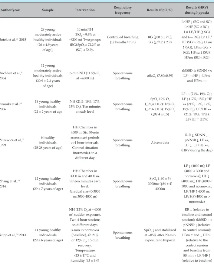
Table 1 – Methodological classification and results of HRV during hypoxia
Author/year: Sample Intervention Respiratory
frequency Results (SpO2%):
Results (HRV) during hypoxia
Botek et al.,21 2015
29 young moderately active healthy individuals
(26 ± 4.9 years of age),
10 min NH (FiO2 = 9.6% at
~6200 m). Two groups: (RG) SpO2 ≥ 72.2% or
(SG) ≤ 72.2%
Controlled breathing (12 breaths/min)
RG (↓80.8 ± 7.0); SG (↓67.2 ± 2.9)
LnHF ↓ (RG and SG); LnHF (SG < RG); Ln LF/HF (↑ SG) and (↔ RG); Ln LF/ HF (SG > RG); LFnu
↑ (SG); LFnu (SG > RG); HFnu ↓ (SG); HFnu (SG < RG)
Buchheit et al.,8
2004
12 young moderately active healthy individuals
(30.9 ± 2.3 years of age)
6-min NH (11.5% O2 at ~4800 m)
Spontaneous
breathing ∆SaO2 (7.80±0.59)
rMSSD ↓; SDNN ↔; LF ↔; HF ↓; LFnu
and HFnu ↔
Iwasaki et al.,10
2006
18 young healthy individuals (22 ± 2 years of age
NH (21%, 19%, 17%, 15% O2). Ten minutes
at each level
Spontaneous breathing
SpO2 19% O2
(↓97,4 ± 0.2); 17% O2 (↓95.6 ± 0.3); 15% O2
(↓92.4 ± 0.5)
LF ↔ (21%, 19% O2);
LF ↑ (17%, 15%); HF
↔ (21%, 19%, 17%, 15% O2); LF/HF ↔
(21%, 19%, 17%); LF/HF ↑ (15%)
Zużewicz et al.,32
1999
6 healthy individuals (25-28 years of age)
HH Chamber in 4500 m. Six 30-min assessment periods at 4-hour intervals. Control situation
(normoxia) on a different day
Spontaneous
breathing Absent data
R-R ↓, SDNN ↓, pNN50 ↓, LF ↔, HF ↓, LF/HF ↔; (HRV during the day)
Zhang et al.,26
2014
12 young healthy individuals (29 ± 7 years of age)
HH Chamber in 3000 m and 4000 m. Fifteen minutes each
level. Gradual rise (0-3000
m; 3000-4000 m)
Spontaneous breathing
SpO2 (↓90 ± 3)
3000m; (↓84 ± 4) 4000m
LF ↓ (4000 m); LF (4000 < 3000 and normoxia); HF ↓ (4000 m); HF (4000 < 3000 and normoxia); LF/HF ↑ 4000 m; LF/HF (4000 m >
normoxia)
Rupp et al.,28 2013
11 young healthy individuals (29 ± 6 years of age)
NH (12% O2 at ~4000 m) sudden exposure. Two 4-hour sessions on different days. 3-min in normoxia (baseline), 4h 21% or 12% O2, 15-min
recovery. Temperature (23 ± 1)°C and humidity (43 ± 9)%
Spontaneous breathing
SpO2↓ and stabilized
at ~85% after 20-min exposure to hypoxia
RR ↓ (relative to baseline and control session); rMSSD ↔; pNN50 ↓ (relative to control session); LFnu ↑ and ↓ HFnu (relative to the control session and baseline from 80 min.); LF/HF ↑ (relative to baseline)
when 4000 m HH was simulated by Zhang et al.26 suggest
impaired cardiac autonomic modulation. Unlike what was found by Iwasaki et al.10 who observed maintenance
of the spectral power of HFms² and increased LFms2,
demonstrating that vagal withdrawal did not occur. In the study by Guger et al.12 after eleven hours' exposure to HH,
rMSSD, SDANN and SDNN components were reduced, indicating cardiac vagal withdrawal.
Continuation
Basualto-Alarcón et al.,11 2012
7 young healthy individuals (22.7 ± 5.8 years
of age)
15-min exposure to HH chamber in 3000 m and 15-min exposure to NH tent
in 3000 m (different days)
Spontaneous breathing
SpO2↓ in both HH
(91.6 ± 4.2) and NH (89.1 ± 3.8)
In HH: rMSSD ↔; pNN50 ↔; LF%
↔; HF% ↓; LFnu
↑;HFnu ↓; LF/HF ↑; In NH: HRV ↔
Bhaumik et al.,14
2013
6 healthy young military servicemen
(24.83 ± 2.93 years of age)
HH (actual altitude 3500 m) after 48 hours and after
5 days
Spontaneous breathing
SpO2↓ after 2 days
(92.83 ± 0.47); ↓ after 5 days (96.5 ± 0.22)
Day 2nd: LF ↔; HF
↓; LFnu ↑; HFnu ↓; LF/HF ↑; 5th day: LF
↔; HF ↓; LFnu ↔; HFnu ↔; LF/HF ↔
Guger et al.,12 2008 10 healthy males
(21-33 years of age)
A rise to 4000 m was reached after 1 hour and kept for 12 hours
in HH chamber
Spontaneous breathing
After 1 hour SpO2
↔; After 5h SpO2
↓ (82.7 ± 6,8); and after 11h SpO2 ↓
(84.9 ± 4.5)
After 1 hour HRV
↔; After 11 hours rMSSD ↓, SDANN
↓; SDNN ↓; LFnu ↑; HFnu ↓; LF/HF ↑
Giles et al.,23 2016
11 physically active males (21.8 ± 0.9 years
of age)
Five visits for 5 levels of FiO2 (20.3%; 17.4%;
14.5%; 12%; 9.8%) in NH, temperature
(20°C) and 50% humidity. The length
of exposure was 10 min
Spontaneous breathing recorded
(RF = 15.6 ± 3.4, 16.2 ± 2.7, 14.8 ± 3.0,
12.4 ± 4.2, 12.9 ± 4.2 breaths/min),
respectively
SpO2 = 20.3% FiO2 (96.8 ± 2.1); 17.4% FiO2 (95.8 ± 1.7);
14,5%FiO2
(91.6 ± 1.7); 12% FiO2
(84.1 ± 4.0); 9.8% FiO2
(77.7 ± 5.8%)
SDNN ↔; Ln rMSSD ↔; Ln VLF
↔; Ln LF ↔; Ln HF
↔; Ln LF/HF ↔ LFnu ↔; HFnu ↔ (in all conditions). The HR ↑ and Ln TP
↓ only in 9.8%FiO2
Krejčí, et al.,24 2016
29 young moderately active healthy individuals
(26 ± 4.9 years of age),
10 min NH (FiO2 = 9.6% at ~6200m), in supine
position
Controlled breathing (12 breaths/min)
SpO2↓ for 10 min:
(96.4 ± 2.3 min in the first minute and reaching 71.9 ± 10% in the tenth minute) without stabilizing
∆HR ↑ (from 1 min);
∆Ln rMSSD ↓ (from
the 2nd min); ∆Ln
SDNN ↓ and ∆Ln
SDNN/rMSSD ↑ (both from the 3rd
min);
Brown et al.,22
2014
10 healthy young individuals (aged
22 – 42)
6 min HN (FiO2 = 10%
at ~5500 m), in supine position
Spontaneous
breathing Absent data
HR ↑; LFnu ↔; HFnu ↔
Haddad et al.,25
2012
10 healthy, physically active males (32.7 ± 4 years
of age),
At rest: 10 min HN (FiO2 = 15.4% at ~2400m), sit down.
At 20-23°C)
Spontaneous
breathing Absent resting SpO2
At rest: RR ↔; Ln rMSSD ↔; Ln HF
↔; LF/HF ↔
NH: normobaric hypoxia; HH: hypobaric hypoxia; RG: hypoxia resistant group; SG: hypoxia sensitive group; FiO2: fraction of inspired oxygen; SpO2: oxygen
saturation pulse; ΔSaO2: delta SpO2; HRV: heart rate variability; R-R: heartbeat in milliseconds (ms); TP: total power; Ln TP: TP in Neperian logarithm;
VLF: very low frequency power; Ln VLF: VLF in Neperian logarithm; LF: low frequency power; HF: high frequency power; LF/HF: sympathovagal balance; LFnu: LF in normalized units; HFnu: HF in normalized units; LnHF: HF in Neperian logarithm; LnLF: LF in Neperian logarithm; Ln LF/HF: LF/HF in Neperian logarithm; rMSSD: square root of the mean of the squared differences between adjacent normal RR intervals; SDNN: standard deviation of all normal RR intervals; SDANN: standard deviation of the means of the normal adjacent RR intervals; pNN50: percentage of adjacent RR intervals that varied
PaO2↓
SaO2↓
Chemoreflex activation ↑
Baroreflex setpoint ↑
Ventilation ↑
Vagal tone ↓ Sympathetic tone ↑
Heart rate ↑ Blood pressure ↑
Thoracic afferent outflow ↑ or ↔
Sympathetic tone ↔
Baroreflex response ↑
or SNA activity ↓ (?)
Vagal tone ↔
Heart rate ↔
Primary Disturb
Acute Hypoxia
Imediate compensatory
response
Steady-state phase
HRV ?
Figure 1 – Regulation of the cardiac autonomic control during acute exposure (10 min) to severe hypoxia.
PaO2: arterial oxygen pressure; SaO2: arterial oxygen saturation; ANS: autonomic nervous system; HRV: heart rate variability.
Buchheit et al.,8 Zuzewicz et al.32 and Giles et al.12 found
no significant difference in sympathovagal balance in hypoxia. However, Buchheit et al.8 and Zuzewicz et al.32
reported reductions in time-domain components. Buchheit et al.8 found decrease in the rMSSD index and
HFms2 and maintenance of SDNN and LFms2. Although
indexes in the time-domain and frequency-domain
absolute values indicate vagal withdrawal, the same was not found in normalized units. In the study by Zuzewicz et al.32 there was a decrease in RR intervals, SDNN,
pNN50, HFms2 and maintenance LFms² and LF/HF, also
suggesting vagal withdrawal. However, Giles et al.23 found
hypoxia — ~4800 m, ~4500 m and ~4450 m, respectively — although the length of exposure and barometric hypoxic conditions were different.
The study by Giles et al.23 experienced 5 different
levels of oxygen (20.3%, 17.4%, 14.5%, 12% and 9.8%) for ten-minute exposure. Each oxygen level was carried out on different days at 24-hour intervals between analyzes in a random manner. Observing the HRV linear data, the authors found no changes in SDNN, rMSSD, VLF, LF and HF (Ln or %) components for all levels of oxygen. However, a significant reduction in Ln TP was found during the most severe level (9.8% O2 to simulated altitude of ~6000 m), indicating a reduction in cardiac autonomic modulation. Although randomization was made, one of the limitations of this study is that reproducibility of HRV measurements were not carried out for the different levels of FiO2. This may be a possible confounding factor to establish the effect of hypoxia on HRV. Furthermore, there may be differences between physically conditioned and unconditioned individuals. Although no increase in LF and no decrease in HF was found, a fall in Ln TP may indicate a decrease in cardiac autonomic modulation. The fall in TP may be associated with cardiac risk events.33
Although there has been no statistical significance for Ln VLF, Ln LF and Ln HF indexes, the effect in severe hypoxia was moderate (9.8% O2). A study24 that examined HRV
dynamics using time-domain indexes at segments per minute, including the transient portion of the opening minutes of sudden exposure to NH (9.6% FiO2 ~6200 m) for 10 min, found that cardiac autonomic modulation responds proportionally to the reduction in SpO2% during the first five-minute exposure to severe hypoxia. The ∆HR had a gradual increase from the first to the fourth minute, where it reached a steady state, while the ∆Ln rMSSD had a reduction from the second to the sixth minute, reaching a steady state. The ∆Ln SDNN had a gradual reduction from the third to the sixth minute and then it stabilized. The ∆Ln SDNN/rMSSD increased from the third to the fifth minute and then it stabilized. However, SpO2 began to decrease from the first to the tenth minute without reaching a steady state. Those results indicate that during the transient time or opening minutes of exposure to severe hypoxia (9.6% FiO2), a vagal withdrawal, reduction in cardiac autonomic modulation and increased sympathetic modulation occur, following the fall in SpO2. Although the study did not conduct such analysis, the authors suggest that stimulation of peripheral chemoreceptors probably overlaps the other autonomic regulation mechanisms during the first five minutes exposure to this level of
hypoxia.24 After this, they suggest two possible routes,
one relates to a possible baroreflex response that may counterbalance the initial disturbance, preventing very high levels of HR31 or even, in the possibility of fall in
SpO2 below a certain threshold, cause a negligible change in ANS activity.24 Although SpO
2 was not been stabilized
during 10-min exposure, the study by Rupp et al.28
indicates SpO2 stabilization only after 20-minute exposure to a level of 12% FiO2 at 4000 m, i.e., less than that achieved by Krejci et al.24
One only study21 separated the sample into two groups,
RG = SpO2 ≥ 72.2% and SG = SpO2 ≤ 72.2%, in addition to controlling RF and using an extreme level of hypoxia of ~6200m. In this study of rapid exposure to NH, the RG group had a decrease in LnHF but with maintenance of Ln LF/HF. While the SG group had a decrease in LnHF and HFnu and an increase in Ln LF/HF and LFnu. The difference between RG and SG (Table 1) indicates that SG has an increased sympathetic modulation associated with decreased vagal modulation during exposure to NH. In the study by Brown et al.22 although the level of
oxygen used was 10% FiO2 ~5500 m and increases in HR were found, in LFnu and HFnu components no differences were found in the normoxic condition. This study conducted a six-minute analysis during normoxia (21% O2), hypercapnia (5% CO2) and hypoxia (10% O2) at 5-minute intervals of normoxia between conditions and in a randomized manner. Few HRV indexes were analyzed in this study, but similar results in LF and HF components during exposure to hypoxia have been reported in other studies.23 Furthermore, during the randomization in some
individuals, a possible residual effect of hypercapnic conditions may have influenced the subsequent hypoxic condition. SpO2 data were not presented.
Haddad et al.25 focused on the impact of hypoxia
on cardiac parasympathetic reactivation measured by HRV after exercise. However, for this review, we considered only the HRV indexes at rest. The section for analysis considered was from the 5th to the 10th min of exposure to 15.4% FiO2 ~2400m. T h e H R V i n d e x e s p r e s e n t e d d i d n o t c h a n g e significantly. Suggesting that the magnitude of hypoxia was not sufficient to cause changes in HRV at rest. However, it was sufficient to cause a delay in cardiac parasympathetic reactivation after a submaximal exercise. In addition, this study sample had good physical capacity, which itself tends to respond to better adjustments in the HRV in stressful situations.17 SpO
The study by Bhaumik et al.14 used an acclimatization to
hypoxia protocol, with assessments after 48 hours and five days, and found that on the second day there was a decrease in the vagal modulation associated with an increase in the LFnu component, leading to an increased sympathovagal balance. However, after the fifth day, despite a reduction in the HFms² component, there was a recovery of sympathovagal balance when compared to normoxia, suggesting a possible acclimatization induced autonomic adaptation. Although the LFnu component is considered a cardiac sympathetic modulation related marker,7 it has
been observed that the LFnu behavior in relation to muscle sympathetic nerve activity (MSNA) converge in specific conditions and this is also associated with the breathing effect.34 Furthermore, in severe hypoxia ~10.5% O
2, the LFnu
component only converged with ANSM when breathing was controlled at 20 breaths/min, which was not the case during spontaneous breathing.34
Breathing effects on autonomic reflexes have been well described by Bernardi et al.3 and generate impact on
HRV indexes, showing changes in spectral components, particularly due to changes in RF.27 In this sense, we believe
that the change in breathing pattern during exposure to hypoxia can impact on HRV. Furthermore, chemoreflex stimulation caused by hypoxia may also affect the autonomic control caused by sympathetic activation, which can be counterbalanced, for example, by either the interaction of baroreceptors or the inhibitory effect of pulmonary-stretch lung afferents.35 Therefore, more
research is needed to explore the record of spontaneous and controlled RF at HRV indexes during exposure to hypoxia associated with interindividual responsiveness and length of exposure. Furthermore, although the effect of hypocapnia on the sympathetic activity is already known,36 the influence of this state, isolated and associated
with hypoxia at HRV indexes, is another factor that needs to be better understood.
Based on the HRV responses observed in this review, we propose a figure with a description of the possible physiological phenomena related to the cardiac autonomic control during acute exposure to severe hypoxia. It is important to note that the figure shows only a general and superficial view of the possible mechanisms in healthy individuals. However, such events can be varied by factors such as the level of FiO2, length of exposure, age, physical capacity, interindividual variations, acclimatization, in addition to physical exercise and postural changes.
Conclusion
This review presented the main studies involving the response of HRV during acute hypoxia in healthy individuals. In general, based on the studies investigated in this review, exposure to hypoxia is capable of changing HRV and result in a decrease in the cardiac autonomic modulation, by either reduction or maintaining of vagal modulation, or by sympathetic predominance or even a combination of these responses. However, the magnitude of the response to hypoxia in unacclimatized healthy young adults at rest seems to be mainly dependent on the altitude level, length of exposure, interindividual variation and barometric pressure.
Study limitations
The overall cardiovascular system functions are vast and cardiac autonomic modulation aims to modulate the heart beat behavior. This study is limited to investigate the cardiac autonomic modulation by HRV, which is a non-invasive and selective method. This method aims to assess the spontaneous fluctuations of HR using several mathematical calculations aimed at indicating the cardiac vagal, sympathetic and sympathovagal drive. This study is also limited to study only the acute hypoxia, rejecting the effects of chronic and intermittent hypoxia on HRV. Furthermore, this study included only healthy individuals and their impact on exposure to hypoxia. This may limit a possible understanding of the effects of hypoxia on HRV in unhealthy individuals, particularly with cardiac disease. However, this review with healthy individuals becomes primary in the investigation of the physiological effects of hypoxia on HRV in humans and can go forward in the investigation of cardiorespiratory interaction. This review is also expected to reflect in future studies and/or reviews, aiming not only healthy but unhealthy individuals such as hypertensive, diabetic, obese and other individuals.
Author contributions
1. Perini R, Veicsteinas A. Heart rate variability and autonomic activity at rest and during exercise in various physiological conditions. Eur J Appl Physiol. 2003;90(3-4):317-25.
2. West JB. Human responses to extreme altitudes. Integr Comp Biol. 2006;46(1):25-34.
3. Bernardi L, Passino C, Wilmerding V, Dallam GM, Parker DL, Robergs RA, et al. Breathing patterns and cardiovascular autonomic modulation during hypoxia induced by simulated altitude. J Hypertens. 2001;19(5):947-58.
4. Halliwill JR, Morgan BJ, Charkoudian N. Peripheral chemoreflex and baroreflex interactions in cardiovascular regulation in humans. J Physiol. 2003;552(Pt 1):295-302.
5. Floras JS. Clinical aspects of sympathetic activation and parasympathetic withdrawal in heart failure. J Am Coll Cardiol. 1993;22(4 Suppl A):72A-84A.
6. Akselrod S, Gordon D, Ubel FA, Shannon DC, Berger AC, Cohen RJ. Power spectrum analysis of heart rate fluctuation: a quantitative probe of beat-to-beat cardiovascular control. Science. 1981;213(4504):220-2.
7. Heart rate variability: standards of measurement, physiological interpretation and clinical use. Task force of The European Society of Cardiology and The North American Society of Pacing and Electrophysiology. Circulation. 1996;93(5):1043-65.
8. Buchheit M, Richard R, Doutreleau S, Lonsdorfer-Wolf E, Brandenberger
G, Simon C. Effect of acute hypoxia on heart rate variability at rest and during exercise. Int J Sports Med. 2004;25(4):264-9.
9. Saito S, Tanobe K, Yamada M, Nishihara F. Relationship between arterial oxygen saturation and heart rate variability at high altitudes. Am J Emerg Med. 2005;23(1):8-12.
10. Iwasaki K, Ogawa Y, Aoki K, Saitoh T, Otsubo A, Shibata S. Cardiovascular regulation response to hypoxia during stepwise decreases from 21% to 15% inhaled oxygen. Aviat Space Environ Med. 2006;77(10):1015-9.
11. Basualto-Alarcon C, Rodas G, Galilea PA, Riera J, Pagés T, Ricart A, et al. Cardiorespiratory parameters during submaximal exercise under acute exposure to normobaric and hypobaric hypoxia. Apunts Med Esport. 2012;47(174):65-72.
12. Guger C, Krausert S, Domej W, Edlinger G, Tannheimer M. EEG, ECG and oxygen concentration changes from sea level to a simulated altitude of 4000 m and back to sea level. Neurosci Lett. 2008;442(2):123-7.
13. Perini R, Miseli S, Biancardi L, Veicsteinas A. Effects of high altitude acclimatization on heart rate variability in resting humans. Eur J Appl Physiol Occup Physiol. 1996;73(6):521-8.
14. Bhaumik G, Dass D, Bhattacharyya D, Sharma YK, Singh SB. Heart rate variability changes during first week of acclimatization to 3500 m altitude in Indian military personnel. Indian J Physiol Pharmacol. 2013;57(1):16-22.
15. Brown SJ, Raman A, Barnes MJ, Mündel T. Autonomic cardiovascular response to acute hypoxia and passive head-up tilting in humans. Eur J Appl Physiol. 2013;113(7):1731-6.
16. Montano N, Ruscone TG, Porta A, Lombardi F, Pagani M, Malliani A. Power spectrum analysis of heart rate variability to assess the changes in sympathovagal balance during graded orthostatic tilt. Circulation. 1994;90(4):1826-31.
17. Gonçalves TR, Farinatti Pde T, Gurgel JL, da Silva Soares PP. Correlation between cardiac autonomic modulation in response to orthostatic stress and indicators of quality of life, physical capacity, and physical activity in healthy individuals. J Strength Cond. Res. 2015;29(5):1415-21.
18. Kuo TB, Lin T, Yang CC, Li CL, Chen CF, Chou P. Effect of aging on gender differences in neural control of heart rate. Am J Physiol Heart Circ Phisiol. 1999;277(6 Pt 2):H2233-9.
19. Yamamoto S, Iwamoto M, Inoue M, Harada N. Evaluation of the effect of heat exposure on the autonomic nervous system by heart rate variability and urinary catecholamines. J Occup Health. 2007;49(3):199-204.
20. Moher D, Liberati A, Tetzlaff J, Altman DG; PRISMA Group. Preferred reporting items for systematic reviews and meta- analyses: The PRISMA Statement. PLoS Med. 2009;6(7):e1000097.
21. Botek M, Krejčí J, De Smet S, Gába A, McKune AJ. Heart rate variability and arterial oxygen saturation response during extreme normobaric hypoxia. Auton Neurosci. 2015;190:40-5.
22. Brown SJ, Barnes MJ, Mündel T. Effects of hypoxia and hypercapnia on human HRV and respiratory sinus arrhythmia. Acta Physiol Hung. 2014;101(3):263-72.
23. Giles D, Kelly J, Draper N. Alterations in autonomic cardiac modulation in response to normobaric hypoxia. Eur J Sport Sci. 2016;16(8):1023-31.
24. Krejcí J, Botek M, McKune A. Dynamics of the heart rate variability and oxygen saturation response to acute normobaric hypoxia within the first 10 min of exposure. Clin Physiol Funct Imaging. 2016 Jul 6. [Epub ahead of print].
25. Haddad H, Mendez-Villanueva A, Bourdon PC, Buchheit M. Effect of acute hypoxia on post-exercise parasympathetic reactivation in healthy men. Front Physiol. 2012;3:289.
26. Zhang D, She J, Zhang Z, Yu M. Effects of acute hypoxia on heart rate variability, sample entropy and cardiorespiratory phase synchronization. Biomed Eng Online. 2014;13:73.
27. Sasaki K, Maruiama R. Consciously controlled breathing decrease the high frequency component of heart rate variability by inhibiting cardiac parasympathetic nerve activity. Tohoku J Exp Med. 2014;233(3):155-63.
28. Rupp T, Leti T, Jubeau M, Millet GY, Bricout VA, Levy P, et al. Tissue deoxygenation kinetics induced by prolonged hypoxic exposure in healthy humans at rest. J Biomed Opt. 2013;18(9):095002.
References
the manuscript: Oliveira ALMB, Rohan PA, Gonçalves TR, Soares PPS. Critical revision of the manuscript for intellectual content: Oliveira ALMB, Rohan PA, Gonçalves TR, Soares PPS.
Potential Conflict of Interest
No potential conflict of interest relevant to this article was reported.
Sources of Funding
This study was funded by CAPES e FAPERJ (E-26/110.166/2014).
Study Association
29. Conkin J, Wessel JH 3rd. Critique of the equivalent air altitude model.
Aviat Space Environ Med. 2008;79(10):975-82.
30. Savourey G, Launay JC, Besnard Y, Guinet A, Travers S. Normo and hypobaric hypoxia: are there any physiological differences? Eur J Appl Physiol. 2003;89(2):122-6.
31. Bernardi L, Passino C, Spadacini G, Calciati A, Robergs R, Greene R, et al. Cardiovascular autonomic modulation and activity of carotid baroreceptors at altitude. Clin Sci (Lond). 1998;95(5):565-73.
32. Zuzewicz K, Biernat B, Kempa G, Kwarecki K. Heart rate variability in exposure to high altitude hypoxia of short duration. Int J Occup Saf Ergon. 1999;5(3):337-46.
33. Tsuji H, Larson MG, Venditti FJ Jr, Manders ES, Evans JC, Feldman CL, et al. Impact of reduced heart rate variability on risk for cardiac events: the Framingham Heart Study. Circulation. 1996;94(11):2850-5.
34. DeBeck LD, Petersen SR, Jones KE, Stickland MK. Heart rate variability and muscle sympathetic nerve activity response to acute stress: the effect of breathing. Am J Physiol Regul Integr Comp Physiol. 2010;299(1):R80-91.
35. Kara T, Narkiewicz K, Somers VK. Chemoreflexes – physiology and clinical implications. Acta Physil Scand. 2003;177(3):377-84.