©Revista Brasileira de Fisioterapia
REDUCTION OF HEART RATE VARIABILITY IN MIDDLE-AGED
INDIVIDUALS AND THE EFFECT OF STRENGTH TRAINING
L
OPESFL
1, P
EREIRAFM
2, R
EBOREDOMM
1, C
ASTROTM
1, V
IANNAJM
2, N
OVOJ
RJM
2& S
ILVALP
1,31 Physical Therapy Departament, Medical School, Juiz de Fora Federal University - UFJF, Juiz de Fora, MG - Brazil
2 Motor Evaluation Laboratory, Physical Education and Sports School - UFJF, Juiz de Fora, MG - Brazil
3 Biomedical Engineering Program, Rio de Janeiro Federal University - UFRJ, Rio de Janeiro, RJ - Brazil
Correspondence to: Lilian Pinto da Silva, Universidade Federal de Juiz de Fora, Departamento de Fisioterapia, Faculdade de Medicina, Centro de Ciências da Saúde, Campus Universitário, Bairro Cidade Universitária, CEP 36036-330, Juiz de Fora, MG – Brazil, e-mail: [email protected]
Received: 31/03/2006 - Revised: 12/09/2006 - Accepted: 01/02/2007
ABSTRACT
Objective: To investigate autonomic modulation of the sinus node, by analyzing heart rate variability (HRV) among young and aged individuals, and to assess the effect of an resistance strength training program on this modulation among middle-aged individuals. Method: Thirty-two healthy nonsmoking men with sedentary lifestyles, of whom 10 were young (22.2 ± 1.5 years) and 22 were middle-aged (49.3 ± 5.3 years), underwent electrocardiogram signal acquisition for time-domain HRV analysis. The middle-aged individuals were divided into two groups: experimental (n= 12) and control (n= 10). The individuals in the experimental group were enrolled in a strength training program lasting three months. The data analysis was carried out using the Wilcoxon and Mann-Whitney tests (p< 0.05). Results: The middle-aged group presented significant reductions (in relation to the young group) for all the variables used in investigating HRV (SDNN= 33.4 vs. 49.7 ms; RMSSD= 29.9 vs. 49.5 ms; pNN50= 6.5 vs. 27%). The training caused a significant increase in muscle strength and resistance for all muscular groups and non-significant increases in the variables SDNN (33.4 vs. 37.6 ms), RMSSD (30.2 vs. 31.3 ms) and pNN50 (7.5 vs. 11.4%). Conclusions: The findings from this study confirm that increased age causes alteration to the autonomic modulation of the sinus node, as demonstrated by reduced HRV in middle-aged individuals, which was not significantly modified by the type of physical training studied.
Key words: autonomic nervous system, physical exercise, heart rate, middle age.
INTRODUCTION
Action of autonomic nervous system (ANS) sympathetic and vagal branches over the pacemaker cells of the sinusal node promotes increase or decrease of heart rate, respectively, and the variation between the successive cardiac beats, obtained in a sinusal rhythm is called heart rate variability (HRV)1,2. Considering that changes in the cardiac rhythm are mediated by the ANS, its measurement represents a non-invasive tool for studying the autonomic modulation of heart rate1,2.
Studies have shown that the process of physiological aging leads to HRV reduction1, 3-5, related to a decrease of the vagal actuation over the sinusal node1,3, which is, in turn, associated to the increase on morbidity and mortality originated from different causes, in addition to coronary arterial disease, in both middle-aged and elderly individuals5. In this context,
some works have analyzed the effects of sedentary life-style changes, through implementation of physical exercise programs in the attempt to minimize or partially reverse the HRV decrease that occurs with aging6-12. Following these investigations, it has been shown that aerobic physical training promotes a HRV increase in elderly individuals7-10, however the studies that assessed anaerobic physical training effects, such as isometric and strength training, over the HRV in individuals of different ages present inconclusive results9,11-12,13.
METHODS
Sample
Thirty-two male (10 young and 22 middle-aged individuals), healthy, sedentary, non-smoking, volunteers were analyzed in this study (Table 1). For sedentary life-style and non-smoking characterization criteria, were selected individuals that did not practice regular physical activity for at least six months and did not smoke for at least a year. All volunteers were instructed and oriented about their participation in the study and, after agreeing on participating, signed an Informed Consent Term, approved by the Research Ethics Committee (process nº 144-013/2001). The volunteers were submitted to physical therapy evaluation, composed by anamnesis, musculoskeletal assessment, resting heart rate and arterial pressure measurements, pulmonary and cardiac auscultation. Middle-age volunteers were divided in two groups: a control group, which was oriented to keep their daily routines during the study’s duration, and a experimental group, in which
participants were referred to a strength-training program (Table 1).
ECG signals acquiring and processing
All middle-aged volunteers were submitted to electrocardiogram (ECG) signal acquisition at the beginning and end of the study, and young volunteers were submitted to a single acquisition at the beginning of the study for future HRV analysis. The ECG signal was obtained using three surface electrodes positioned to obtain MC5 derivation, at resting condition, in supine position, during a period of one hour. The volunteers remained with respiratory rhythm controlled, with the aid of a metronome, at a frequency of 12 respirations per minute (0.2 Hz). The signal was captured using a Lab-PC+ A/D converter board (National Instruments), which constitutes of an interface between the cardiac monitor of an one channel TC-500 (Funbec) and a microcomputer (AMD K6, 200 MHz), at a sampling frequency of 250Hz. Automatic detection of the R waves of the ECG signal was made, using
Table 1. Description of the groups and characterization of the sample in terms of size (N), age and values of heart rate (HR), systolic arterial
pressure (SAP) and diastolic arterial pressure (DAP) at rest. Values are mean ± standard deviation.
Groups N Age (years) HR (bpm) SAP (mmHg) DAP (mmHg)
Young 10 22.2 ± 1.5 70.6 ± 10.1 124.2 ± 7 76.4 ± 8.8
Middle aged 22 49.3 ± 5.3 70.4 ± 6.9 133.2 ± 12 88.7 ± 9.7
Control 10 48.5 ± 6.4 72.9 ± 6.2 134 ± 13 90.7 ± 8.9
Experimental 12 50 ± 4.3 68 ± 6.9 132.5 ± 11.5 86.8 ± 10.5
Figure 1. A and B: Histogram with the frequency of occurrence of samples of the collected ECG signal and the initial stretch of the same signal
for each signal a fixed threshold, chosen by the operator (Figure 1 - A and B). The values of the intervals between the ECG R waves (IRR) were calculated and, after visual analysis, the normal IRR values were considered (INN) for the tachogram construction (Figure 1C). Considering that the procedures above were not robust, resulting in flaws detection and T wave erroneous detections, in addition to the absence of stationarity on the collected signal, and observing the orientations of the European and American Cardiology Societies2 regarding the length of the short duration signal, we have opted to automatically select, for HRV analysis, only the five-minute uninterrupted tachogram interval that presented the lowest variance (Figure 1 – C and D). These selected segments were submitted again to visual analysis and, in the presence irregularities on the R-R intervals, the procedure described above was repeated, with the establishment of a new threshold for automatic detection of R waves.
For the HRV measurement, on the time domain, the recommended variables by the European and American Cardiology Societies2 were calculated. These following variables were extracted from the INN tachograms and were based in statistical relations:
SDNN – INN standard deviation;
RMSSD – Root mean square of the successive heart beats differences, given by:
chest muscles (bench press), elbow flexors (biceps curls) and elbow extensors (triceps on the pulley).
Maximal dynamic strength and endurance of the eight cited muscular groupings were assessed before and after the 12 weeks. For assessment of the maximal dynamic strength it was employed the one-repetition maximum weight test (1RM), the muscle resistance was assessed using the repetition test (RT) with the load of 50% of 1RM, obtained on the first 1RM test. Before the strength assessments, three training sessions were made on the exercising equipment, with light loads, for familiarization of the subjects with the equipments and with the proper techniques for each exercise.
The initial workload was established as 50% of 1RM and for the loading adjustments it was used Baechle’s14 protocol. Before each training session it was measured the arterial pressure at sitting position. A warm-up session was performed with total length of 15 minutes; consisting of a 10-minute pedaling in a cyclergometer without load and a 5-minute active stretching of the muscles that would be trained. The ordering of the exercises started with the largest muscular grouping and followed to the smallest ones, alternating the trained body segments. Each training session was constituted of eight exercises executed with an interval of one minute between the series. Training was divided into 12 micro-cycles of three sessions each, as described in Table 2.
Statistical analysis
The calculated variables from the INN tachogram, previously described, and the 1RM test and repetition test, were compared before and after training, by means of the Wilcoxon and Mann-Whitney tests, with a significance level of 5%.
RESULTS
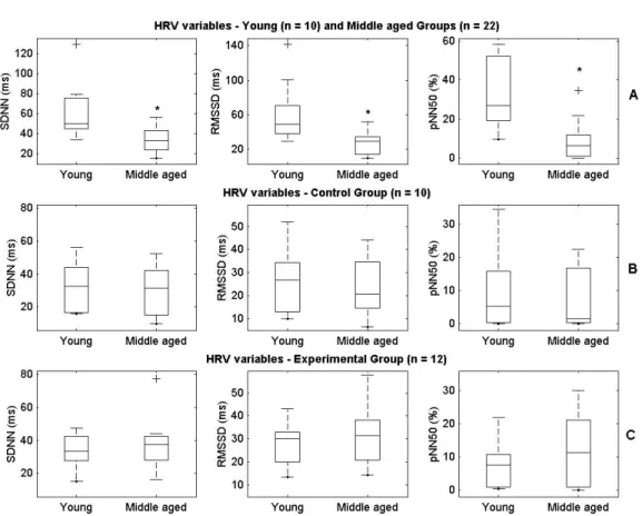
The middle-aged volunteers presented significantly lower values, in relation to the young volunteers, in all variables used on the HRV analysis (Figure 2).
No statistically significant differences was found by comparing the variables used for HRV investigation between the controls and experimental groups, before and after training. The control group presented a decrease of all variables used for HRV analysis, after the follow-up period, however the differences were not statistically significant, as illustrated in Figure 2.
At the end of the training period an increase in all the variables used for HRV analysis was observed on the experimental group, however the differences were not statistically significant (Figure 2). Moreover, statistically significant gains were observed, on the loading reached on the 1RM assessments and on the numbers of repetitions
(
)
1 1 1 2 1 − − =∑
− = + n NN NN RMSSD n i i i (1)In which n indicates the total INN number on the analyzed signal and i, the duration of the i-eth interval;
NN50 – counter of the number of times that successive INN present duration difference higher than 50 ms;
pNN50 – proportion obtained by the NN50/n ratio. The SDNN variable reflects the participation of all rhythmic components responsible for the variability. It is related to the contribution of both the branches of the ANS. The RMSSD, NN50 and pNN50 reflect the contributions of variations in high frequencies, which are related to the vagal action.
Training prescription and execution
Table 2. The subdivision in microcycles of the resistance strength program. * The values of loads for each exercise were specific for each volunteer
and depended on the test of 1MR (one maximal repetition). ** Adjustment: adjustment of the load (no overload).
Figure 2. Boxplots of the HRV variables of young group and middle-aged group (panel A), control group before and after 3 months of attendance
in the study (panel B), and experimental group before and after 3 months of training (panel C). Each boxplot represents the values minimum, first and third quartiles and maximum, with median in black color in the interior of the box and outliers represented by a cross. SDNN = standard deviation of all NN intervals; RMSSD = square root of the sum of the squares of differences between adjacent NN intervals; NN50 count = number of pairs of adjacent NN intervals differing more than 50 ms in the entire recording; pNN50 = NN50 count divided by the total number of all NN intervals. * The difference between young and middle aged group is significant at p< 0.05, p-value obtained for the Mann-Whitney test.
Week Microcycles
(3 weekly sessions)
Number of sets
for exercise
Number of
repetitions
Load (Kg)*
(50% 1 RM)
Assessment of the muscular strength
1 Adaptation I 1 12 - 20
2 Adaptation II 2 12 - 20
3 Stabilization 2 12 - 20
4 Development 2 12 - 20 adjustment **
5 Stabilization 2 12 - 20
6 Stabilization 2 12 - 20
7 Development 2 12 - 20 adjustment
8 Stabilization 2 12 - 20
9 Stabilization 2 12 - 20
10 Development 2 12 - 15 adjustment
11 Stabilization 2 12 - 20
12 Recovery 1 12 - 20
reached on the RT in all the trained muscular groupings (Table 3).
DISCUSSION
According to the obtained results at the reassessment after training of the maximal dynamic load (1RM) and of the repetition test, it can be observed that the training was effective in promoting the increase in maximal dynamic strength and muscle resistance. Since aging leads to a decrease of both muscular strength and resistance15, the type of physical training made was adequate to minimize these losses.
The smaller values of the variables obtained in the HRV time domain analysis, found on the Middle-aged volunteers when compared to the youngsters, demonstrate that the reduction of the HRV that occurs with aging3-4 is already evident at the age between 40 and 60 years old. Catai et al.6 and Marães et al.16, investigating subjects of this age range, found similar results to those in the present study.
After the physical training program, there was a non-significant increase of all variables for HRV analysis. Madden et al.9 have found similar results with a exercise program of muscular strengthening for healthy elderly women applied at 85% of 1RM, during six months. These authors have observed that the benefits of the strength training, regarding cardiac autonomic modulation, are less evident when compared to an aerobic training program. In this study, a similar group of elderly women submitted to a program of cyclergometer exercises during the same period presented a significant increase of the SDNN variable.
Forte et al.12 assessed the effects of an endurance training program, in women aged between 65 and 74 years old, and after 16 weeks of training they did not find changes regarding autonomic modulation, assessed using HRV at the time and
frequency domains. Similarly, Cooke et al.13 investigated if a high intensity strength-training program, applied during eight weeks, could increase vagal cardiac control and baroreflexive sensitivity in healthy youngsters of both genders. Their results have showed that the applied strength training did not affect the cardiac autonomic modulation, investigated using the HRV analysis at the time and frequency domains, or the cardio-vagal baroreflexive sensitivity.
Taylor et al.17, on the other hand, have observed an increase on the vagal modulation, demonstrated by the spectral analysis of the tachogram, in elders with arterial hypertension that were submitted to an isometric handgrip training for 10 weeks. Selig et al.18 have verified that three months of resisted exercise of moderate intensity, produced modifications on the autonomic modulation of patients with cardiac insufficiency. Such modification was evidenced by a significant increase of the high frequency (HF) spectral component and a significant decrease of the low frequency (LF) component and of the LF/HF ratio. Considering that the HF component represents vagal action, the performed training resulted in a HRV increase.
The information described in the literature, added to the results obtained in the present study, corroborates with the finding that the strength training does not seem to promote significant effects regarding autonomic modulation exerted over the sinusal node in healthy individuals, independently of age and gender. However, this is not true for individuals whose autonomic modulation over the cardio-vascular system is modified not only because of the physiological aging process, but also by the presence of diseases such as arterial hypertension17 and cardiac failure18.
Sample size, as well as the utilization of variables limited to the HRV analysis at the time domain, may have limited the observation of significant results on the study. However, such
Table 3. Loads of the 1MR test e number of repetitions reached in the repetition test, performed before (1st test) and after (2nd test) the resistance
strength training. Values are medians. * The difference between first and second test is significant at p< 0.05. The p-values were obtained by the Wilcoxon test.
Tests
1 RM (Kg) Weight for repetition
(number of repetitions)
Machine lifts 1
st
Test 2nd Test 1st Test 2nd Test
Leg extensions 47 70.4 * 17.5 31 *
Leg curls 52 56.4 * 17 30.5 *
Hip adduction 112.2 154 * 24 100.5 *
Hip abduction 96.8 120.8 * 22 51 *
Dorsal pulley 60 68.3 * 22.5 41.5 *
Bench press 80 90 * 22 40 *
Biceps curls 62.5 72.5 * 14.5 24.5 *
results allowed establishing a new experimental protocol that allows the continuity of the present work, in order to seek for more consistent results.
CONCLUSIONS
Healthy middle-aged individuals presented alteration on autonomic modulation of the heart rate, demonstrated by a HRV decrease, which was not minimized in a significant way by the applied physical training program. The physical training protocol was effective for strength and endurance muscle gains, however, it is not totally clear if this training can modify the autonomic modulation exerted over the sinusal node.
Acknowledgments: This work has counted with the collaboration of the graduate students Mariana D. M Fonseca and Rogério B. Bergamaschine, and of the Professors Fernando M. A. Nogueira, Jorge R. Perrout de Lima and Jurandir Nadal.
REFERENCES
1. Malik M, Camm AJ, editors. Heart Rate Variability. New York: Futura; 1996.
2. Task Force of The European Society of Cardiology and The North American Society of Pacing and Electrophysiology. Heart rate variability: Standards of measurement, physiological interpretation and clinical use. Eur Heart J. 1996;17(3):354-81.
3. Barbosa PR, Barbosa FJ, Sá CAM. Influência da idade, sexo e doença coronária sobre a modulação autonômica do coração. Arq Bras Cardiol. 1996;67(5):325-9.
4. Jensen-Urstad K, Storck K, Bouvier F, Ericson M, Lindblad LE, Jensen-Urstad M. Heart rate variability in healthy subjects is related to age and gender. Acta Physiol Scand. 1997;160:235-41.
5. Dekker JM, Schouten EG, Klootwijk P, Pool J, Swenne CA, Kromhout D. Heart rate variability from short electro-cardiographic recordings predicts mortality from all causes in middle-aged and elderly men. Am J Epidemiol. 1997;145(10): 899-908.
6. Catai AM, Chacon-Mikahil MPT, Martinelli FS, Forti VAM, Silva E, Golfetti R, et al. Effect of aerobic exercise training on heart rate variability during wakefulness and sleep and cardiorespiratory responses of young and middle-aged healthy men. Braz Med Biol Res. 2002;35(6):741-52.
7. Schuit JA, Van Amelsvoort LG, Verheij TC, Rijneke RD, Maan AC, Swenne CA, et al. Exercise training and heart rate variability in older people. Med Sci Sports Exerc. 1999;31(6):816-21.
8. Stein PK, Ehsani AA, Domitrovich P, Kleiger RE, Rottman JN. Effect of exercise training on heart rate variability in healthy older adults. Am Heart J. 1999;138(Pt 1):567-76.
9. Madden KM, Levy WC, Stratton JR. Exercise training and heart rate variability in older adult female subjects. Clin Invest Med. 2006;29(1):20-8.
10. Levy CW, Cerqueira MD, Harp GD, Johannessen KA, Abrass IB, Schwartz RS, et al. Effect of endurance exercise training on heart rate variability at rest in healthy young and older men. Am J Cardiol. 1998;82:1236-41.
11. Paschoal MA. Variabilidade da freqüência cardíaca: estudo das influências autonômicas sobre suas características temporal e espectral em halterofilistas e sedentários [dissertação]. Campinas (SP): Universidade Estadual de Campinas; 1999.
12. Forte R, De Vito G, Figura F. Effects of dynamic resistance training on heart rate variability in healthy older women. Eur J Appl Physiol. 2003;89(1):85-9.
13. Cooke WH, Carter JR. Strength training does not affect vagal-cardiac control or cardiovagal baroreflex sensitivity in young healthy subjects. Eur J Appl Physiol. 2005;93:719-25.
14. Baechle TR, Earle RW. Essentials of strength and conditioning. 1ª ed. Champaign: Human Kinetics; 1994.
15. Fleck SJ, Kraemer WJ. Fundamentos do treinamento de força muscular. 2a ed. Porto Alegre: ArtMed; 1999.
16. Marães VRFS, Santos MDB, Catai AM, Moraes FR, Oliveira L, Gallo Jr L, et al. Modulação do sistema nervoso autonômico na resposta da frequência cardíaca em repouso e à monobra de valsalva com o incremento da idade. Rev Bras Fisiot. 2004; 8(2):97-103.
17. Taylor AC, Mccartney N, Kamath MV, Wiley RL. Isometric training lowers resting blood pressure and modulates autonomic control. Med Sci Sports Exerc. 2003;35(2):251-6.