Ten cases with 46,XX testicular disorder of sex development:
single center experience
_______________________________________________
Emre Can
Akinsal
1, Numan Baydilli
1, Abdullah Demirtas
1, Cetin Saatci
2, Oguz Ekmekcioglu
11 Department of Urology, Erciyes University Medical Faculty, Kayseri, Turkey; 2 Department of Genetics,
Erciyes University Medical Faculty Medical, Kayseri, Turkey
ABSTRACT
ARTICLE
INFO
______________________________________________________________ ______________________
Objective: To present clinical, chromosomal and hormonal features of ten cases with SRY-positive 46,XX testicular disorder of sex development who were admitted to our infertility clinic.
Cases and Methods: Records of the cases who were admitted to our infertility clinic between 2004 and 2015 were investigated. Ten 46,XX testicular disorder of sex devel-opment cases were detected. Clinical, hormonal and chromosomal assessments were analized.
Results: Mean age at diagnosis was 30.4, mean body height was 166.9cm. Hormonal data indicated that the patients had a higher FSH, LH levels, lower TT level and normal E2, PRL levels. Karyotype analysis of all patients confirmed 46,XX karyotype, and FISH analysis showed that SRY gene was positive and translocated to Xp. The AZFa, AZFb and AZFc regions were absent in 8 cases. In one case AZFb and AZFc incomplete deletion and normal AZFa region was present. In the other one all AZF regions were present.
Conclusion: Gonadal development disorders such as SRY-positive 46,XX testicular dis-order of sex development can be diagnosed in infertility clinics during infertility work-up. Although these cases had no chance of bearing a child, they should be protected from negative effects of testosterone deficiency by replacement therapies.
Keywords:
Chromosome Aberrations; Infertility; 46, XX Testicular Disorders of Sex Development
Int Braz J Urol. 2017; 43: 770-5
_____________________ Submitted for publication: September 09, 2016 _____________________ Accepted after revision: February 09, 2017 _____________________ Published as Ahead of Print: March 24, 2017
INTRODUCTION
Forty Six,XX
testicular disorder of sex development (DSD) is a rare clinical condition with a reported incidence of 1:20.000 in newborn males (1). It was first described by De la Chapelle et al. in 1964 (2) By 1996, only 150 patients with classical 46,XX testicular DSD syndrome have been reported (3); however, more than 100 cases were described in the next ten years (4).The sex-determining region Y gene (SRY) located in Y chromosome plays a major role in encoding a testis determining factor (TDF) (5, 6).
About 90% of 46,XX testicular DSD have Y chro-mosomal material including the SRY gene, that is usually translocated to the distal tip of the short arm of X chromosome or autosomal chromoso-mes. About 10% of 46,XX testicular DSD cases are negative for SRY gene, which could carry diffe-rent degrees of masculinization (4, 7).
In this retrospective study, we analized clinical, chromosomal and hormonal features of ten cases with SRY-positive 46,XX testicular DSD who were admitted to our infertility clinic.
CASES AND METHODS
Records of the cases who were admitted to our infertility clinic between 2004 and 2015 were investigated. Ten 46,XX testicular DSD ca-ses were detected.
Medical/family history and detailed phy-sical examination records, body mass indexes, presence of parenteral consanguinity, records of each testicular volume measured with Prader or-chidometer, serum levels of follicle-stimulating hormone (FSH), luteinizing hormone (LH), estra-diol (E2), prolactin (PRL), total testosterone (TT), semen analysis results, karyotype and molecular analysis results and Dual-energy X-ray absorp-tiometry (DEXA) reports, if present, were asses-sed retrospectively.
Samples of peripheral blood (3mL) for chromosomal analysis were collected into 0.3mL heparin containing injectors. Conventional me-thod was used on the lymphocyte cultures for karyotype analysis. By G band staining techni-que 550 level bands were obtained and twenty metaphases were counted.
The probe mix containing SRY gene, Yp11.31 locus specific probe, labelled in red, and control probes for the X centromere (DXZ1), la-belled in blue, and for chromosome Y (DYZ1, the heterochromatic block at Yq12), labelled in green which was used for FISH analysis was purcha-sed from Cytocell (Oxford Gene Technology, UK). After the harvest of cultured blood samples and FISH slide preparation, probe mix was applied according to the manufacturer’s instructions, and materials were examined by using the Nikon, ECLIPSE E1000 fluorescent microscope (Tokyo, Japan) and analized with the CytoVision softwa-re (CytoVision, AB Imaging, Germany).
Samples of peripheral blood for sequen-ce analysis were collected into commercially available EDTA-treated tubes. DNA was isolated from peripheral blood samples drawn from con-trol, and study groups using High Pure
Polyme-rase chain reaction (PCR) Template Preparation Kit (Roche Applied Science). After amplifying AZF regions by using PCR mixture containing 10xPCR Buffer 5µL, 2.5mM dNTP 3µL, 25mM MgCl 3µL, 10pmol Primer-1 3µL, 10pmol Pri-mer-2 3µL, Taq DNA polymerase 0.5µL, DNA 5µL and completed to total volume of 50µL by adding water. Primers are specific to AZFa (sy81p1, sy81p2, sy82p1, sy82p2, sy84p1, sy84p2), AZFb (sy127p1, sy127p2, sy142p1, sy142p2, sy164p1, sy164p2, rbm1p1, rbm1p2), and AZFc (sy254p1, sy254p2, sy255p1, sy255p2, sy277p1, sy277p2, cdyp1, cdyp2, bpy2p1, bpy2p2) regions and were used separately in mixture. After 40 cycles of PCR by using “94ºC 40 seconds, 58ºC 40 seconds, 72ºC 45 seconds” program, final product was lo-aded to 2% agarose gel. After electrophoresis, gel was examined by using UV transilluminator for absence or presence, location and, size of bands.
The medical ethics committee of Erciyes University approved this study and informed consent was obtained from all patients.
RESULTS
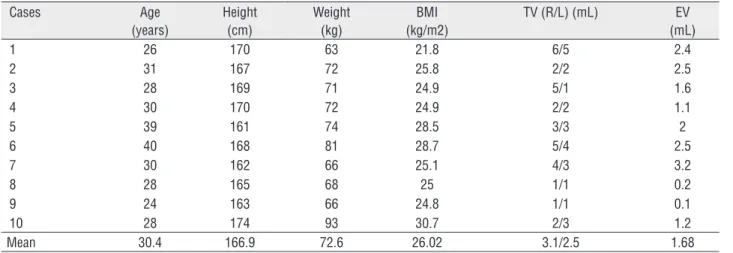
In our ten cases, mean age at diagnosis was 30.4, mean body height was 166.9cm (lo-wer than general population), mean body wei-ght was 72.6kg, mean BMI was 26.02. Semen analysis showed azoospermia and average se-men volume was 1.68mL. All cases had small testicular volumes. Mean testicular volume was 3.1mL for right testes and 2.5mL for left testes. Detailed general characteristics of the cases are presented in Table-1.
Two of the patients (patients 3, 10) had prior orchiopexy operation in their medical his-tory and one of them (patient 3) had parental consanguinity. All patients had no family his-tory for genetic disorders.
Four patients (patients 1, 5, 9, 10) had de-creased axillary and pubic hair and one patient (patient 5) had gynecomastia Tanner stage III.
Hormonal data indicated that the pa-tients had a higher FSH, LH levels, lower TT level and normal E2, PRL levels (Table-2).
(Patient 5) was assessed osteopenic for lumbar vertebraes and femoral neck; second one (Pa-tient 8) was assessed osteopenic for lumbar ver-tebraes and third one (Patient 7) was assessed osteoporotic for lumbar vertebraes and osteo-penic for femoral neck.
Initial karyotyping analysis of all pa-tients were considered as 46,XX with some he-sitation because of derivative X chromosomes observed in metaphase fields. After fluores-cence in situ hybridization (FISH) analysis, it was revealed that all patients were SRY
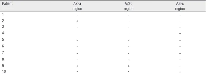
posi-tive. Next step which was the determination of the presence or the deletion of AZFa, AZFb, and AZFc regions revealed that all these regions were deleted in eight patients, in one patient (patient 9) none of the regions were deleted and in one patient (patient 2) AZFb and AZFc regions were deleted and AZFa was present.
Final karyotype for patients was as 46,XX, final FISH report was written as 46,XX ish der (X) t(X;Y) (p22.3;p11.3) (SRY+) and AZF region dele-tion summarized in Table-3. Presumable Ideogram of the chromosomes is given in Figure-1.
Table 1 - Clinical data and semen volume analysis.
Cases Age (years)
Height (cm)
Weight (kg)
BMI (kg/m2)
TV (R/L) (mL) EV (mL)
1 26 170 63 21.8 6/5 2.4
2 31 167 72 25.8 2/2 2.5
3 28 169 71 24.9 5/1 1.6
4 30 170 72 24.9 2/2 1.1
5 39 161 74 28.5 3/3 2
6 40 168 81 28.7 5/4 2.5
7 30 162 66 25.1 4/3 3.2
8 28 165 68 25 1/1 0.2
9 24 163 66 24.8 1/1 0.1
10 28 174 93 30.7 2/3 1.2
Mean 30.4 166.9 72.6 26.02 3.1/2.5 1.68
BMI = Body Mass Index; TV = Testicular Volume; EV = Ejaculate Volume; R = Right; L = Left
Table 2 - Hormonal status of the patients.
Cases FSH (mIU/mL)
LH (mIU/mL)
E2 (pg/mL)
TT (ng/dL)
PRL (ng/mL)
1 58.1 42.2 52.0 183.0 5.6
2 36.0 16.8 57.9 376.0 3.5
3 35.4 9.5 48.6 94.4 8.1
4 17.9 10.3 24.6 272.7 7.9
5 37.5 16.3 24.5 96.6 6.9
6 36.8 9.8 53.8 290.0 5.6
7 57.3 16.9 49.2 153.6 6.4
8 50.5 11.3 39.1 103.0 9.0
9 43.1 17.9 21.2 242.0 8.6
10 31.3 23.5 27.6 203.0 9.0
Mean and 95% CI 40.4 (31.5-49.2) 17.4 (10.4-24.4 ) 39.8 ( 29.7-49.9) 201.4 (134.0-268.7) 7.1 (5.8-8.3)
Table 3 - AZF region analysis of the patients.
Patient AZFa region
AZFb region
AZFc region
1
-
-
-2 + -
-3
-
-
-4 - -
-5
-
-
-6
-
-
-7
-
-
-8
-
-
-9 + + +
10 - -
-- deletion of gene region; + presence of gene region
Figure 1 - Presumable Ideogram of chromosomes.
1 - Normal Y Chromosome; 2 - Normal X Chromosome; 3 - Derivative X Chromosome of cases number 1, 3-8, and 10; 4 - Derivative X Chromosome of case 2; 5 - Derivative X Chromosome of case 9.
DISCUSSION
46,XX testicular DSD is a rare sex rever-sal syndrome characterized by a female karyo-type in discordance with a male phenokaryo-type (4). Although 46,XX male DSD is frequently spora-dic (8), familial cases have also been reported
(9). All our patients were considered sporadic based on their family history.
Vorona et al. stated that 46,XX men tend to be shorter than men with Klinefelter syndrome (7). In a previous study, Y chromosome growth-control gene had a possible impact on growth (14). Mean body height was relatively lower than normal po-pulation in our cases. Absence of specific growth genes in the Y chromosome may have some effects for this situation.
Classical 46,XX testicular DSD have nor-mal testosterone level and free testosterone level during adolescence, but may decrease in adulthood, leading to hypergonadotropic hypogonadism (15). Testicular volumes are usually lower than 5mL in these cases. While testis morphology is normal in infancy, hyalinization of the seminiferous tubules in early childhood causes loss of spermatogonia (16, 17). Gunes et al. showed hyalinization of the seminiferous tubules by testıcular biopsies of a pa-tient with 46,XX testicular DSD (18). Low testicu-lar volume and hypergonadotropic hypogonadism are constant findings in our patients. But testicular biopsies were not performed because these patients had no chance of bearing a child by assisted repro-ductive techniques.
Imaging of the pelvis is required to look for remnants of mullerian ducts that may cause morbi-dity in the form of repeated infections or hematuria and require surgical removal (19, 20). Differential diagnosis with other genetic conditions such as Persistent Mullerian Duct Syndrome in which viri-lization is achieved despite having low testosterone levels may be a challenging issue in some clinical presentations.
Recent epidemiologic studies have sugges-ted that hypogonadal concentrations of TT are as-sociated with an increased risk of fragility fracture (21, 22). Replacement therapies may protect these patients from bone fracture risk. Prior to initiating therapy, a baseline bone density scan should be per-formed to look for osteopenia or frank osteoporosis (23). Patients with a T score of <-1.0 would be-nefit from treatment with Vitamin D and calcium, bisphosphonates, or calcitonin, and require annual repeats of DEXA scan until results are normal (16). Our three patients had DEXA assay and all of them were assessed as osteopenic or osteoporotic. Bone mineral density measurements were absent in the records of our initial patients. We began to perform
these measurements and apply replacement thera-pies as our clinical experience increased. Important part of individuals with SRY-positive 46,XX testi-cular DSD are diagnosed at adulthood in infertility clinics. But testosterone replacement should be gi-ven to patients which have clinical and/or labora-tory signs of androgen deficiency in puberty.
During genetic counselling, it was advised to patients that this report must not affect their life and they must continue to live as before. While ex-plaining inheritance it was stated that SRY-positive 46,XX testicular DSD is generally not inherited be-cause of infertility of patients and de novo occur-rence of Y and X chromosome translocation. But as there is always possibility of paternal balanced translocation or gonadal mosaicism, chromosome analyses were advised to father and brothers.
In conclusion, chromosomal abnormalities such as SRY-positive 46,XX testicular DSD can be diagnosed in infertility clinics during infertility work-up. Although these patients had no chance of bearing a child, they should be protected from negative effects of testosterone deficiency by repla-cement therapies.
CONFLICT OF INTEREST
None declared.
REFERENCES
1. Zenteno-Ruiz JC, Kofman-Alfaro S, Méndez JP. 46,XX sex reversal. Arch Med Res. 2001;32:559-66.
2. Delachapelle A, Hortling H, Miemi M, Wennstroem J. XX sex chromosomes in a human male. First case. Acta Med Scand. 1964;175:(Suppl 412):25-8.
3. Rego A, Margarit E, Estivill X, Regal M, García-Mayor RV. Development in a 46 XX boy with positive SRY gene. J Pediatr Endocrinol Metab. 1996;9:623-6.
4. Ergun-Longmire B, Vinci G, Alonso L, Matthew S, Tansil S, Lin-Su K, et al. Clinical, hormonal and cytogenetic evaluation of 46,XX males and review of the literature. J Pediatr Endocrinol Metab. 2005;18:739-48.
5. Anık A, Çatlı G, Abacı A, Böber E. 46,XX male disorder of sexual development:a case report. J Clin Res Pediatr Endocrinol. 2013;5:258-60.
7. Vorona E, Zitzmann M, Gromoll J, Schüring AN, Nieschlag E. Clinical, endocrinological, and epigenetic features of the 46,XX male syndrome, compared with 47,XXY Klinefelter patients. J Clin Endocrinol Metab. 2007;92:3458-65. 8. Fechner PY, Marcantonio SM, Jaswaney V, Stetten G,
Goodfellow PN, Migeon CJ, et al. The role of the sex-determining region Y gene in the etiology of 46,XX maleness. J Clin Endocrinol Metab. 1993;76:690-5.
9. Zenteno JC, López M, Vera C, Méndez JP, Kofman-Alfaro S. Two SRY-negative XX male brothers without genital ambiguity. Hum Genet. 1997;100:606-10.
10. Ferguson-Smith MA, Cooke A, Affara NA, Boyd E, Tolmie JL. Genotype-phenotype correlations in XX males and their bearing on current theories of sex determination. Hum Genet. 1990;84:198-202.
11. Wang T, Liu JH, Yang J, Chen J, Ye ZQ. 46, XX male sex reversal syndrome: a case report and review of the genetic basis. Andrologia. 2009;41:59-62.
12. Bianco B, Christofolini DM, Ghersel FR, Gava MM, Barbosa CP. XX testicular disorder of sex differentiation: case report. Einstein (Sao Paulo). 2011;9:394-6.
13. McLachlan RI, O’Bryan MK. Clinical Review#: State of the art for genetic testing of infertile men. J Clin Endocrinol Metab. 2010;95:1013-24.
14. Kirsch S, Weiss B, Schön K, Rappold GA. The definition of the Y chromosome growth-control gene (GCY) critical region: relevance of terminal and interstitial deletions. J Pediatr Endocrinol Metab. 2002;15 Suppl 5:1295-300. 15. Velasco G, Savarese V, Sandorfi N, Jimenez SA, Jabbour S.
46, XX SRY-positive male syndrome presenting with primary hypogonadism in the setting of scleroderma.Endocr Pract. 2011;17:95-8.
16. Boucekkine C, Toublanc JE, Abbas N, Chaabouni S, Ouahid S, Semrouni M, et al. Clinical and anatomical spectrum in XX sex reversed patients. Relationship to the presence of Y specific DNA-sequences. Clin Endocrinol (Oxf). 1994;40:733-42.
17. Abbas NE, Toublanc JE, Boucekkine C, Toublanc M, Affara NA, Job JC, et al. A possible common origin of “Y-negative” human XX males and XX true hermaphrodites. Hum Genet. 1990;84:356-60.
18. Gunes S, Asci R, Okten G, Atac F, Onat OE, Ogur G, et al. Two males with SRY-positive 46,XX testicular disorder of sex development. Syst Biol Reprod Med. 2013;59:42-7. 19. Morgan RJ, Williams DI, Pryor JP. Müllerian duct remnants
in the male. Br J Urol. 1979;51:488-92.
20. Da Aw L, Zain MM, Esteves SC, Humaidan P. Persistent Mullerian Duct Syndrome: a rare entity with a rare presentation in need of multidisciplinary management. Int Braz J Urol. 2016;42:1237-1243.
21. Cawthon PM, Ensrud KE, Laughlin GA, Cauley JA, Dam TT, Barrett-Connor E, et al. Sex hormones and frailty in older men: the osteoporotic fractures in men (MrOS) study. J Clin Endocrinol Metab. 2009;94:3806-15.
22. Meier C, Nguyen TV, Handelsman DJ, Schindler C, Kushnir MM, Rockwood AL, et al. Endogenous sex hormones and incident fracture risk in older men: the Dubbo Osteoporosis Epidemiology Study. Arch Intern Med. 2008;168:47-54. 23. Majzoub A, Arafa M, Starks C, Elbardisi H, Al Said S, Sabanegh
E. 46 XX karyotype during male fertility evaluation; case series and literature review. Asian J Androl. 2017;19:168-172.