“Total reconstruction” of the urethrovesical anastomosis
contributes to early urinary continence in laparoscopic
radical prostatectomy
_______________________________________________
Xiaoxing Liao
1, 2, Peng Qiao
1, Zhaohui Tan
3, Hongbin Shi
4, Nianzeng Xing
11 Department of Urology, Beijing Chao-Yang Hospital, Capital Medical University, Beijing, China; 2
Department of Urology, Beijing Aerospace General Hospital, Beijing, China; 3 Department of Urology, Inner Mongolia People’s Hospital, Inner Mongolia, China; 4 Department of Urology, General Hospital of Ningxia Medical University, Yinchuan, China
ABSTRACT
ARTICLE
INFO
______________________________________________________________ ______________________
Purpose: To demonstrate the effect of total reconstruction technique on postoperative urinary continence after laparoscopic radical prostatectomy (LRP).
Material and Methods: LRP was performed using a standard urethrovesical anastomo-sis in 79 consecutive patients (Group-A) from June 2011 to October 2012, and a total reconstruction procedure in 82 consecutive patients (Group-B) from June 2012 to June 2013. The primary outcome measurement was urinary continence assessed at 1, 2, 4, 12, 24 and 52 weeks after catheter removal. Other data recorded were patient age, body mass index, International Prostate Symptoms Score, prostate volume, preopera-tive PSA, Gleason score, neurovascular bundle preservation, operation time, estimated blood loss, complications and pathology results.
Results: In Group-A, the continence rates at 1, 2, 4, 12, 24 and 52 weeks were 7.59%, 20.25%, 37.97%, 58.22%, 81.01% and 89.87% respectively. In Group-B, the continence rates were 13.41%, 32.92%, 65.85%, 81.71%, 90.24% and 95.12% respectively. Group--B had significantly higher continence rates at 4 and 12 weeks after surgery (P<0.001 and P=0.001). There were no significant differences between the groups with respect to patient’s age, body mass index, prostate-specific antigen level, prostate volume, IPSS, estimated blood loss, number of nerve-sparing procedures and postoperative complications.
Conclusions: Total reconstruction technique in the procedure of urethrovesical anasto-mosis during LRP improved early recovery of continence.
Key words:
Laparoscopy; Prostatectomy; Urinary Incontinence; Prostatic Neoplasms; Reconstructive Surgical Procedures
Int Braz J Urol. 2016; 42: 215-22
_____________________
Submitted for publication: December 24, 2014
_____________________
Accepted after revision: May 03, 2015
INTRODUCTION
Radical prostatectomy (RP) is the standard surgical treatment for localized prostate cancer (1). Postoperative urinary incontinence is one of the drawbacks after RP, especially early inconti-nence, which has a major impact on patient’s
neck preservation (5), sparing or reconstruction of the puboprostatic ligament (6-8), posterior re-construction of the rhabdomyosphincter (9, 10), and anterior retropubic suspension (11, 12). These techniques are associated with different improve-ment on early continence. More studies on surgi-cal modifications are still needed to improve pos-toperative early continence.
In an effort to improve early urinary conti-nence, we applied a total reconstruction technique during laparoscopic radical prostatectomy (LRP). In the present study, we compared the periopera-tive and urinary continence outcomes of LRP with and without total reconstruction technique.
MATERIALS AND METHODS
From June 2011 to June 2013, a total of 161 consecutive patients that underwent LRP were reviewed, of which 79 patients (Group-A) were treated with standard anastomosis technique from June 2011 to October 2012, and 82 patients (Group-B) were managed with total reconstruction technique from June 2012 to June 2013. This stu-dy was approved by the Ethical Committee of our hospital. All surgeries were performed by a single surgeon (NX), highly experienced in LRP, having performed more than 100 LRP previously to this study.
All patients received a standardized pre-operative evaluation including measurement of body mass index, preoperative serum prostate specific antigen (PSA), transrectal ultrasound of the prostate for prostate volume, digital rectum examination (DRE), prostate magnetic resonance imaging (MRI) and ultrasound guided transrec-tal prostate biopsy. In selected cases (totransrec-tal PSA level>10ng/mL and/or Gleason score≥7, or clinical T3 prostate cancer), we also obtained a bone scan. Patient’s characteristics are shown in Table-1. Baseline urinary symptoms were measured using the IPSS and the Expand Prostate Cancer Index Composite validated health-related QOL (HRQOL) instruments.
Urinary continence was assessed using the self-administrated validated Expanded Prostate Cancer Index Composite (EPIC) questionnaire at 1, 2, 4, 12, 24 and 52 weeks after catheter
remo-val (13). The questionnaire was performed either at our outpatient or by telephone interview. The definition of continence was based on patient’s responses to the items selected to reflect the range of incontinence severity. The items were (‘4 weeks’ was changed to ‘1 or 2 weeks’ for the question-naire at 1 or 2 weeks after catheter removal): (i) Over the past 4 weeks how often have you leaked urine? (ii) Which of following best describes your urinary control during the last 4 weeks? (iii) How many pads or adult diapers per day did you usu-ally use to control leakage? Patients were consi-dered continent if they answered ‘Not at all’ to (i), ‘total control’ to (ii), and ‘No pads’ to (iii). Patients were considered incontinent when they were lost for follow-up.
SURGICAL TECHNIQUE
In Group-A, the urethrovesical anastomo-sis was performed using a 3/0 monocryl absor-bable suture. The procedure was completed using one suture technique. The first suture was perfor-med at the right posterior area of the bladder neck from outside to the inside, and placed this suture from inside to outside at the corresponding section of the urethral stump, and one knot was made. Then the procedure of outside to inside at bladder neck and inside to outside at urethral stump was repeated in the left side area of the bladder neck. An 18F Silastic Foley catheter was placed gently
into the bladder. The urethrovesical anastomosis was accomplished when the suture was continued around from left side to right side of the bladder neck.
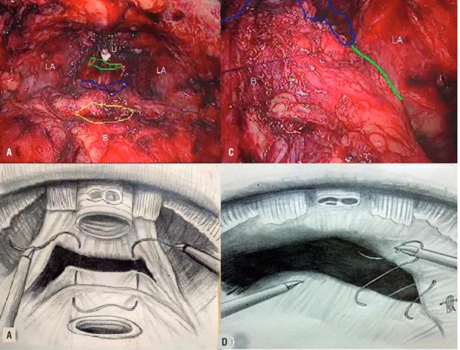
In Group-B, the total reconstruction was started with posterior reconstruction (15). Poste-rior reconstruction was accomplished by suturing the bladder musculature, Denonvillier’sfascia and the musculofascial plate posterior to the urethra (Figures 1A and B). The second step of the recons-truction was the urethrovesical anastomosis, whi-ch was made in the manner than in Group-A. The
Table 1 - Patient characteristics and perioperative parameters.
Group A Group B P value
Patient, n 79 82
Age, yr 67.88±6.65 65.79±7.27 0.08
BMI, kg/m2 25.35±2.13 24.26±2.88 0.77
Preoperative PSA, ng/mL 26.86±31.95 33.07±40.65 0.13
Prostate weight, g 41.92±14.76 42.06±19.34 0.24
IPSS score, n 6 (0~19) 7 (0~18) 0.79
Biopsy Gleason score, n (%) 0.42
≤6 45.8% 82.9%
7 45.8% 35.1%
≥8 8.5% 7.0%
Operative time, min (range) 130.81±21.66 147.33±29.89 0.001
Estimated blood loss, mL 225.42+164.96 232.63±217.93 0.38
Transfusion rate (%) (3/79) 3.79% (5/82) 6.09% 0.50
Duration of Catheter, d 16.13±16.47 13.96±2.20 0.18
Nerve-sparing procedure, n (%) 0.11
Bilateral nerve sparing (16/79) 20.25% (27/82) 32.92%
Unilateral nerve sparing (28/79) 35.4% (19/82) 23.17%
Non-nerve sparing (35/79) 44.3% (36/82) 43.90%
Complications, n (%) 8/79 (10.13%) 6/82 (7.32%) 0.53
Complications grade I 7/79 (8.9%) 4/82 (4.88%) 0.32
Complications grade II 1/79 (1.3%) 2/82 (2.4%) 0.58
third step was anterior reconstruction consisting of reattachment of the arcus tendineus and pubo-prostatic plate to the bladder neck (Figures 1C and D). A 3/0 monocryl absorbable suture was used to approximate the remaining arcus tendineus and distal triangular plate anterior to the urethra (in-cluding the residual of the endopelvic fascia and puboprostatic ligaments, rhabdosphincter, dorsal venous complex) to the bladder neck (16).
Statistical analysis
The categorical variables were summarized in frequency Tables. Continuous variables were
presented as the mean±standard deviation. Two--group t-tests were used to compare numerical variables. Pearson’s chi-squared test was used to compare categorical variables. Data was processed using SPSS 17.0. Statistical significance was defi-ned as P<0.05.
RESULTS
There was no significant difference between Group A and B regarding patient age (67.88±6.65 versus 65.79±7.27, P=0.08), BMI (25.35±2.13 versus 24.26±2.88kg/m2, P=0.77), preoperative
serum PSA (26.86±31.95 versus 33.07±40.65ng/
Figure 1 - A) The first layer of reconstruction (approximating the musculofascial plate posterior to the urethra (green line region) and the Denonvillier’s fascia posterior to the bladder (blue line region) and the bladder musculature. U, urethra, LA, levator ani, B, bladder neck, (the yellow region). B) Illustration shows a separate “U” type of suture that is used for the first layer reconstruction. C) A 3/0 monocryl absorbable suture is used to approximate the remaining arcus tendineus and distal triangular plate anterior to the urethra (including the residual of the endopelvic fascia and puboprostatic ligaments, rhabdosphincter, dorsal venous complex) to the bladder neck. D) Illustration shows the anterior reconstruction.
A
A
C
ml, P=0.13), prostate weight (41.92±14.76 versus 42.06±19.34g, P=0.24), IPSS (6 versus 7, P=0.79), Gleason score (P=0.42), mean estimated blood loss (225.42±164.96 versus 232.63±217.93mL, P=0.38), transfusion rates (3.79% versus 6.09%, P=0.50), nerve-sparing procedure (P=0.11) and duration of catheter (16.13±16.47 versus 13.96±2.20d, P=0.18) (Table-1). The operative time was on ave-rage 17 minutes longer in the procedure of LRP with total reconstruction technique (130.81±21.66 versus 147.33±29.89 min, P=0.001) (Table-1).
Early postoperative complications were encountered in both groups. In Group-A, 4/79 pa-tients with anastomotic site leakage were treated with prolonged catheterization for 1 additional week. In Group-B, 4/82 patient with anastomotic site leakage were dealt with the same procedure. In Group-A, 3/79 patients with acute urinary
re-tention after catheter removal were treated with re-catheterization for 1 week, and one patient with postoperative anastomotic stenosis was trea-ted with urethral dilation 4 times (once per week). In Group-B, 2 patients with anastomotic stenosis were treated with urethral dilation. No significant difference was noted between the two groups with respect to anastomotic site leakage, postoperative urethral stenosis and the severity of complications (based on the Clavien-Dindo classification, Grade I and II) (Table-1) (17).
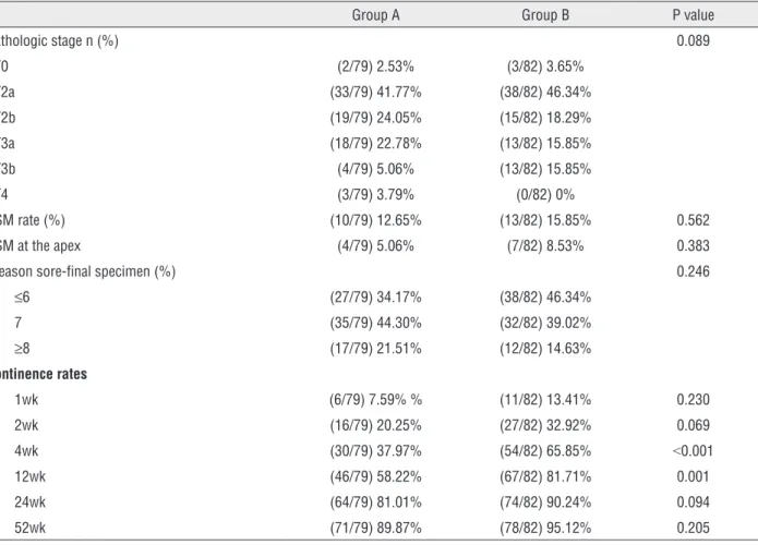
The two groups had no significant differen-ces in their pathologic stage, positive surgical mar-gin (PSM), or Gleason score of the surgical specimen (Table-2). The incidence of PSM at the prostate apex was 4/79 in Group-A and 7/82 in Group-B. There was no difference in the number of PSM at the pros-tate apex in the two groups (P=0.383).
Table 2 - Pathologic stage and continence rates between groups.
Group A Group B P value
Pathologic stage n (%) 0.089
pT0 (2/79) 2.53% (3/82) 3.65%
pT2a (33/79) 41.77% (38/82) 46.34%
pT2b (19/79) 24.05% (15/82) 18.29%
pT3a (18/79) 22.78% (13/82) 15.85%
pT3b (4/79) 5.06% (13/82) 15.85%
pT4 (3/79) 3.79% (0/82) 0%
PSM rate (%) (10/79) 12.65% (13/82) 15.85% 0.562
PSM at the apex (4/79) 5.06% (7/82) 8.53% 0.383
Gleason sore-final specimen (%) 0.246
≤6 (27/79) 34.17% (38/82) 46.34%
7 (35/79) 44.30% (32/82) 39.02%
≥8 (17/79) 21.51% (12/82) 14.63%
Continence rates
1wk (6/79) 7.59% % (11/82) 13.41% 0.230
2wk (16/79) 20.25% (27/82) 32.92% 0.069
4wk (30/79) 37.97% (54/82) 65.85% <0.001
12wk (46/79) 58.22% (67/82) 81.71% 0.001
24wk (64/79) 81.01% (74/82) 90.24% 0.094
52wk (71/79) 89.87% (78/82) 95.12% 0.205
We evaluated the questionnaires (EPIC) from 76 patients in Group A and 80 patients in Group B. Five patients (3 patients in Group A and 2 patients in Group B) were lost to follow-up, who were considered to be urinary incontinent. Group B had significantly higher continence rates at 4 and 12 weeks after catheter removal (P<0.001 and P=0.001). The continence rate of the two groups at 1, 2, 24 and 52 weeks were not significantly different (Table-2, Figure-2).
LRP, a significant improvement was found at 1 month, but not at 3 months (15). In a well desig-ned but not randomized trial including 803 pa-tients, Coelho et al. (19) showed that post recons-truction shortened continence recovery time and improved early continence in patients undergoing robot-assisted radical prostectomy.
Tewari and coworkers (16) evaluated groups of patients undergoing anterior recons-truction alone, anterior and posterior
reconstruc-Figure 2 - Postoperative urinary continence rate of both groups.
DISCUSSION
Urinary incontinence is a major quality--of-life concern for patients undergoing radical prostatectomy.The causes of urinary incontinence after radical prostatectomy are likely multifacto-rial and include both functional and anatomical changes related to removal of the prostate gland and alterations in the pelvic floor musculature and the urinary sphincter complex (3, 17).
Attempts have been made to modify Walsh’s anatomic radical prostatectomy to pre-vent injury or to reconstruct the rhabdo-urinary sphincter. Rocco et al. (18) first described post reconstruction in a study and found continence improved in the reconstruction group at 1 and 3 months. When this technique was transferred to
re-construction technique included two concepts in-volving posterior and anterior reconstructions. In posterior reconstruction, Denonvillier’s fascia was approximated to the bladder neck and the median dorsal raphe by slipknot. The anterior surface of the bladder-neck was approximated to the ante-rior detrusor apron and the puboprostatic liga-ment collar for anterior reconstruction.
After the prostate specimen was remo-ved, we performed the total reconstruction tech-nique for patients in Group B. The first layer of the anastomosis was the posterior reconstruction. A separate “U” type of suture was used for the entire anastomosis. This was based on the prin-ciples of Rocco’s posterior reconstruction tech-nique. But, unlike his technique, we performed a single-layer reconstruction, approximating the musculofascial plate posterior to the urethra, the Denonvillier’s fascia posterior to the bladder and the bladder musculature. This modified posterior reconstruction technique was to provide support to the urethra, restore it to a more anatomic posi-tion and facilitate the tension-free approximaposi-tion of bladder neck to the urethral stump (Figures 1 A and B). During the procedure of posterior re-construction, we decreased the pressure of CO2 to 8mmHg, so it was easy to approximate the bladder neck to the urethra. Then the pressure was increa-sed to 20mmHg to make space for the urethrove-sical anastomosis. When the posterior wall of the bladder neck and urethra stump were sutured, the pressure decreased to 15mmHg.
We performed an anterior reconstruction after the urethrovesical anastomosis. Our anterior reconstructive technique was modified from the Tewari technique. The third-layer of reconstruc-tion was performed by suturing the arcus tendi-neus and distal triangular plate anterior to the urethra to the anterior musculofascial of bladder neck, which was different from the anterior re-construction technique described by Hoshi. This technique allowed the anatomic structure of the bladder neck and urethra to be reconstructed as much as possible (Figures 1C and D).
We found that patients who underwent total reconstructive repair had significantly im-proved urinary control at 4 and 12 weeks
com-pared to those undergoing a standard LRP (Figu-re-2). Total reconstruction provides anatomical support to the urethra, and stabilizes the urethra and striated sphincter in the normal anatomical position. The posterior reconstruction enabled a tension free anastomosis and recreated the pos-terior support for the sphincter. The complication rates were similar in the total reconstruction and control Groups (P=0.53). Moreover, our study con-cluded that the total reconstruction technique was not associated with a higher incidence of positi-ve surgical margin (PSM) (12.65% positi-versus 15.85%, P=0.562).
There were some flaws in this study. It was a retrospective case-controlled study rather than a prospective randomized trial. Another limita-tion was that 5 patients in our study were lost to follow-up. We evaluated these patients as all in-continent and found that the findings are still sig-nificant at 4 and 12weeks (P=0.002 and P=0.001). The third limitation was the longer term main-tenance of continence could not be evaluated. Urodynamics were not performed in this study to evaluate bladder stability and its contribution to continence. The fourth limitation was the techni-ques employed in the study were sequential, thus patients from Group B may have benefited from increased surgeon experience since they were tre-ated sequentially to patients from Group A.
CONCLUSIONS
We employed a total reconstruction tech-nique supporting the posterior and anterior struc-ture of urethra, which improved early continence of patients undergoing laparoscopic radical pros-tatectomy in our study. This technique was easy and simple to perform, and resulted in a signifi-cant improvement in early continence at 4 and 12 weeks. Our findings support the need for further studies on technical refinements for earlier urina-ry continence in LRP.
CONFLICT OF INTEREST
REFERENCES
1. Boorjian SA, Eastham JA, Graefen M, Guillonneau B, Karnes RJ, Moul JW, et al. A critical analysis of the long-term impact of radical prostatectomy on cancer control and function outcomes. Eur Urol. 2012;61:664-75.
2. Lombraña M, Izquierdo L, Gómez A, Alcaraz A. Impact of a nurse-run clinic on prevalence of urinary incontinence and everyday life in men undergoing radical prostatectomy. J Wound Ostomy Continence Nurs. 2013;40:309-12.
3. Song C, Doo CK, Hong JH, Choo MS, Kim CS, Ahn H. Relationship between the integrity of the pelvic floor muscles and early recovery of continence after radical prostatectomy. J Urol. 2007;178:208-11.
4. Choi WW, Freire MP, Soukup JR, Yin L, Lipsitz SR, Carvas F, et al. Nerve-sparing technique and urinary control after robot-assisted laparoscopic prostatectomy. World J Urol. 2011;29:21-7.
5. Dal Moro F. Athermal bladder neck dissection during robot-assisted radical prostatectomy. Int Braz J Urol. 2014;40:433. 6. Porpiglia F, Fiori C, Grande S, Morra I, Scarpa RM. Selective
versus standard ligature of the deep venous complex during laparoscopic radical prostatectomy: effects on continence, blood loss, and margin status. Eur Urol. 2009;55:1377-83. 7. Patel VR, Coelho RF, Palmer KJ, Rocco B. Periurethral
suspension stitch during robot-assisted laparoscopic radical prostatectomy: description of the technique and continence outcomes. Eur Urol. 2009;56:472-8.
8. Daouacher G, Waldén M. A simple reconstruction of the posterior aspect of rhabdosphincter and sparing of puboprostatic collar reduces the time to early continence after laparoscopic radical prostatectomy. J Endourol. 2014;28:481-6.
9. Ficarra V, Gan M, Borghesi M, Zattoni F, Mottrie A. Posterior muscolofascial reconstruction incorporated into urethrovescical anastomosis during robot-assisted radical prostatectomy. J Endourol. 2012;26:1542-5.
10. Rocco B, Cozzi G, Spinelli MG, Coelho RF, Patel VR, Tewari A, et al. Posterior musculofascial reconstruction after radical prostatectomy: a systematic review of the literature. Eur Urol. 2012;62:779-90.
11. Noguchi M, Kakuma T, Suekane S, Nakashima O, Mohamed ER, Matsuoka K. A randomized clinical trial of suspension technique for improving early recovery of urinary continence after radical retropubic prostatectomy. BJU Int. 2008;102:958-63.
12. Hurtes X, Rouprêt M, Vaessen C, Pereira H, Faivre d’Arcier B, Cormier L, et al. Anterior suspension combined with posterior reconstruction during robot-assisted laparoscopic prostatectomy improves early return of urinary continence: a prospective randomized multicentre trial. BJU Int. 2012;110:875-83.
13. Wei JT, Dunn RL, Litwin MS, Sandler HM, Sanda MG. Development and validation of the expanded prostate cancer index composite (EPIC) for comprehensive assessment of health-related quality of life in men with prostate cancer. Urology. 2000;56:899-905.
14. Stolzenburg JU, Andrikopoulos O, Kallidonis P, Kyriazis I, Do M, Liatsikos E. Evolution of endoscopic extraperitoneal radical prostatectomy (EERPE): technique and outcome. Asian J Androl. 2012;14:278-84.
15. Rocco B, Gregori A, Stener S, Santoro L, Bozzola A, Galli S, et al. Posterior reconstruction of the rhabdosphincter allows a rapid recovery of continence after transperitoneal videolaparoscopic radical prostatectomy. Eur Urol. 2007;51:996-1003.
16. Tewari A, Jhaveri J, Rao S, Yadav R, Bartsch G, Te A, et al. Total reconstruction of the vesico-urethral junction. BJU Int. 2008;101:871-7.
17. Dindo D, Demartines N, Clavien PA. Classification of surgical complications: a new proposal with evaluation in a cohort of 6336 patients and results of a survey. Ann Surg. 2004;240:205-13.
18. Rocco F, Carmignani L, Acquati P, Gadda F, Dell’Orto P, Rocco B, et al. Early continence recovery after open radical prostatectomy with restoration of the posterior aspect of the rhabdosphincter. Eur Urol. 2007;52:376-83.
19. Coelho RF, Chauhan S, Orvieto MA, Sivaraman A, Palmer KJ, Coughlin G, et al. Influence of modified posterior reconstruction of the rhabdosphincter on early recovery of continence and anastomotic leakage rates after robot-assisted radical prostatectomy. Eur Urol. 2011;59:72-80. 20. Menon M, Muhletaler F, Campos M, Peabody JO. Assessment
of early continence after reconstruction of the periprostatic tissues in patients undergoing computer assisted (robotic) prostatectomy: results of a 2 group parallel randomized controlled trial. J Urol. 2008;180:1018-23.
21. Hoshi A, Nitta M, Shimizu Y, Higure T, Kawakami M, Nakajima N, et al. Total pelvic floor reconstruction during non-nerve-sparing laparoscopic radical prostatectomy: impact on early recovery of urinary continence. Int J Urol. 2014;21:1132-7.
_______________________ Correspondence address: