Signiicant Alterations of Serum Cytokine Levels in Patients with
Peyronie’s Disease
Reinhold P. Zimmermann, Gerhard Feil, Conny Bock, Lorenz Hoeltl, Arnulf Stenzl
Department of Urology (RPZ, LH), Elisabethinen Hospital, University Afiliated Hospital, Universities
of Vienna and Innsbruck, Linz, Austria, and Department of Urology (GF, CB, AS),
Eberhard-Karls-University of Tuebingen, Tuebingen, Germany
ABSTRACT
Objective:To determine the expression of the cytokines transforming growth factor-β1 (TGF-β1), interferon-γ (IFN-γ), interleukin-6 (IL-6), and tumor necrosis factor-α (TNF-α) in serum from patients with Peyronie’s disease (PD) compared
to healthy controls.
Materials and Methods: Ninety-one consecutive PD patients aged 20 - 74 years were included in this study. All patients were diagnosed with symptomatic PD for the irst time and had a palpable penile plaque. The patients previously had the disease for 6 - 72 months. None of the patients had a severe infectious disease or known systemic illness. For cytokine analyses, peripheral venous blood samples were obtained before treatment. Fifty healthy male blood donors aged 22 - 64 years served as the control group. TGF-β1, IFN-γ, Il-6, and TNF-α were analyzed quantitatively with commercial im -munoassays.
Results: Mean cytokine levels in serum from patients were increased for TGF-β1 and IFN-γ compared to healthy controls. The difference for TGF-β1 was considered statistically signiicant (p < 0.001). IL-6 was not detectable in PD patients (p < 0.01) and TNF-α was decreased (p < 0.0001).
Conclusion: The signiicantly elevated serum level of the proibrotic TGF-β1 cytokine underscores the effect of cytokines in the pathophysiology of PD. The signiicantly decreased TNF-α serum level suggested no acute immunomodulatory process. Therefore, the relevance for therapeutic administration of TNF-α should be further investigated. Quantiication of TGF-β1 in serum of PD patients provides a possible diagnostic tool and target for therapy. The data on altered cytokine levels in PD patients also provide a new understanding for etiopathogenesis of PD, which warrants further investigation.
Key words: Peyronie’s disease; pathophysiology; cytokines; IL-6; TNF-α; TGF-β; IFN-γ Int Braz J Urol. 2008; 34: 457-66
INTRODUCTION
Peyronie’s disease (PD) has been known for a very long time and even the ancient Egyptians reported PD-like symptoms. In 1743, the French surgeon and court physician François de la Peyronie was the irst to scientiically describe penile deviation due to tumors and penile nodes (1). He separated the syndrome from more common sexually transmitted diseases. PD was
previously thought to be associated with unnatural or excessive sexual behavior. Degenerative alterations
like ageing of the penile fascia layers or penile trauma
were considered as possible causes of PD as well as systemic diseases like hyperuricemia, diabetes, rheu
the corpora cavernosa and the tunica albuginea (TA) still remains unknown (2,3).
The anatomical-pathological correlation
of PD might be a traumatically caused inlamma
-tion followed by prolifera-tion of ibroblasts and the formation of scar areas such as ibrous plaques and, in some cases, bone formation. The indurations are
primarily found on the dorsal surface of the penis
and can extend as far as the corpora cavernosa and the deep penile fascia. Smith, in his histological in
-vestigations, explained that PD was a form of ibrosis resulting from chronic vasculitis (2). The hypothesis
that penile micro-traumata and disorders resulting
from the healing of wounds can cause PD has been reported as the most likely explanation (4).
The involvement of the immune system in triggering ibroblast proliferation or an autoimmune reaction to infectious agents, as well as an alteration in the collagen metabolism, have been reported as possibilities due to the fact that erectile tissue in PD patients exhibits a signiicant increase of collagen ibers (5-7).
Consistently due to the important role of
cytokines in mediating inlammation and ibrosis,
the identification of appropriate mediators could
lead to a better understanding of pathophysiological mechanisms in PD. Accordingly, inlammatory cyto
-kines like IL-6 and TNF-α and the ibrosis-associated cytokines TGF-β and IFN-γ are of particular interest. TGF-β is known to induce ibrosis and IFN-γ is a marker for ibroblast proliferation.
MATERIALS AND METHODS
Ninety-one consecutive patients initially diagnosed with symptomatic PD were included in
this study. Age range of the patients was 20 - 74 (average age 52) years. All patients had a palpable penile plaque, which was veriied by means of magnet resonance tomography (MRT) (detection rate 100%). The plaques led to penile deviation during erection in all patients, 70% of these showed a deviation angle between 30 – 60 degrees. The patients previously had the disease for at least 6 months (range 6 - 72 months). None of the patients had a severe infec
-tious disease or known systemic illness. All patients were intended to be treated by extracorporeal shock wave therapy (ESWT) in the context of a prospective randomized placebo controlled study. All additional treatment procedures were stopped at least 12 weeks prior to inclusion in the study. For cytokine analyses, peripheral venous blood samples were obtained be
-fore treatment. Fifty healthy male blood donors aged 22 - 64 (average age 41) years with no systemic or local disease served as a control group. Serum samples were immediately stored at -20°C and defrosted for cytokine determination at regular intervals. Cytokine levels were determined quantitatively by commer
-cially available immunoassays (Table-1). Cytokines
were measured in duplicates. The assays were
per-formed according to the manufacturer’s instructions. Statistical analyses were calculated by t-test using JMP software for personal computers (Version 3.2.6., SAS Institute Inc., Cary, NC)
RESULTS
Analyses of the ibrosis-associated cytokines TGF-β1 and IFN-γ revealed increased levels in serum from patients compared to healthy male blood donors. TGF-β1 was increased by 14%, whereas IFN-γ was not expressed in the healthy controls (Figures-1 and
Table 1 – Immunoassays used for cytokine detection.
Parameter Method
2). The difference for TGF-β1 was statistically highly signiicant (p < 0.001). The inlammatory cytokine IL-6 was not detectable in PD patients (Figure-3).
Interestingly, TNF-α was 5-fold higher in serum of healthy blood donors (Figure-4). Differences in
comparison to the control group were statistically
Figure 1 – TGF-β1 serum levels in PD patients and healthy blood donors.
signiicant for IL-6 (p < 0.01) and highly signiicant
for TNF-α (p < 0.0001). The mean values (± SEM) and ranges of TGF-β1, IFN-γ, IL-6, and TNF-α are shown in Table-2.
Figure 3 – IL-6 serum levels in PD patients and healthy blood donors.
COMMENTS
Cytokines promote communication between immunocompetent cells. Thus, if illness activates the immune system, cytokine levels are locally or - despite a short half life - systemically measurable. Previous investigations have opened a possible link between various types of cytokines and PD provoking
factors.
All patients with PD over a long period of time, i.e. at least more than 12 months, who were scheduled to be treated within our placebo-controlled prospectively randomized ESWT study, had to meet the strict inclusion criteria. The patients who previ -ously had undergone different medical therapy
sched-ules without exception, had their therapy stopped at least three months before treatment and therefore also prior to cytokine investigation. The majority had been treated by vitamin E or para-aminobenzoic acid. However, at the beginning of our investigation
no patients were allowed to undergo any additional
therapy either pharmacological or physical. Due to the extremely varying treatment schemes, it made
no sense in our opinion to stratify the patients with
regards to duration or type of previous treatments. In fact, patient’s medical history continued the same course in the vast majority of cases for more than 12 months. This was established by the fact that the
“stable disease” was one of the crucial inclusion crite
-ria for our prospectively randomized ESWT study. Subdividing the patients based on age did not seem reasonable because the particular length of PD
medical history was a crucial factor for this study.
Therefore, we could not explain why, for example, a patient at the age of 30 with PD for a certain period
of time showed different results from a patient of
60 with PD.
However, the majority of our patients were between 40 to 60 years of age.
Cytokines levels after ESWT, to our knowl
-edge, have not been previously investigated. This is regrettable from the current point of view and at the time the ESWT study was completed we did not know the results of the cytokines investigation. Therefore, we did not conclude that it could be of interest to repeat the investigations after the end of ESWT treat -ment.
The major proibrotic cytokines are IL-4, TGF-β1 and platelet derived growth factor, while IL-6 and TNF-α can act as potent promoters of inlamma
-tory and destructive processes in ibrotic diseases. In contrast, IFN-γ is known as the most potent antii
-brotic agent. This was the rational basis for making
this choice of cytokines.
Fibrosis is a pathologic process including scar formation and overproduction of extracellular
Table 2 – Mean values (± SEM, standard error of the mean) and range of cytokine levels in serum of PD patients and
male blood donors.
Patients (N = 91) Blood Donors (N = 50)
Parameter Mean Value
(± SEM) Range Mean Value(± SEM) Range p Value TGF-β1 (ng/mL) 41.84 (± 1.15) 21.70-61.80 36.75 (± 0.72) 27.31-51.39 < 0.0002
IFN-γ (pg/mL) 1.72 (± 0.89) 0.00-63.75 0.00 (± 0.00) 0.1540
IL-6 (pg/mL) 0.00 (± 0.00) 0.58 (± 0.29) 0.00-8.90 0.0068
matrix by connective tissue as a response to tissue damage. It involves inlammation and disruption of normal tissue architecture followed by tissue repair
and accumulation of mesenchymal cells. The molecu-lar process is not different from normal formation of
connective tissue and extracellular matrix in organs. Pathogenesis of ibrosis has not yet been completely understood. Evidence has been accumulating which
suggests that immunologically and cytokine mediated
mechanisms are pivotal.
Cytokines may have a causal connection with ibrotic diseases, leading to typical and inal
alterations of organs and their functionality. They are
well known to be involved in many ibrotic diseases
(Table-3) of lung, liver, kidney, pancreas, and systemic aggressive ibromatosis as well as colloid and hyper
-trophic scar formation (8-13). With the exception of
a reported series from our group including a smaller
patient population (14) to our knowledge there have been no further investigations of serum cytokine levels in PD.
TGF-β can be considered as an immuno
-regulatory and strong proibrotic factor, which is antagonized in part by IFN-α and -γ. The wound healing process in TA in particular involves TGF-β1, showing a clear correlation to other ibrotic diseases. Pulmonary or liver ibrosis also show elevated local expression of ibrotic factors causing alterations in
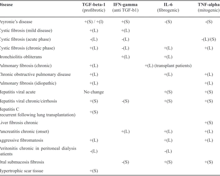
Table 3 – Local and systemic cytokine alterations in various ibrosis-associated diseases.
Disease TGF-beta-1
(proibrotic) (anti TGF-b1)IFN-gamma (ibrogenic)IL-6 TNF-alpha(mitogenic)
Peyronie’s disease +(S) / +(l) +(S) -(S) -(S) Cystic ibrosis (mild disease) +(L) +(L)
Cystic ibrosis (acute phase) -(L) -(L) -(L)/(S) Cystic ibrosis (chronic phase) +(L) -(L) +(L) +(L)
Bronchiolitis obliterans +(L) +(L)
Pulmonary ibrosis (chronic) +(L) +(L) (transplant patients)
Chronic obstructive pulmonary disease +(L) +(L) +(L)
Pulmonary ibrosis (idiopathic) +(L) +(L)
Hepatitis viral acute No change +(S) +(S)
Hepatitis viral chronic/cirrhosis +(S) -(S) +(S) +(S) Hepatitis C
(recurrent following lung transplantation) +(S)
Liver ibrosis chronic +(S)
Pancreatitis chronic (onset) +(L) +(L) +(L)
Aggressive ibromatosis +(L) +(L) +(L)
Peritonitis chronic in peritoneal dialysis
patients -(L) -(L)
Oral submucosis ibrosis -(S) +(S) +(S)
Hypertrophic scar tissue +(S)
the composition of connective tissue. For example, patients who had peritonitis during continuous ambu
-latory peritoneal dialysis showed increased levels of TGF-β1 in the peritoneal dialysate efluent in relation to non-infectious patients, which has been considered as an active release of these proinlammatory cyto
-kines (15).
Idiopathic pulmonary ibrosis, the adult respi
-ratory distress syndrome, and the focal tumor stroma in lung cancer have also revealed a local increase of TGF-β1 (9). Serum concentrations of TNF-α and TGF-β1 have shown to be signiicantly increased in the majority of patients with chronic viral hepatitis and liver cirrhosis (8). In severely wounded patients elevated serum levels of TGF-β1 have been reported. These authors concluded that this could be due to secretion of wound ibroblasts (16). In the develop
-ment of renal ibrosis, TGF-β1 is considered to play
a key role as regards cytokines and growth factors
(10). In contrast, in an animal model the progression of chronic liver ibrosis could have been prevented by inhibition of TGF-β1 (17). In a major series con
-ducted by Hauck and co-workers it was shown that genetic alterations of the TGF-β1 gene could inlu
-ence the predisposition to PD (18). In the rat penis of a PD animal model (19), as well as in TA of PD patients, local TGF-β1-expression was reported to be 5-fold higher than in healthy subjects. Injections of TGF-β1 or the very similar cytomodulin in TA of rat penis produced chronic cellular iniltration, elas
-tosis, thickening, and clumping of collagen bundles. These PD-like histological alterations resulted in a signiicant reduction of erectile function (20,21). Incision and suture repair of the rat penis revealed inlammatory reactions similar to those observed in an acute phase of PD, in particular a transient up-regulation of TGF-β1 protein expression. Thus, TGF-β1 has been clearly shown to be an initiation factor of wound healing as well as ibrosis. Possibly, the transformation of the inactive TGF-β1 molecule into an active one is already induced even by minimal tissue lesions with consecutive cellular reparative and ibrotic mechanisms. This fact supports the theory of a traumatic genesis of PD. Based on these indings TGF-β1 may be considered to be one of the
central agents in the predisposition and manifestation
of PD.
Additionally, other plaque-inducing sub
-stances like ibrin (22) might be involved. The signii
-cantly higher TGF-β1 levels in serum of PD patients revealed major systemic factors of well known local alterations, particularly as none of the patients showed further signs of ibrotic alterations, but revealed the “typical” pro-ibrotic cytokine constellation in serum. Our results signiicantly demonstrated a critical role for TGF-β1 in the formation of PD and suggest that anti-TGF-β1 intervention might have a therapeutic effect on ibrotic penile tissue, not only by suppress
-ing ibrosis but also by facilitat-ing regeneration of TA
cells.
IL-6 is a multifunctional cytokine and acts
as the main synthesis mediator for many acute phase
proteins involved in proinlammatoric and cytotoxic conditions. Acute phase proteins may also contribute to the regulation of ibrosis by inhibition of proteases and by binding of cytokines. IL-6 serum levels were increased in the acute inlammation phase of viral hepatitis (23) and in the airways of patients with chronic ibrosis (CF) (24,25). Tissue mRNA expres
-sion of IL-6 in patients not only with acute but also with chronic viral hepatitis, was slightly elevated (8,23). In a rat model for chronic pancreatitis IL-6 tissue expression was able to be established (11). IL-6 gene polymorphism led to an increased number of chronic ibrotic lung diseases in transplanted patients (26). In addition, patients with peritoneal dialysis related chronic peritonitis exhibited increased levels of IL-6 in the peritoneal dialysate efluent (15). In contrast, it has been demonstrated in our previously reported investigation that the acute-phase proteins alpha-1-antitrypsin and alpha-2-macroglobulin were not detectable in serum of patients with PD (14). In this study, IL-6 could not be demonstrated in serum of PD patients. Therefore, acute immunological defense
mechanisms do not appear to play any particular role
in PD. This has been conirmed by the inding that other systemic indicators of acute inlammation like CRP (C-related protein) were only slightly increased in serum of PD patients (14).
TNF-α influences the production of col
the damage and the repair process by regulating additional mediators. Immunological processes or initial perivasculitis might explain the involvement of TNF-α in the etiology of PD. In animals with artiicial cochlear inlammation TNF-α induced an ampliication of the immune response leading to an increase of inlammation and disease progression (28). In patients with acute exacerbation of cystic ibrosis, the secretion of TNF-α was signiicantly lower in
comparison with healthy controls and returned to
normal secretion after treatment (24,25,30). In the airways of CF patients signiicantly elevated levels of circulating proinlammatory cytokines, in particular TNF-α have been reported (24). Other pulmonary diseases harboring diffuse ibrotic alterations like id
-iopathic pulmonary ibrosis, adult respiratory distress syndrome, lung cancer and chronic pancreatitis have revealed elevated TNF-α levels (9,11). In serum of patients with severe chronic hepatitis and liver cir
-rhosis signiicantly higher TNF-α levels were found compared to healthy controls, whereas patients with mild chronic hepatitis did not exhibit such alterations. In our study, serum TNF-α levels were signiicantly decreased. This inding does not indicate a systemic involvement of TNF-α in etiopathogenesis of PD. In general, TNF-α seems to be involved in acute and highly active phases of ibrotic diseases. The major -ity of patients included in our study had a duration
of PD longer than 12 months. The decreased TNF-α levels found in our study therefore may possibly be an indicator for the chronic phase of PD in our patients (stable disease). Due to these low TNF-α-levels, it could be concluded that an anti-ibrotic therapy of PD based on TNF-α does not appear to be promising.
IFN-γ can reduce collagen synthesis, pro
-liferation of fibroblasts with subsequent fibrotic activity and increase collagenase production. In the airway epithelium of CF patients increased levels of IFN-γ mRNA should be veriied (30). The mRNA of both IFN-γ and IL-6 mRNA were elevated for liver
specimens of a transgenic mouse model for chronic
hepatitis C-virus liver disease. In spontaneous chronic pancreatitis in a rat model, IFN-γ was related to the progression of chronic pancreatitis (11). These ind
-ings are in agreement with the signiicantly increased IFN-γ serum levels detected in our series of PD pa -tients.
Anti-TGF-β1 agents have been considered to be a therapeutic option in various ibrotic diseases (31,32). The basis of that assumption is a down regu
-lation of the ibrotic protein expression induced by IFN-γ. On the one hand, our results may explain that therapeutically administered local IFN-γ was seen to be ineffective as PD plaques barely responded to local interferon therapy. On the other hand, elevated IFN-γ levels in PD patients might be a type of physiological defense mechanism as in antiibrotic activities.
CONCLUSION
No straightforward description of the patho -physiological process that leads to the formation of
PD is currently known. To our knowledge, the present study is the irst reported series with special emphasis on ibrogenic factors in patient’s serum.
Cytokines, in particular TGF-β1 and IFN-γ play a local and systemic role in the formation of PD. Local or systemic cytokine patterns might be crucial in different stages of PD. Micro traumata in the tunica albuginea could be the triggering incident.
TGF-β1 may be a target for an antiibrotic PD therapy as well as the irst marker for PD therapy response. TGF-β1 serum levels should be integrated
in future therapy studies. The role of cytokines in
PD, in particular TGF-β1, should be followed-up in clinical as well as in investigative trials.
ACKNOWLEDGEMENT
Drs. Reinhold P. Zimmermann and Gerhard Feil have both contributed equally to this work.
CONFLICT OF INTEREST
The authors have nothing to disclose.
REFERENCES
1. Nöske HD, Hauck EW: Morbus Lapeyronie aus histo
2. Smith BH: Peyronie’s disease. Am J Clin Pathol. 1966; 45: 670-8.
3. Smith BH: Subclinical Peyronie’s disease. Am J Clin Pathol. 1969; 52: 385-90.
4. Devine CJ Jr, Somers KD, Jordan SG, Schlossberg SM: Proposal: trauma as the cause of the Peyronie’s lesion. J Urol. 1997; 157: 285-90.
5. Leffell MS: Is there an immunogenetic basis for Peyronie’s disease? J Urol. 1997; 157: 295-7.
6. Bichler KH, Lahme S, Mattauch W, Petri E: Collagen metabolism in induratio penis plastica (IPP). Urologe A. 1998; 37: 306-11.
7. Somers KD, Sismour EN, Wright GL Jr, Devine CJ Jr, Gilbert DA, Horton CE: Isolation and characterization of collagen in Peyronie’s disease. J Urol. 1989; 141: 629-31.
8. Mammaev SN, Lukina EA, Shul’pekova IuO, Levina AA, Ivashkin VT: Cytokine regulation of liver inlam
-mation and ibrosis during chronic hepatic diseases. Klin Lab Diagn. 2001; 12: 37-40.
9. Martinet Y, Menard O, Vaillant P, Vignaud P, Vignaut JM, Martinet N: Cytokines in human lung ibrosis. Arch Toxicol Suppl. 1996; 18: 127-39.
10. Eddy AA: Molecular basis of renal ibrosis. Pediatr Nephrol. 2000; 15: 290-301.
11. Xie MJ, Motoo Y, Su SB, Sawabu N: Expression of tumor necrosis factor-alpha, interleukin-6, and inter -feron-gamma in spontaneous chronic pancreatitis in
the WBN/Kob rat. Pancreas. 2001; 22: 400-8. 12. Haque MF, Meghji S, Khitab U, Harris M: Oral
submucous ibrosis patients have altered levels of cytokine production. J Oral Pathol Med. 2000; 29: 123-8.
13. Mills BG, Frausto A, Brien E: Cytokines associated with the pathophysiology of aggressive ibromatosis. J Orthop Res. 2000; 18: 655-62.
14. Feil G, Bichler KH, Lahme S, Götz T: Investigations on cytokines in serum of patients with Peyronie’s disease. Akt Urol. 2001; 32 (Suppl 1): 33-6.
15. Lai KN, Lai KB, Lam CW, Chan TM, Li FK, Leung JC: Changes of cytokine proiles during peritonitis in patients on continuous ambulatory peritoneal dialysis. Am J Kidney Dis. 2000; 35: 644-52.
16. Tredget EE, Wang R, Shen Q, Scott PG, Ghahary A: Transforming growth factor-beta mRNA and protein in hypertrophic scar tissues and ibroblasts: antagonism by IFN-alpha and IFN-gamma in vitro and in vivo. J Interferon Cytokine Res. 2000; 20: 143-51.
17. Nakamura T, Sakata R, Ueno T, Sata M, Ueno H: In
-hibition of transforming growth factor beta prevents progression of liver ibrosis and enhances hepatocyte
regeneration in dimethylnitrosamine-treated rats.
Hepatology. 2000; 32: 247-55.
18. Hauck EW, Hauptmann A, Schmelz HU, Bein G, Wei
-dner W, Hackstein H: Prospective analysis of single
nucleotide polymorphisms of the transforming growth
factor beta-1 gene in Peyronie’s disease. J Urol. 2003; 169: 369-72.
19. Lin CS, Lin G, Wang Z, Maddah SA, Lue TF: Up
-regulation of monocyte chemoattractant protein 1 and effects of transforming growth factor-beta 1 in Peyronie’s disease.Biochem Biophys Res Commun. 2002; 295: 1014-9.
20. El-Sakka AI, Hassoba HM, Chui RM, Bhatnagar RS, Dahiya R, Lue TF: An animal model of Peyronie’s-like
condition associated with an increase of transforming
growth factor beta mRNA and protein expression. J Urol. 1997; 158: 2284-90.
21. Bivalacqua TJ, Diner EK, Novak TE, Vohra Y, Sikka SC, Champion HC, et al.: A rat model of Peyronie’s disease associated with a decrease in erectile activity and an increase in inducible nitric oxide synthase protein expression. J Urol. 2000; 163: 1992-8. 22. Davila HH, Ferrini MG, Rajfer J, Gonzalez-Cadavid
NF: Fibrin as an inducer of ibrosis in the tunica al
-buginea of the rat: a new animal model of Peyronie’s disease. BJU Int. 2003; 91: 830-8.
23. Missale G, Ferrari C, Fiaccadori F: Cytokine media
-tors in acute inlammation and chronic course of viral hepatitis. Ann Ital Med Int. 1995; 10: 14-8.
24. Bonfield TL, Konstan MW, Berger M: Altered
respiratory epithelial cell cytokine production in
cystic fibrosis. J Allergy Clin Immunol. 1999; 104: 72-8.
25. Osika E, Cavaillon JM, Chadelat K, Boule M, Fitting C, Tournier G, et al.: Distinct sputum cytokine proiles in cystic ibrosis and other chronic inlammatory air
-way disease. Eur Respir J. 1999; 14: 339-46.
26. Lu KC, Jaramillo A, Lecha RL, Schuessler RB, Aloush A, Trulock EP, et al.: Interleukin-6 and interferon-gamma gene polymorphisms in the development of bronchiolitis obliterans syndrome after lung transplan
-tation. Transplan-tation. 2002; 74: 1297-302.
27. Elborn JS, Norman D, Delamere FM, Shale DJ: In vitro tumor necrosis factor-alpha secretion by monocytes from patients with cystic ibrosis. Am J Respir Cell Mol Biol. 1992; 6: 207-11.
28. Satoh H, Firestein GS, Billings PB, Harris JP, Keithley EM: Tumor necrosis factor-alpha, an initiator, and etanercept, an inhibitor of cochlear inlammation. Laryngoscope. 2002; 112: 1627-34. Erratum in: La
EDITORIAL COMMENT
Despite the prevalence of Peyronie’s disease is increasing fast, until now we do not know the exact
pathophysiological mechanism of this disease. The
main hypothesis is an inlammatory reaction followed by ibroblasts proliferation and scar tissue formation. However, even this mechanism was not proven yet. So far, how and why ibrosis takes place is not clear. Anyway, cytokines and in particular TGF-beta seem to play a crucial role in Peyronie’s disease. If it is so, any information about these very mediators is very welcome. Furthermore, until we do understand why and how ibrosis and Peyronie’s disease occur we will
not really treat the disease itself.
In our opinion, it is time to search the patho
-physiological process that causes Peyronie’s disease, in the same that the authors did in this valuable article. In the era that aggressive surgeries to man
-age Peyronie’s disease are being reviewed, probably understanding inlammatory reaction and ibrogenic factors are way to go. In this way, I really believe that cytokines, mainly TGF-beta are the keys to un
-derstand what really happens in the tunica albuginea due to Peyronie’s disease.
Only after knowing this mechanism, we would treat and cure the disease, instead of manage
its complications with poor results.
Dr. Joaquim A. Claro
Division of Urology University of Sao Paulo, USP Sao Paulo, Brazil E-mail: [email protected] 29. Wang X, Chen YX, Xu CF, Zhao GN, Huang YX,Wang QL: Relationship between tumor necrosis factor-alphaand liver ibrosis. World J Gastroenterol. 1998; 4: 18.
30. Wojnarowski C, Frischer T, Hofbauer E, Grabner C, Mosgoeller W, Eichler I, et al.: Cytokine expression in bronchial biopsies of cystic ibrosis patients with and without acute exacerbation. Eur Respir J. 1999; 14: 1136-44.
31. Oldroyd SD, Thomas GL, Gabbiani G, El Nahas AM: Interferon-gamma inhibits experimental renal ibrosis. Kidney Int. 1999; 56: 2116-27.
32. Takahara T, Sugiyama K, Zhang LP, Ando O, Fujii M, Yata Y, et al.: Cotreatment with interferon-alpha and -gamma reduces liver ibrosis in a rat model. Hepatol Res. 2004; 28: 146-154.
Accepted after revision: May 13, 2008
Correspondence address:
Dr. Gerhard Feil Department of Urology
Eberhard-Karls-University of Tuebingen Hoppe-Seyler-Str. 3
72076 Tuebingen, Germany