Spontaneous isolated dissection of the superior
mesenteric artery – Which is the best therapeutic choice?
Dissecção espontânea isolada da artéria mesentérica
superior – Qual a melhor abordagem terapêutica?
Ricardo de Alvarenga Yoshida1, Winston Bonetti Yoshida2, Ralf Kolvenbach3,
Paulo Roberto Bahdur Vieira4, Rodolfo Lellis Zuppardo4,Osvaldo Lunardi5
C H A L L E N G E
1 Angiovalle, São José dos Campos, SP, Universidade Estadual Paulista – UNESP, Faculdade de Medicina de Botucatu – FMB, Departamento de Cirurgia e Ortopedia,
Botucatu, SP, Brazil.
2 Universidade Estadual Paulista – UNESP, Faculdade de Medicina de Botucatu – FMB, Botucatu, SP, Brazil. 3 Hospital Augusta, Dusseldorf, Germany.
4 Angiovalle, São José dos Campos, SP, Brazil. 5 Hospital de Clínicas Antônio Alfonso, Jacareí, SP, Brazil.
Financial support: None.
Conlicts of interest: No conlicts of interest declared concerning the publication of this article. Submitted: 02.04.12. Accepted: 07.11.12.
he study was carried out at Hospital de Clínicas Antônio Alfonso, Jacareí, SP.
INTRODUCTION
Spontaneous isolated dissection of superior
mesenteric artery (SIDSMA) is a rare and dificult to
diagnose cause of acute abdominal pain1. A previous
study analyzing 6,666 autopsies found an incidence
of 0.06% for this condition. Notwithstanding,
advances in imaging technology in general and multichannel computed tomography (AngioCT) in particular have allowed to improve the diagnostic
accuracy ofSIDSMA2-4.
A variety of treatment options have been described for SIDSMA, including conservative treatment, anticoagulation therapy, endovascular repair with stent placement, and open surgical repair.3
The objective of this article is to discuss the best treatment approach in cases of SIDSMA, based on literature review and a case description.
PART I - CLINICAL SITUATION
A 45-year-old male patient with no comorbidities presented at the emergency room complaining of severe acute abdominal pain, resistant to common analgesic and antispasmodic drugs. Physical
examination revealed a laccid abdomen, hydro-aerial
sounds, no palpable masses or visceromegalies, and
absence of any other inlammatory signs.
The patient was clinically treated with fasting and hydration and was subjected to laboratory tests (blood cell count, urea, creatinine, TGO, TGP, FA, GGT, amylase, glucose), all normal. Subsequently, AngioCT images were obtained and showed isolated
dissection of the superior mesenteric artery (SMA), starting at 1.7 cm from its origin and extending for approximately 6.5 cm, with slight local dilation to a maximum caliber of 1.4 cm, a discreetly reduced true lumen in medium and distal portions, and blood
low present in both lumens, reentering in the distal
portion (Figure 1).
After 5 hours of fasting and hydration, full remission of symptoms was observed.
PART II - WHAT WAS DONE?
Some treatment options were available for this case:
• Conservative treatment, with clinical observation and support measures; in the case of nonresponse, hospital admission would be indicated;
• Anticoagulant treatment, with clinical observation and support measures; in the case of nonresponse, hospital admission and maintenance anticoagulant therapy;
• Endovascular treatment, repairing the dissection with stent placement;
• Surgical treatment, with open repair of the SMA. Due to remission of symptoms and AngioCT
indings suggestive of an uncomplicated dissection
(according to criteria discussed below), the therapeutic option was immediate anticoagulation therapy with low-molecular-weight heparin.
A new AngioCT performed 3 days after the event evidenced maintenance of dissection, without any change in extent when compared with previous AngioCT. Therefore, the patient was discharged and advised to maintain oral anticoagulant therapy.
After 3 months (latest follow-up), a new control AngioCT scan did not reveal any abnormality in relation to previous exams that might suggest worsening. The patient remains asymptomatic under oral anticoagulant therapy. He will be submitted to new CT exams after 6 and 12 months, and yearly thereafter.
DISCUSSION
The etiology of SIDSMA has not been completely understood. Even though atherosclerosis,
fibromuscular dysplasia, cystic medial necrosis, and conditions affecting elastic tissues (Marfan and Ehlers-Danlos syndromes) may be possible causes of SIDSMA, usually they are not mentioned in the
cases described in the literature3. The presence
of uncontrolled hypertension, observed in about 30% of the cases, may be an additional risk factor
for the occurrence of this condition4. Hemodynamic
abnormalities caused by the curvature at the origin of the SMA could be another etiologic possibility, according to Park et al.5.
The natural history of the disease is unclear and varies among cases. Notwithstanding, the following events may occur: 1) limited progression of the dissection, with false lumen thrombosis; 2) progressive dissection towards distal branches of
the SMA; 3) artery rupture through the adventitia; 4) rapid expansion of the false lumen, causing narrowing or obliteration of the true lumen and potentially leading to ischemia and intestinal necrosis; 5) aneurysmatic degeneration caused by the dissection or dissection progression with hemorrhage due to rupture of the SMA3,4.
Some authors2-4,6 advocate four treatment options
for this condition: 1) clinical observation; 2) conservative treatment with or without anticoagulant therapy; 3) endovascular repair; and 4) open surgery.
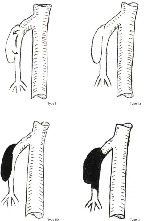
In an attempt to establish a relationship between type of dissection (based on AngioCT
and angiographic indings) and clinical evolution,
Sakamoto7 and Yun1 proposed a classiication based
on radiological indings (Figure 2). However, neither of the authors were able to ind a clear relationship between these indings and clinical course2.
At present, there is not a protocol defining indications for conservative or surgical treatment,
endovascular repair, or conventional treatment2.
Some algorithms have been proposed2,6 to assist in
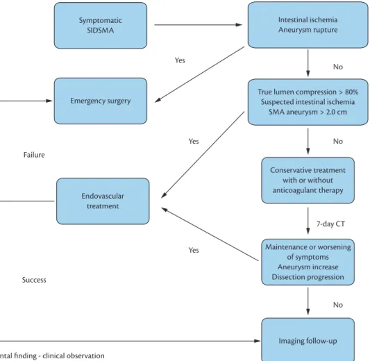
clinical decision-making. In the presence of intestinal necrosis, artery rupture, or any other complication, surgery is mandatory; otherwise, clinical observation with or without anticoagulant therapy seems to be the best therapeutic choice. Primary or secondary endovascular treatment should be reserved for cases with an unfavorable evolution, and conventional surgery to those in whom endovascular treatment fails6 (Figure 3).
Some authors have proposed that conservative treatment should be followed by anticoagulation
therapy2,3. This recommendation is reinforced by
experience in the treatment of spontaneous dissection of the carotid artery, where anticoagulation is effective in preventing thrombus formation2,3. Other
authors,4,5 however, claim that anticoagulation can
help prevent thrombosis and obliteration of the false lumen, but may increase the possibility of SMA rupture.
Patients treated in an acute phase with conservative therapy should be closely monitored and underwent tomographic evaluation weekly. In symptomatic cases showing no remission within 7 days, or showing progression of the dissection, surgical treatment should be considered3 (Figure 3).
Park et al.4 observed that most SIDSMA lesions
improved or remained stable over the 20-month
follow-up period according to AngioCT indings,
with clinical assessments showing patients free of symptoms after conservative treatment.
Figure 3. Algorithm proposed by the authors for the treatment of SIDSMA, based on the recommendations of Min et al.3 and
In endovascular repair, the aim of placing stents in the SMA is covering the intimal detachment and preventing blood flow through the false lumen, which could lead to obliteration of the false lumen by thrombosis. The stent should undergo minimal
shortening, present good lexibility, adequate radial
force, to ensure that it will not migrate as a result of the continuous displacement of the SMA3,8,9. The use
of self-expanding stents is recommended in these cases, as their radial force is enough to stand the
pressure of the false lumen, which is usually illed
with fresh thrombus, and also because these stents adequately accommodate to the diameter of the true lumen.3 Nevertheless, dificulties may be faced in
correctly locating entry and reentry fenestrations; in addition, not rarely does the dissection expand to
the distal portion of the SMA, causing dificulties
to endovascular repair2. There may also be a risk of
occlusion of the SMA with stent placement, as well as possible obstruction of side branches located along
to this treatment segment2. Although endovascular
treatment may be indicated even for patients with no mesenteric ischemia or peritonitis, long-term results should be evaluated.
In relation to conventional surgical treatment, several procedures have been described, including thrombectomy, endoaneurysmorraphy, intimectomy, patch angioplasty, and bypasses with vein or
prostheses1,6. Given the invasive nature of these
procedures, open surgery does not seem to be
justiied as a irst option, but should be performed in
patients who do not respond to endovascular repair. AngioCT scans should be regularly obtained, after both conservative and surgical treatment, after 1 and 6 months, and yearly thereafter3.
Considering there is no consensus regarding the best treatment approach in SIDSMA, in the present case the conservative treatment approach was chosen, as most authors agree in that only symptomatic patients should be surgically treated. Anticoagulant therapy was also adopted, at least initially, based on the fact it was a young and active patient, in whom intestinal ischemic complications could cause several adverse effects, including a negative impact on quality of life.
CONCLUSIONS
There is no consensus regarding the best treatment approach in cases of SIDSMA. Even though indications for surgery are still controversial, this option should be reserved for symptomatic patients, whereas endovascular repair should be the first choice. Nonsurgical approach requires rigorous
abdominal AngioCT follow-up, focusing on clinical signs of mesenteric ischemia and blood supply to
the SMA, including collateral low from the celiac
artery and inferior mesenteric artery. In the absence of intestinal necrosis or other complications with a clear indication for surgery, conservative treatment, with or without anticoagulation, seems to be the best choice.
REFERENCES
1. Yun WS, Kim YW, Park KB, et al. Clinical and angiographic follow-up of spontaneous isolated sfollow-uperior mesenteric artery dissection. Eur J Vasc Endovasc Surg. 2009;37:572-577. PMid:19208448. http:// dx.doi.org/10.1016/j.ejvs.2008.12.010
2. Katsura M, Mototake H, Takara H, Matsushima K. Management of spontaneous isolated dissection of the superior mesenteric artery: Case report and literature review. World J Emerg Surg. 2011;6:16. PMid:21549001 PMCid:3108290. http://dx.doi. org/10.1186/1749-7922-6-16
3. Min SI, Yoon KC, Min SK, et al. Current strategy for the treatment of symptomatic spontaneous isolated dissection of superior mesenteric artery. J Vasc Surg. 2011;54:461-466. PMid:21571493. http://dx.doi.org/10.1016/j.jvs.2011.03.001
4. Park YJ, Park KB, Kim DI, Do YS, Kim DK, Kim YW. Natural history of spontaneous isolated superior mesenteric artery dissection derived from follow-up after conservative treatment. J Vasc Surg. 2011;54:1727-1733. PMid:21944909. http://dx.doi. org/10.1016/j.jvs.2011.07.052
5. Park YJ, Park CW, Park KB, Roh YN, Kim DI, Kim YW. Inference from clinical and luid dynamic studies about underlying cause of spontaneous isolated superior mesenteric artery dissection. J Vasc Surg. 2011;53:80-86. PMid:20855179. http://dx.doi.org/10.1016/j. jvs.2010.07.055
6. Cho BS, Lee MS, Lee MK, et al. Treatment guidelines for isolated dissection of the superior mesenteric artery based on follow-up ct findings. Eur J Vasc Endovasc Surg. 2011;41:780-785. PMid:21333559. http://dx.doi.org/10.1016/j.ejvs.2010.12.022 7. Sakamoto I, Ogawa Y, Sueyoshi E, Fukui K, Murakami T,
Uetani M. Imaging appearances and management of isolated spontaneous dissection of the superior mesenteric artery. Eur J Radiol. 2007;64:103-110. PMid:17628380. http://dx.doi. org/10.1016/j.ejrad.2007.05.027
8. Nasser F, Affonso BB, Silva-Jesus SG, et al. Endovascular treatment for superior mesenteric artery pseudoaneurysm: Case report. J Vasc Bras. 2010;9:182-185. http://dx.doi.org/10.1590/ S1677-54492010000300016
9. Gobble RM, Brill ER, Rockman CB, et al. Endovascular treatment of spontaneous dissections of the superior mesenteric artery. J Vasc Surg. 2009;50:1326-1332. PMid:19782510. http://dx.doi. org/10.1016/j.jvs.2009.07.019
Correspondence
Ricardo de Alvarenga Yoshida FMB-UNESP Av. Bento Lopes, 679 – Rubião Junior CEP 18618-970 – Botucatu (SP), Brazil Fone: (14) 3811-6269 E-mail: [email protected]
Author information
Faculdade de Medicina de Botucatu (FMB), Universidade Estadual Paulista (UNESP); PhD, Department of Surgery and Orthopedics, Faculdade de Medicina de Botucatu (FMB), Universidade Estadual Paulista (UNESP). WBY is full professor and chief of the Service of Vascular and Endovascular Surgery at Faculdade de Medicina de Botucatu (FMB), Universidade Estadual Paulista (UNESP). RK is full professor and chief of the Service of Vascular and Endovascular Surgery at Hospital Augusta PRBV and RLZ are vascular and endovascular surgeons at Angiovalle. OL is a gastric surgeon at Hospital de Clínicas Antônio Alfonso.
Author contributions