Pseudoaneurysm of common carotid due to trauma:
treatment option for open sky surgery
Pseudoaneurisma de carótida comum secundário a trauma contuso: opção de tratamento
por cirurgia a céu aberto
Edson Pedroza dos Santos Junior1, Rodolfo Rogers Américo Machado Batista1, Maykon Brescancin de Oliveira2, Remy Faria
Alves2, Ricardo Russi Blois2
Introduction
Cervicothoracic vascular trauma represents a challenge for surgeons, irrespective of the latest advances in diagnos-tic and surgical techniques. Lesions from penetrating or blunt vascular trauma have low prevalence, but morbidity and mortality rates remain high1.
Carotid artery rupture caused by blunt trauma ac-counts for 3 to 10% of all carotid lesions2. The incidence
of carotid artery lesions due to blunt trauma has been reported to vary from 0.08 to 0.33%, and up to half the patients do not present signs of cervical trauma or neurologic deficit. The known mechanism of lesions are: cervical hyperextension–rotation (most common);
direct trauma to the neck; intraoral trauma and basilar skull fracture1.
Cervical trauma is the leading cause of carotid artery pseudoanerysms (PA)3. he mechanism of injury is
rup-ture of the arterial wall, leading to local hemorrhage that is contained by adjacent structures, forming a pulsatile he-matoma, or pseudoaneurysmal sac. Clinically, they present as: a pulsatile mass on the neck, symptoms of compression of adjacent structures, bleeding or neurologic deicits1,2.
Traditionally, the treatment of choice is open surgical re-pair3. Nevertheless, endovascular techniques, such as
stent-ing, endogratstent-ing, and balloon angioplasty have been increas-ingly used to repair the carotid lesions, making it possible to treat those lesions with a lesser risk of complications3,4.
Abstract
Case report of a female patient, 44 years-old, victim of cervical trauma in a traic accident, who had painful cervical mass, associated with hoarseness and dysphagia three weeks after trauma. Additional tests identiied the pseudoaneurysm of common carotid artery in zone II. We opted for treatment through the open conventional surgery with excellent immediate result. Control examination was performed seven months after surgery, and the results conirmed the therapeutic success.
Keywords: pseudoaneurysm;extracranial carotid; surgery.
Resumo
Relato de caso de paciente feminina, com 44 anos de idade, vítima de trauma cervical em acidente de trânsito, que apresentou massa cervical dolorosa, rouquidão e disfagia associados, três semanas após o trauma. Exames complementares identiicaram pseudoaneurisma de carótida comum em zona II. Optou-se pelo tratamento por meio de cirurgia convencional a céu aberto com excelente resultado imediato. Foi realizado um exame de controle após sete meses do procedimento cirúrgico, e os resultados conirmaram o sucesso terapêutico.
Palavras-chave: pseudoaneurisma;carótida extracraniana; cirurgia.
Study carried out at the Service of Vascular and Haemodynamic Surgery Hospital e Maternidade Dom Orione – Araguaína (TO), Brazil.
1 Physicians of Instituto Tocantinense Presidente Antonio Carlos – Araguaína (TO), Brazil.
2 Physician; Vascular surgeon; Professor of Clinical Surgery at Instituto Tocantinense Presidente Antonio Carlos – Araguaína (TO), Brazil. Conlict of interests: nothing to declare.
Case report
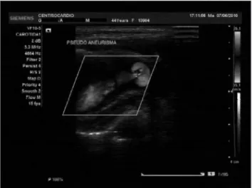
A 44 year-old female patient, victim of motorcycle accident, was admitted with blunt craniocervical trau-ma, without loss of consciousness or neurologic deficit. Three weeks after conservative treatment for her cran-ioencephalic trauma, she was seen in the Outpatient Clinic. She was classified as 15 in the Glasgow scale, had no impairment of cranial nerves and complained of a painful tumor in the left lateral aspect of the neck, with hoarseness and dysphagia. Doppler ultrasonogra-phy showed a pseudoaneurysm with turbulent flow out-side the left common carotid artery, medially and below the carotid bulb (Figure 1 to 2). Digital angiography was
Figure 1. Collection at the distal portion of the common carotid artery
measuring 25 x 30 mm with low, and artery lateralization.
Figure 2. Communication of the carotid low with the aneurysmal sac.
Figure 3. Carotid pseudoaneurysm and arterial lateralization.
Figure 4. Intraoperative arterial control and identiication of the
aneu-rysmal sac.
then performed to confirm the diagnosis, for surgical planning, and to exclude associated lesions. It confirmed a carotid pseudoaneurysm, with lateral displacement of the distal common carotid artery (Figure 3).
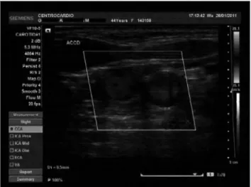
The patient was discharged from hospital on the third postoperative day in good condition, with no neu-rologic deficits or signs of local bleeding. An antiplate-let agent was prescribed. The patient was seen at the outpatient clinics 15 days after the operation and pre-sented no complications. At the follow-up visit seven months after the operation, Doppler ultrasonography was normal (Figures 5 to 6) and clinical examination did not show any sequelae.
Discussion
Patients with extracranial carotid pseudoanerysms present variable clinical pictures: from a palpable and
Figure 5. Follow-up Doppler ultrasonography seven months after
procedure.
Figure 6. Doppler ultrasonography: common carotid artery near the
bulb without lesions seven months later.
pulsatile cervical mass to strokes with definitive neu-rological deficits5. Ischemic symptoms are caused
by impaired carotid artery flow or by distal embo-lism3. Dysphagia and respiratory distress can also be
observed1,5, as in the case reported.
The diagnosis of carotid pseudoaneurysm is impor-tant because of the drastic consequences of its rupture or distal embolization. There is also the possibility of mistaking its clinical picture for a tonsillar abscess or arteriovenous fistula, whose managements are quite different. The differential diagnosis must include ca-rotid body tumors, tortuosity and kinking of the sub-clavian and carotid arteries, lymphadenomegaly or ar-terial tumors6.
The imaging exam of choice for diagnosis of carotid pseudoanerysm is digital arteriography, that is recom-mended when the clinical presentation or Doppler ul-trasonography suggest this entity. CT angiography has been described as a good choice for the diagnosis of pseudoaneurysm, with sensitivity of 100% and speci-ficity of 90%. It is a fast, non-invasive, and low-cost method that has high-spatial-resolution and is not op-erator-dependent. It can be performed in critically ill patients, such as those with multiple trauma with vas-cular involvement5,7.
Conventional angiography is the classic method for diagnosis of pseudoaneurysms8. However, some authors
question this statement, and state that conventional an-giography may fail and is not indispensable, for it is in-vasive and has the limitation of not evaluating bones and adjacent tissues7.
Historically, the treatment of pseudoaneurysm has been quite discussed. Some studies have described open repair as the treatment of choice for traumatic carotid pseudoaneurysms, especially extracranial ones, because they present low rates of spontaneous resolution, when treated medically7,9. Extracranial pseudoaneurysms,
when are small, inaccessible or located distally to the origins of brain vessels, may be treated with antico-agulation and medical follow-up. Surgical procedures recommended for arterial pseudoaneurysms include arterial wall repair using venous patch in most cases, and also end-to-side anastomosis, tangential repair, en-dovascular stenting and extra-anatomic bypass10.
time, compared to conventional open repair1,3,6,11.
Stenting excludes the pseudoaneurysm permanently from the circulation. However, this technique is not free of complications: it may cause thrombosis and low local blood flow which favors platelet aggregation and microembolic events, stenosis1,9,12 and some other
prob-lems, such as the need for anticoagulation for life, stent deformities and kinking, and neointimal hyperplasia6.
The conventional surgical treatment of penetrating arterial lesions at the base of the skull remains the standard approach. This technique, however, is extremely chal-lenging13. Sencer et al. reported cases of patients with
in-ternal carotid pseudoaneurysm, associated with carotid-cavernous fistula, who presented with massive epistaxis and ophthalmic signs and symptoms. Endovascular em-bolization was successful14. Transarterial embolization
with detachable balloons has been accepted as treatment of choice for carotid-cavernous fistula. This technique, however, results in a pseudoaneurysm formation rate of 30 to 40%15. New generations of stents could make
en-dovascular therapy the treatment of choice for such dif-ficult lesions in the long-term16,17.
Injury or compression of adjacent structures, as seen in the present case, plus the location near the ca-rotid bifurcation, led us to choose open surgical repair. Placement of a stent at the orifice of the pseudoaneu-rysm, which was close to origin of the internal carotid artery, could have caused impairment of cerebral blood low. Preservation of carotid perfusion was the desirable goal in this case.
Conclusion
Trauma to the neck may result in blood vessel in-jury and rarely, to arterial pseudoaneurysm formation. However, this event must be promptly diagnosed and treated due to its high risk of morbidity and mortality.
The dearth of clinical studies in the literature com-paring conventional and endovascular treatment of pseudoaneurysms make it difficult to compare those two methods.
In the case reported, the choice of conventional open repair was based on the pseudoaneurysm features, and a good immediate result was achieved, as normal flow in the vessel was shown at Doppler ultrasonogra-phy. As the patient presented no neurologic deficits, the choice of treatment was considered adequate.
References
1. Rutherford. Cirurgia Vascular. Lesões das Artérias Carótidas e Vertebrais. Cap. 70. Rio de Janeiro: Dilivros; 2007. p.1006-16. 6ed.
2. Sá PM, Machado MC, Silva DA, Brito LL, El Hassan S. Epistaxe tardia secundária a pseudoaneurisma intracavernoso de caróti-da interna. Rev Bras Otorrinolaringol. 2003;69(5):715-8.
3. Oliveira AF, Kajita D, Garzon RGA, Centola APC, Bosnardo CAP, Neto MF. Tratamento endovascular de pseudo-aneurisma de carótida interna em criança. J Vasc Br. 2006;5(1):67-70.
4. Zanini MA, Tahara A, dos Santos GS, de Freitas CCM, Jory M, Caldas JGMP, et al. Pseudoaneurysm of the Internal Carotid Artery Presenting with Massive (recurrent) Epistaxes. Arq Neuropsiquiatr. 2008;66(2-A):268-71.
5. Väärämämäki S, Pimimenoff G, Heikkinen M, Suominen V, Saarinen J, Zeitlin R, et al. Ten-year outcomes after endo-vascular aneurysm repair (EVAR) and magnitude of additional procedures. Scand J Surg. 2007;96(3):221-8.
6. Bredt CFG, França LHG, Back LA, Machado EL, Jacobovicz C, Júnior HJS. Aneurisma Gigante de Carótida Interna: Tratamento Cirúrgico, Hospital de Clínicas da UFPR. J Vasc Surg. 2003.
7. Romanus AB, Mazer S, Carvalho AN, Liu CB, De Toni FS, Jacob GVV, et al. Pseudo-aneurismas – relato de dois casos e revisão da literatura. Radiol Bras. 2002;35(5):303-6.
8. Bush RL, Lin PH, Dodson TF, Dion JE, Lumsden AB. Endoluminal stent placement and coil embolization for the management of carotid artery pseudoaneurysms. J Endovasc Ther. 2001;8:53-61.
9. Kasthoori JJ, Nawawi O. Endoluminal Stent Placement in the Management of Recurrent Carotid Blowout Syndrome. J HK Coll Radiol. 2007;10:62-5.
10. Coldwell DM, Novak Z, Ryu RK, Brega KE, Biffl WL, Offner PJ, et al. Treatment of posttraumatic internal carotid arterial pseudo-aneurysms with endovascular stents. J Trauma. 2000;48:470-2.
11. Yi AC, Palmer E, Luh GY, Jacobson JP, Smith DC. Endovascular treatment of Carotid and Vertebral Pseudoaneurysms with Covered Stents. Am J Neuroradiol. 2008;29:983-7.
12. Zhang YS, Yang XJ, Wang SZ, Qiao AK, Chen JL, Zhang KY, et al. Hemodynamic effects of stenting on wide-necked intracranial aneurysms. Chin Med J (Engl). 2010;123(15):1999-2003.
13. Garcia MRT, Chammas MC, Caiado AHM, Juliano AG, Leite CC, Cerri GG. Aneurisma da artéria carótida interna extracraniana: Relato de caso. Radiol Bras. 2004;37(4):295-7.
14. Sencer S, Minareci O, Poyanli A. Posttraumatic Damage To The Internal Carotid Artery. Pseudo aneurysm Presenting With Epistaxis And Direct Carotid Cavernous Fistula. Turkish Neurosurgenj. 2002;12:128-34.
16. Mcneil CJD, Chiou MAC, Gunlock MMG, Grayson CDE, Soares MG, Hagino MRT. Successful endovascular therapy of a penetrat-ing zone III internal carotid injury. J Vasc Surg. 2002;36:187-90.
17. Baum AR, Stavropoulos SW, Fairman RM, Carpenter JP. Endoleaks after Endovascular Repair of Abdominal Aortic Aneurysms. JVasc Interv Radiol. 2003;14:1111-7..
Correspondence
Edson Pedroza dos Santos Junior Rua Alfredo Nascer, 828, Centro CEP 77600-000 – Paraíso do Tocantins (TO), Brasil
Author’s contribution