Endotelium in Turner syndrome with capillaroscopy
Endotélio na síndrome de Turner com capilaroscopia
Simone Cristina da Silva Coelho1, Marília Martins Guimarães2, Terezinha Jesus Fernandes3
Abstract
Objective: he aim of this study was to assess the endothelium function in patients with Turner syndrome using videocapillaroscopy and to compare the results with healthy control.
Methods: Subjects and controls were studied in a temperature-controlled room, 20 days after no nailfold manipulations. he capillaries were visualized by a microscope connected to a television and a computer. he test of post-occlusive reactive hyperemia was performed using a sphygmomanometer attached to the fourth left inger, 20mmHg above maximum arterial pressure during 1 minute, and the following patterns were studied: area of transverse segment, maximal post-ischemia area and time to reach maximal post-ischemia area.
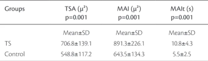
Results: he value of measure of transverse segment projected area , the maximal postischemia area of hand nailfold capillary loops using computerized videophotometry and the time to reach maximal post ischemia area were studied in 40 patients with Turner syndrome and 26 healthy women controls of comparable age (20±7.5 versus 18±8.1 years old; p=0.57). here were diferences between transverse segment area (706.8±139.1 versus 548.8±117.2; p=0.001). Maximal post-ischemia area (891.3±226.1 versus 643.5±134.3; p=0.001) and the time to reach it (10.8±4.3 versus5.5±2.5; p=0.001) were diferent between patients and controls.
Conclusions:Changes of capillary response to ischemia could be observed in patients with Turner syndrome using videocapillaroscopy when they were compared to a healthy control group.
Keywords: Turner syndrome; microscopic angioscopy; endothelium.
Resumo
Objetivos:O objetivo deste estudo foi avaliar a função endotelial de pacientes com síndrome de Turner, utilizando a videocapilaroscopia e comparar os resultados com grupo controle saudável.
Métodos: Pacientes e controles foram estudados em sala com temperatura controlada, após 20 dias, sem a manipulação das cutículas. Os capilares foram visualizados por microscópio conectado a televisão e computador. O teste de hiperemia reativa pós-oclusiva foi realizado utilizando-se esigmomanômetro ixado no quarto quirodáctilo da mão esquerda, 20mmHg acima da pressão arterial máxima durante 1 minuto, e os seguintes parâmetros foram estudados: área do segmento transverso, área máxima pós-hiperemia e tempo para alcançar a área máxima pós-hiperemia. Resultados: O valor da área do segmento transverso, área máxima pós-hiperemia dos capilares da região da mão usando-se a videocapilaroscopia computadorizada e o tempo para alcançar a área pós-hiperemia foram estudadas em 40 pacientes com síndrome de Turner e 26 controles do sexo feminino pareados para idade (20±7,5 versus 18±8,1 anos; p=0,57). Houve diferenças nos grupos quanto à área do segmento transverso (706,8±139,1
versus 548,8±117,2; p=0,001). A área máxima pós-hiperemia (891,3±226,1 versus 643,5±134,3; p=0,001) e o tempo para alcançá-la (10,8±4,3 versus
5,5±2,5; p=0,001) foram signiicativamente diferentes entre pacientes e controles.
Conclusão:Houve alterações na resposta do capilar à isquemia em pacientes com síndrome de Turner utilizando-se videocapilaroscopia quando comparados ao grupo controle saudável.
Palavras-chave: Síndrome de Turner; angioscopia microscópica; endotélio.
Study carried out at Universidade Federal do Rio de Janeiro (UFRJ), Rio de Janeiro (RJ), Brazil.
1PhD Student of Endocrinology Service of the Universidade Federal do Rio de Janeiro (UFRJ), Rio de Janeiro (RJ), Brazil. 2Adjunct Professor of Endocrinology Service of the UFRJ, Rio de Janeiro (RJ), Brazil.
3Adjunct Professor of Angiology of the UFRJ, Rio de Janeiro (RJ), Brazil.
No conlicts of interest declared concerning the publication of this article. Received on: 04.12.10. Accepted on: 12.04.11
Introduction
Turner syndrome (TS) is characterized by total or par-tial loss of a sex chromosome1.
It occurs in approximately 1/2,500 live births with fe-minine phenotype2, which represents more than 1/500,000 women worldwide2. he two most common features, which afect over 90% of recognized patients, are short stature and premature ovarian failure. here are also a series of phe-notypical alterations that occur with variable frequency3. Morbidity is clearly increased and these patients demons-trate a greater prevalence of arterial hypertension4, central obesity, reduction of insulin sensitivity5, impaired glucose tolerance6 and diabetes mellitus7,8.
Cardiovascular complications in Turner syndrome are the most common cause of early mortality, with a life ex-pectancy that may be reduced by more than ten years9.
he dilatations of ascending aorta are oten described and may be an isolated factor, suggesting a vasculopathy speciic to the syndrome, probably related to extracardiac factors such as estrogen deiciency, diabetes, dyslipidemia and overweight10.
Endothelium disturbance is present in groups with in-creased risk for diabetes and insulin resistance11.
Human microcirculation can be studied in vivo,
using diferent methods, such as plethysmography and capillaroscopy12,13.
he aim of this study was to assess the endothelium function in these patients by means of videocapillaroscopy and to compare it to healthy control.
Methods
A cross-sectional study was designed with no diabetic TS patients, with diagnosis conirmed by karyotype and
26 healthy control in 2008. Efective use of vascular drugs, hepatopathy, renal failure, vascular disorders, diabetes and hand lesions were exclusion criteria. his study was appro-ved by Ethical Comitte of the institution where it was per-formed, under the number 085/06.
Videocapillaroscopy was performed in a temperatu-re controlled room (24-26°C), in the morning following a night fasting, 20 days after no nailfold manipulations. The patients and control subjects were comfortably se-ated in a chair with observed hand at heart level with forearm and hand bent at the elbow. All subjects had their arm blood pressure measures in this position using auscultatory method. Capillaries were visualized by mi-croscope Wild Leitz GLS100 connected to a television monitor and computer.
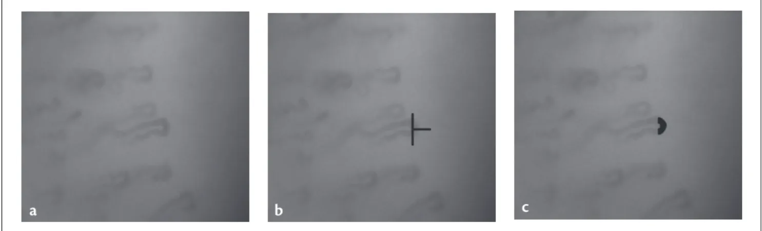
Post occlusive reactive hyperemia was performed using a sphygmomanometer attached to the fourth let hand inger, 20mmHg above maximum arterial pressure during 1 minu-te. Images were captured by a computer through Pinnacle 5.0 sotware, each 2 seconds, during 1 minute ater releasing pressure. Measures were determined through Studio2.0 sof-tware by at least two investigators blind to clinical data an in two diferent moments by each investigator to establish the concordance (k>0.7 in both). A perpendicular line tan-gent to internal limit of a capillary loop transverse segment deined the transverse segment area (TSA) to be measured (Figure 1). Zero time point ater ischemia was considered just ater releasing inger pressure. Maximum post ischemia area (MAI) and time to get the maximum post ischemia area (MAIt) was determined in TS and controls.
Statistical analysis
Data were expressed as mean±standard deviation (SD). he statistical analysis was obtained with Student t-test and
Figure 1 - Image with: a) capillary, b) transverse segment area (TSA) and c) maximum post-ischemia area.
Mann-Whitney test. A value of p<0.05 was considered sta-tistically signiicant.
Results
Forty patients with TS and 26 health controls were stu-died, aged between 8-38 years. he subjects were classiied in Groups 1 (TS) and 2 (control). Both groups had com-parable ages (20±7.5 versus 18±8.1 years old; p=0.57). he
karyotype of 23 (58%) patients was 45,X and of 17 (42%) ones were mosaic. Nine (23%) patients with TS were prepu-bertal and 30 (77%) were postpuberal, meanwhile 5 (19%) controls were prepubertal and 21 (81%) were postpuberal with no statistical diferences (p=0,79). Spontaneus puberty occurred in 5 (12%) patients with TS and all continued to present regular menses. Both groups had comparable body mass index (p=0.45).he diabetes familial history was si-milar between groups (p=0.45).
Table 1 shows that there were diferences in measures between TS and controls before thepost occlusive reactive hyperemia,and in both groups in MAI (891.3±226.1 versus
643.5±134.3; p=0.001) and the time to reach it (10.8±4.3
versus 5.5±2.5; p=0.001).
Discussion
Nailfold videocapillaroscopy is one of the best non-invasive imaging techniques to evaluate microcirculation
in vivo and has been shown to be applicable to the study of
many rheumatic diseases, specially in systemic sclerosis14. Patients with rheumatic diseases presented with morpho-logical abnormalities, such as increase in the vascular loop diameter and tortuosity. his pattern was also observed in diseases such as dermatomyositis and presclerodermic Raynaud’s phenomenon15. hese patients had also decrease of blood low speed14. More recently, this method has been used to evaluate microcirculation in extra-rheumatic dise-ases such as arterial hypertension, diabetes mellitus, Crohn’s
disease and psoriasis.
Capillary nailfold low responses to ischemia are well studied among diabetes through dynamic videocapillaros-copy (VC). Most studies in diabetic patients show redu-ced low velocity and more time to get maximum velo-city ater induced ischemia than among controls. Other studies in diabetic individuals showed the same response to heat stimulus and nitrous acid intravenous infusion and ater 3-minute arterial occlusion using laser Doppler anemometry16.
Lu et al. have reported advantages in the use of post occlusive reactive hyperemia (PRH) as a means of standar-dizing assessment of microcirculatory low regulation17.
Tooke highlighted the role of endothelium as the main responsible for capillary low regulations through its abili-ty to release several mediators that promote vasodilatation in response to ischemia and vasoconstriction to restore ca-pillary area ater adequate oxygen ofer18. He emphasized the role of endothelium to promote platelet adherence, ag-gregation and control of homeostasis. hose factors allied to blood viscosity are important to cause low abnormali-ties among diabetic patients and even in insulin resistance syndrome19.
TS is associated to insulin resistance and the cardio-vascular disease is the most prevalent cause of morbidity in these patients9. Coelho et al. showed morphological chan-ges in patients with TS when compared to control group, specially tortuosity20.
here was not a study evaluating low response to is-chemia using capillary nailfold dynamic videocapillarosco-py among TS patients. Our study was compared to studies with diabetic patients.
Videocapillarocopy is unable to show capillary wall, but the erythrocyte images relect wall thickness because its diameter is higher than capillary diameter and it has to be deformed to low inside it. Meyer, Pfohl and Schatz showed positive correlation between capillary apex diameter and low velocity among diabetic patients21.
he maximum post ischemia area was increased in TS when they were compared to control. Halfoun el al. studied diabetic patients using videocapillaroscopy and their results were similar. hey found no diferences in basal areas of transverse segments, but there was a signiicant increment between basal and maximum area among diabetic patients when compared to control group22.
Chang et al. studied microcirculation in patients with
diabetes mellitus using dynamic capillaroscopy and
evalu-ated peak blood cell velocity (pCBV) ater post occlusive reactive hyperemia response and found that pCBV was slo-wed down in diabetic patients with or without retinopathy when compared with controls23.his functional parameter
Table 1 - Projected area of transverse segment (TSA) before ischemia, maximum post-ischemia area and time to reach maximum post-ischemia area
Groups TSA (µ²)
p=0.001 MAI (µ²) p=0.001 MAIt (s) p=0.001 TS Control
Mean±SD 706.8±139.1 548.8±117.2
Mean±SD 891.3±226.1 643.5±134.3
Mean±SD 10.8±4.3
5.5±2.5
shows more sensitivity and detects changes earlier than morphologic abnormalities; this response is independent of neural mechanisms and the deviation of normal response curve in diabetic patients may be due to impaired myogenic mechanism23.
he time to reach MAI was diferent among TS and controls in our study. Halfoun et al. found that MAIt was signiicantly increased among diabetic individuals when compared to healthy control patients22. his impairment of cutaneous microcirculation in TS with a 1-minute digital arterial occlusion may be compared with the slow blood cell velocity found by Chang et al. in diabetic patients. his diference in MAIt is very important because studies of mi-crocirculation consider that the time-to-peak velocity ater a 1-minute occlusion of the low to one of the ingers is the most stable variable during PRH23-25.
he prolonged maximum vasodilatation in response to ischemia among TS patients suggests abnormal adaptation to hypoxia event and this fact is concordant with studies that show the same characteristics in diabetes mellitus and a
reduced increment of low velocity during reperfusion. We conclude that patients with TS showed changes of capillary response to ischemia compared to control group through bi-dimensional measures of projected area of ca-pillary loops transverse segment in videocapillaroscopy. here were diferences among TS and control in maximum area increments and time to reach, suggesting abnormali-ties in reperfusion and capillary low, but there are few re-cent studies to be compared with our results and this is a limitation of our research.
We suggest that further studies studies that use other techniques to assess endothelium function in TS producing results comparable to ours.
References
1. Bondy, CA; Turner Syndrome Study Group. Care of girls and wo-men with Turner Syndrome: a guidline of Turner Syndrome Study Group. J Clin Endocrinol Metab. 2007;92:10-25.
2. Stochholm K, Juul S, Juel K, Naeraa RW, Gravholt CH. Prevalence, incidence, diagnostic delay, and mortality in Turner Syndrome. J Clin Endocrinol Metab. 2006;91:3897-902.
3. Bondy CA. Turner syndrome 2008. Horm Res. 2009;71:52-6.
4. Dulac Y, Pienkowski C, Abadir S, Tauber M, Acar P. Cardiovascular abnormalities in Turner’s syndrome: what prevention? Arch Cardiovasc Dis. 2008;101:485-90.
5. Gravholt CH. Epidemiological, endocrine and metabolic features in Turner syndrome. Eur J Endocrinol. 2004;151:657-87.
6. Bakalov VK, Cooley MM, Quon MJ, et al. Impaired insulin secre-tion in the Turner metabolic syndrome. J Clin Endcrinol Metab. 2004;89:3516-20.
7. Alves STF, Gallichio CT, Guimarães MM. Insulin resistance and body composition in Turner syndrome: Efect of sequential chan-ge in the route of estrochan-gen administration. Gynecol Endocrinol. 2006;22:590-4.
8. Gravholt CH. Epidemiology of Turner syndrome. Lancet Oncol. 2008;9:193-5.
9. Ho VB, Bakalov VK, Cooley M, et al. Major vascular anomalies in Turner Syndrome: prevalence and magnetic resonance angiogra-phic features. Circulation. 2004;110:1694-700.
10. Bannink EM, van der Palen RL, Mulder PG, de Muinck Keizer-Schrama SM. Long-term follow-up of GH-treated girls with Turner syndrome: metabolic consequences. Horm Res. 2009; 71:343-9.
11. Tooke JE, Hannemann MM. Adverse endothelial function and the insulin resistance syndrome. J Intern Med. 2000;247:425-31.
12. Gravholt CH, Nyholm B, Saltin B, Schmitz O, Christiansen JS. Muscle iber composition and capillary density in Turner syndro-me: evidence of increased muscle iber size related to insulin resis-tance. Diabetes Care. 2001;24:1668-73.
13. Halfoun VLRC, Fernandes TJ, Pires MLE, Braun E, Cardozo MGT, Bahbout GC. Estudos morfológicos e funcionais da microcircu-lação da pele no diabetes mellitus. Arq Bras Endocrinol Metab. 2003;47:271-9.
14. Gallucci F, Russo R, Buono R, Acampora R, Madrid E, Uomo G. Indications and results of videocapillaroscopy in clinical practice. Adv Med Sci. 2008;53:149-57.
15. Lambora SN, Müller-Ladner U. he speciicity of capillarosco-pic pattern in connective autoimunne diseases. A comparison with microvascular changes in diseases of social importance: arterial hypertension and diabetes mellitus. Mod Rheumatol. 2009;19:600-5.
16. Meyer MF, Rose CJ, Schartz H, Klein HH. Efects of a short-term improvement in glycaemic control on skin microvascular dys-function in Type 1 and Type 2 diabetic patients. Diabet Med. 2009;26:880-6.
17. Lu Q, Freyschuss A, Jonsson AM, Björkhem I, Henriksson P. Post-occlusive reactive hyperemia in single nutritive capillaries of the nail fold: methodological considerations. Scand J Clin Lab Invest. 2002;62:537-9.
18. Tooke JE. Endothelium: the main actor or choreographer in remo-delling of the retinal microvasculature in diabetes? Diabetologia. 1996;39:745-6.
19. Golster H, Hyllienmark L, Ledin T, Ludvigsson J, Sjöberg F. Impaired microvascular function related to poor metabolic con-trol in young patients with diabetes. Clin Physiol Funct Imaging. 2005;25:100-5.
20. Coelho SCS, Ramos AD, Pinheiro VS, et al. Nailfold video ca-pillaroscopy in Turner syndrome: a descriptive study. J Vasc Bras. 2007;6:325-31.
21. Meyer MF, Pfohl M, Schatz H. Assessment of diabetic alterations of microcirculation by means of capillaroscopy and laser-Doppler anemometry. Med Klin (Munich). 2001;96:71-7.
23. Chang CH, Tsai RK, Wu WC, Kuo SL, Yu HS. Use of dynamic ca-pillaroscopy for studying cutaneous microcirculation in patients with diabetes mellitus. Microvasc Res. 1997;53:121-7.
24. Tooke JE, Ostergren J, Lins PE, Fagrell B. Skin microvascular blood low control in long duration diabetics with and without compli-cation. Diabetes Res. 1987;5:189-92.
25. Fredriksson I, Larsson M, Nyströn FH, Länne T, Ostgren CJ, Strömberg T. Reduced arteriovenous shunting capacity after local heating and redistribution of baseline skin blood low in type 2 diabetes assessed with velocity-resolved quantitative laser Doppler lowmetry. Diabetes. 2010;59:1578-84.
Correspondence:
Simone Cristina Coelho Rua Castro Alves, 74, apto 101 − Méier CEP 20775-040 − Rio de Janeiro (RJ), Brazil E-mail: [email protected]