Acute blood volume expansion delays
the gastrointestinal transit of a charcoal
meal in awake rats
Departamento de Fisiologia e Farmacologia, Faculdade de Medicina, Universidade Federal do Ceará, Fortaleza, CE, Brasil
G.R. de-Oliveira, F. de-A.A. Gondim, J.R.V. da-Graça, J. Xavier-Neto, R.P. Dantas, R.B.M. Gondim, A.A. Santos and F.H. Rola
Abstract
The present study evaluates the effect of blood volume expansion on the gastrointestinal transit of a charchoal meal (2.5 ml of an aqueous suspension consisting of 5% charcoal and 5% gum arabic) in awake male Wistar rats (200-270 g). On the day before the experiments, the rats were anesthetized with ether, submitted to left jugular vein cannulation and fasted with water ad libitum until 2 h before the gastrointestinal transit measurement. Blood volume expansion by iv
infusion of 1 ml/min Ringer bicarbonate in volumes of 3, 4 or 5% body weight delayed gastrointestinal transit at 10 min after test meal admin-istration by 21.3-26.7% (P<0.05), but no effect was observed after 1 or 2% body weight expansion. The effect of blood volume expansion (up to 5% body weight) on gastrointestinal transit lasted for at least 60 min (P<0.05). Mean arterial pressure increased transiently and central venous pressure increased and hematocrit decreased (P<0.05). Sub-diaphragmatic vagotomy and yohimbine (3 mg/kg) prevented the delay caused by expansion on gastrointestinal transit, while atropine (0.5 mg/kg), L-NAME (2 mg/kg), hexamethonium (10 mg/kg), prazo-sin (1 mg/kg) or propranolol (2 mg/kg) were ineffective. These data show that blood volume expansion delays the gastrointestinal transit of a charcoal meal and that vagal and yohimbine-sensitive pathways appear to be involved in this phenomenon. The delay in gastrointesti-nal transit observed here, taken together with the modifications of gastrointestinal permeability to salt and water reported by others, may be part of the mechanisms involved in liquid excess management.
Correspondence
F.H. Rola
Departamento de Fisiologia e Farmacologia, UFC
Rua Coronel Nunes de Melo, 1127 Caixa Postal 3157
60430-270 Fortaleza, CE Brasil
Fax: 55 (085) 243-9333 Presented at the 8th European Symposium on GI Motility, Copenhagen, Denmark, June, 1996 and reported in abstract form (Neurogastroenterology and Motility (1996) 8: 168). Research supported by CAPES, CNPq, UNIMED and UFC.
Received February 14, 1997 Accepted March 17, 1998
Key words
•Hypervolemia •Blood volume •Gastrointestinal transit •Rats
•Gastrointestinal motility
Introduction
The gastrointestinal (GI) tract, besides carrying out its function of nutrient digestion and absorption/secretion, is vitally impor-tant in maintaining water, electrolyte and acid-base regulation (1,2). Small intestine fluid absorption capacity in humans is about
8-9 liters per day (3) and the intestinal lumen fluid is a potential liquid reservoir readily accessible to body needs (4). However, its functional coupling with the other body sys-tems involved in liquid homeostasis and its role in acute volume homeostasis are not well understood.
intestinal fluid/electrolyte absorption and increases secretion (5) while intestinal water and sodium absorption increases during BV retraction (6). Hypervolemia may also in-crease uretero-pelvic pressure, which has been shown to decrease jejunal absorption (7). This reflex pattern has been considered to be important for the management of in-creased body fluid (7). Since GI motility is coupled to absorption and secretion (8), GI motility changes may be important for the management of liquid volume imbalances.
We have shown that BV changes modify the gastrointestinal tonus in anesthetized dogs: volume overload is followed by de-creased gastric and jejunal compliances while bleeding increases them (9,10). In a baro-statically perfused system we have also ob-served, in anesthetized dogs and rats, that the resistance offered by the gastroduodenal (GD) segment to the flow of liquid is modu-lated by BV levels: BV expansion decreased the GD flow while BV retraction increased it (11,12).
This evidence comes from experiments carried out on anesthetized animals after rather invasive surgical procedures. Search-ing for an experimental model that avoids the influence of anesthesia on the autonomic nervous system, in the present study we evalu-ated the effect of BV expansion on the GI transit of a charcoal meal in awake rats and the neural mechanisms involved in this pro-cess.
Material and Methods
Surgical procedures
On the day before the experiment, male Wistar rats (N = 181, 200-270 g) were anes-thetized with ether and a polyethylene cath-eter (PE 50) was inserted into the left exter-nal jugular vein. The catheter was passed under the skin and its free end fixed outside through a dorsal skin incision between the shoulders. The rats were fasted for 24 h but
received water ad libitum until 2 h before the
experiment. All surgical procedures and ani-mal treatments were conducted in accor-dance with the “Guide for the Care and Use of Laboratory Animals”, NIH, Bethesda, MD.
GI transit measurement
For GI transit measurements, we used a modification of the method reported by Green (13). First, 2.5 ml of an aqueous suspension of 5% charcoal and 5% gum arabic was administered by gavage. After 10, 15, 30, 45 and 60 min, the animals were sacrificed with
an iv thiopental overdose. The small
intes-tine was removed after laparotomy, its length measured (cm) from the pyloric sphincter to the ileocecal junction and the distance trav-eled by the front of the meal was recorded as percent of the small intestine length. Since the intestines of all animals were of quite similar length (mean 110.3 ± 4.1 cm), the GI transit index is defined as distance the marker traveled/total length of intestine x 100 and is reported as % at a specified time after receiv-ing the test meal by gavage. Unless other-wise identified 10 min was used for GI tran-sit measurement.
Treatments and experimental design
The animals were randomly assigned to one of the two following protocols: normo-volemic control or BV expansion. Groups consisting of 5-8 animals were used for each subset of experiments. GI transit index was determined 10 min after receiving the test meal in normovolemic control animals and
in animals previously submitted to an iv
infusion of Ringer bicarbonate (140 mEq/l
Na+, 4 mEq/l K+, 124 mEq/l Cl-, 20 mEq/l
HCO3-), at the rate of 1 ml/min in volumes of
Investigation of neural mechanisms
Atropine sulfate (0.5 mg/kg; Sigma
Chem-ical Co., St. Louis, MO), Nω
-nitro-L-argi-nine methyl ester (2 mg/kg; L-NAME, Sigma), prazosin chloride (1 mg/kg; Pfizer, Guarulhos, SP, Brazil), yohimbine hydro-chloride (3 mg/kg; Sigma), propranolol hy-drochloride (2 mg/kg; Sigma) or hexametho-nium bromide (10 mg/kg; Sigma), were
in-jected iv for investigation of neural
mecha-nisms. GI transit measurements were per-formed in drug controls or in animals sub-mitted to 5% BV expansion, after drug pre-treatment. The time elapsed between drug pretreatment and test meal administration was 10 (prazosin, yohimbine, L-NAME) or 30 min (atropine, propranolol or hexametho-nium). All animals were sacrificed 10 min after administration of the test meal.
In another group, submitted to a 6-h fast-ing period, a subdiaphragmatic vagotomy was performed 24 h prior to the experiments. A complete esophageal transection was per-formed 1 cm above the gastroesophageal junction and the esophageal lumen was re-constituted by inserting and fixing a 0.7-cm plastic tube (0.4 cm ID) (14). Sham opera-tion for this group consisted of laparotomy.
Hemodynamic data
In separate groups of 4-5 awake rats, mean arterial pressure (MAP) was moni-tored for 45 min before and after the submis-sion to the different experimental treatments, including drug administration. For this pur-pose, a catheter was placed into the carotid artery and connected to a mercury (Hg) ma-nometer. Central venous pressure (CVP) was monitored in awake animals before and after 5% BV expansion (N = 5). A catheter was inserted into the right external jugular vein, positioned in the right atrium and connected to a low pressure transducer (Statham P23) which was connected to a Mark IV Phy-siograph (Narco Byo-Systems, Houston, TX).
Intracardiac blood samples from controls (N = 5) and animals submitted to 5% BV expan-sion (N = 5) were also collected for hemato-crit (Ht) determination after sacrifice.
Statistical analysis
The results are reported as mean ± SEM. Descriptive statistics were applied to each group of experiments. One-way analysis of variance and the Student-Newman-Keuls test were used to compare the various groups. Differences were considered significant at P<0.05.
Results
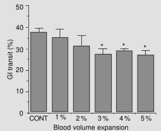
Figure 1 shows the effect of BV expan-sion on the GI transit of a charcoal meal when measured 10 min after test meal ad-ministration. BV expansion up to 1 and 2% body weight caused a slight but nonsignifi-cant decrease in GI transit rates while BV expansion of 3, 4 and 5% body weight signif-icantly decreased GI transit (P<0.05). Figure 2 shows that GI transit rates were decreased for at least 60 min after 5% BV expansion (P<0.05).
Table 1 shows GI transit rates in drug-pretreated or vagotomized animals submit-ted or not (drug controls) to 5% BV expan-sion. As can be seen, either subdiaphrag-matic vagotomy or yohimbine prevented the effect of BV expansion on GI transit (Figure
Figure 1 - Effect of acute blood volume expansion on gas-trointestinal (GI) transit rates, 10 min after intragastric adminis-tration of a charcoal meal (2.5 ml of an aqueous suspension of 5% charcoal and 5% gum ara-bic) to controls (CONT; N = 6) and animals submitted to blood volume expansion by iv infusion of Ringer-bicarbonate, 1 ml/min, in volumes of 1, 2, 3, 4 and 5% body weight (N = 5, 5, 5, 5 and 6, respectively).*P<0.05 com-pared to control (Student-Newman-Keuls test).
GI transit (%)
50
40
30
20
10
0
CONT 1 % 2 % 3 % 4 % 5 %
Blood volume expansion
3). Despite preventing the BV expansion effect on GI transit, we still observed a trend for a decrease in GI transit after BV expan-sion in vagotomized animals, which how-ever was not statistically significant. Hexa-methonium, atropine, L-NAME, prazosin or propranolol pretreatments, however, were ineffective (Table 1).
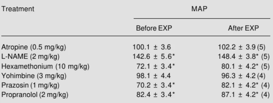
BV expansion (5%) transiently increased MAP during the expansion period (from 108.2 ± 3.2 to 119.7 ± 4.1, P<0.05, N = 5). However, MAP levels were not significantly modified after expansion was completed (ex-panded period, 116.1 ± 3.1). Table 2 shows that atropine and yohimbine decreased MAP. L-NAME increased while hexamethonium, propranolol and prazosin significantly de-creased MAP values (P<0.05). BV expan-sion also increased CVP values (from 3.6 ±
1.6 to 9.6 ± 3.2 cmH2O, P<0.05, N = 5) while
decreasing the mean hematocrit (from 49.6 ± 1.6 to 34 ± 1.1, P<0.05, N = 5).
Discussion
We have observed that acute BV changes modify the GI motility in anesthetized rats and dogs: gastric and jejunal compliance (9,10), as well as the resistance offered by the gastroduodenal segment to the flow of liquid (11,12). We have also recently ex-tended these observations to the ileocolonic region (15). The present study, which has been reported in abstract form (16), extend these observations from anesthetized to awake rats, avoiding the interference of an-esthesia on the autonomic nervous activity. Furthermore, we evaluated the relationship between the volume infused and the GI tran-sit delay and studied the neural mechanisms involved in this phenomenon.
The results show that BV expansion de-lays the GI transit of an aqueous charcoal meal in awake rats. BV expansion of 1 and 2% body weight had no significant effect on GI transit but BV expansion of 3, 4 and 5% body weight significantly decreased GI
tran-GI transit (%)
80
0 60
40
20
CONT 5% EXP
10 15 30 45 60
Time (min) Figure 2 - Comparison of
gas-trointestinal (GI) transit rates of an aqueous charcoal meal (2.5 ml of an aqueous suspension of 5% charcoal and 5% gum ara-bic) in controls (CONT; N = 6, 5, 7, 6 and 6, respectively) and in animals submitted to 5% blood volume expansion (EXP; N = 5, 5, 8, 7, and 6, respectively) by iv infusion of Ringer-bicarbonate, 1 ml/min, 10, 15, 30, 45 and 60 min after the intragastric test meal administration. *P<0.05 compared to control (Student-Newman-Keuls test).
Figure 3 - Effect of subdiaphrag-matic vagotomy (VAG) and yo-himbine (YOH) administration (3 mg/kg) on gastrointestinal (GI) transit delay of an aqueous char-coal meal due to 5% blood vol-ume (BV) expansion (EXP) by iv infusion of Ringer-bicarbonate, 1 ml/min in awake rats. VAG and VAG + EXP refer to vagotomized animals submitted or not to BV expansion (N = 5 in each group) and YOH and YOH + EXP to yo-himbine-pretreated animals sub-mitted or not to BV expansion (N = 5 in each group). NS, Not significant (Student-Newman-Keuls test).
GI transit (%)
60
50
20
0 40
30
10
VAG VAG + EXP YOH YOH + EXP
Groups
Table 1 - GI transit indexes 10 min after test meal administration in drug-pretreated or vagotomized controls (Control) and in pretreated or vagotomized animals submitted to 5% blood volume expansion (Expansion) with iv infusion of Ringer-bicarbonate, 1 ml/ min.
The number of animals is given within parentheses. *P<0.05 compared to None-Control (Student-Newman-Keuls test); +P<0.05 compared to the respective treatment
control (Student-Newman-Keuls test).
Treatment Control Expansion
None 38.1 ± 1.6 (6) 27.5 ± 1.8*+ (6)
Vagotomy 50.2 ± 4.6* (5) 44.6 ± 3.3* (5)
Hexamethonium (10 mg/kg) 45.9 ± 3.1 (6) 30.1 ± 3.4+ (5)
Atropine (0.5 mg/kg) 30.1 ± 2.3* (5) 22.2 ± 2.1*+ (5)
L-NAME (2 mg/kg) 31.1 ± 1.8* (5) 21.7 ± 2.6*+ (5)
Prazosin (1 mg/kg) 35.7 ± 3.1 (5) 26.4 ± 2.4*+ (5)
Yohimbine (3 mg/kg) 41.3 ± 3.5 (5) 42.2 ± 4.9 (5)
Propranolol (2 mg/kg) 44.1 ± 2.5 (5) 25.7 ± 2.6*+ (5)
* *
*
* *
NS
sit rates. This effect persisted for at least 60 min after expansion of 5% body weight.
The factors that influence the rate of propulsion of a meal through the small intes-tine are not completely understood. It has been demonstrated that a delay in gastric emptying may determine a delay in GI transit (17). In fact, we have reported that BV ex-pansion delays the gastric emptying of liquid in awake rats (14,16). However, changes in small intestine transit time can occur inde-pendently of changes in gastric emptying (17) and the rate of gastric emptying can influence the transit of food down the first 70 cm of the human intestine, but have little or no influence down the entire small intes-tine in normal subjects (17).
In this respect, GI transit delay due to BV expansion may not be essentially determined by a gastric emptying delay, since BV ex-pansion modifies jejunal compliance (10) and in animals submitted to fundectomy, BV expansion also did not delay gastric empty-ing, while the final GI transit was markedly delayed, thus indicating the activation of small bowel resistance by BV expansion (14). Subdiaphragmatic vagotomy prevented the effect of BV expansion on GI transit. These findings indicate that vagal pathways are necessary for the full expression of the phenomenon. However, the effect of va-gotomy may be incomplete since we still observed a tendency to a delay in GI transit after BV expansion in animals submitted to subdiaphragmatic vagotomy. Cholinergic pathways appear not to be involved since atropine did not block the effect of BV ex-pansion on GI transit. L-NAME was also unable to prevent the effect of BV expansion on GI transit.
Yohimbine (an α2 antagonist) prevented
the effect of BV expansion on GI transit, while hexamethonium (a ganglion blocker), propranolol (a ß blocker) and prazosin (an
α1 antagonist) were ineffective. Alpha-2
re-ceptors are located both in the central ner-vous system and peripherally (18). Our
find-ings suggest that central rather than
periph-eral α2 receptors are activated by BV
expan-sion, since peripheral activation would be associated with an increased sympathetic activity and thus would be blocked by hexa-methonium and/or by prazosin and
propran-olol. Central α2 activation causes a
signifi-cant decrease in sympathetic activity (19), similar to that observed after BV expansion (20), and mediates GI motility inhibition (18). However, it has been demonstrated that yohimbine can also modulate vagal ac-tivity and act upon non-adrenergic receptors (21).
Our findings point to an involvement of neural pathways. However, increased CVP also leads to a release of several hormones and autacoids, such as the atrial natriuretic peptide (ANP), which could interfere with GI motility, absorption and secretion. In fact, it has been demonstrated that ANP can in-crease the magnitude of spontaneous duode-nal phasic contractions (22) and reduces fluid and electrolyte intestinal absorption (23).
Hepatorenal and hepatointestinal reflex systems are of paramount importance for
Na+ and ultimately liquid homeostasis (24).
BV expansion causes important homeostatic modifications to manage increased body liq-uid volume and attain a new steady state. Diuretic and natriuretic responses following BV expansion have been extensively studied (25), as well as decreased intestinal sodium/
Table 2 - Effect of drug pretreatment on mean arterial pressure (MAP) before and after 5% blood volume expansion (EXP) with iv infusion of Ringer-bicarbonate, 1 ml/min.
The number of animals is given within parentheses. *P<0.05 compared to basal MAP levels (Student-Newman-Keuls test).
Treatment MAP
Before EXP After EXP
Atropine (0.5 mg/kg) 100.1 ± 3.6 102.2 ± 3.9 (5)
L-NAME (2 mg/kg) 142.6 ± 5.6* 148.4 ± 3.8* (5)
Hexamethonium (10 mg/kg) 72.1 ± 3.4* 80.1 ± 4.2* (5)
Yohimbine (3 mg/kg) 98.1 ± 4.4 96.3 ± 4.2 (4)
Prazosin (1 mg/kg) 70.2 ± 3.4* 82.1 ± 4.2* (4)
water absorption and increased secretory rates (6). Even subtle postural changes (tilt-ing and stand(tilt-ing up) simulat(tilt-ing BV changes might modify intestinal salt and water per-meability rates in man (26).
Since GI motility is related to absorption and secretion (8), the delay in GI transit of liquid observed here, taken together with the absorptive and secretory modifications
de-scribed by others (5,6), may be part of the body strategies involved in the management of liquid excess.
Acknowledgments
We thank Fernando Antonio A. Gondim and R. Fogaça for statistical support and Dr. G.B. Viana for providing research facilities.
References
1. Sawchenko PE & Fridman MI (1979). Sen-sory functions of the liver. American Jour-nal of Physiology, 236: R5-R20.
2. Michel AR (1986). The gut: the unobtru-sive regulator of sodium balance. Perspec-tives in Biology and Medicine, 29: 203-213.
3. Guyton A & Hall J (1996). Digestion and absorption in the gastrointestinal tract. In: Textbook of Medical Physiology. 9th edn. W.B. Saunders, Philadelphia, PA, 833-851. 4. Miller WL & Dale HE (1978). Restoration of hemorrhaged plasma volume by gas-trointestinal fluid in the dog. American Journal of Physiology, 234: H80-H87. 5. Duffy PA, Granger DN & Taylor AE (1978).
Intestinal secretion induced by volume expansion in the dog. Gastroenterology, 75: 413-418.
6. Levens NR (1985). Control of intestinal absorption by the renin-angiotensin sys-tem. American Journal of Physiology, 249: G3-G15.
7. Suzuki S, Khanchowdhury MR, Uemura N, Morita H & Hosomi H (1992). Renojeju-nal reflex controlling jejuRenojeju-nal absorption of fluid and NaCl. Journal of the Autonomic Nervous System, 39: 219-228.
8. Lee JS (1983). Relationship between in-testinal motility, tone, water absorption and lymph flow in the rat. Journal of Phys-iology, 345: 489-499.
9. Capelo LR, Cavalcante DM, Leitão IA, Cristino Filho G & da-Silva EAT (1983). Modifications of gastric compliance in dogs related to changes of extracellular fluid volume. Brazilian Journal of Medical and Biological Research, 16: 73-76. 10. Rola FH, dos-Santos AA, Xavier-Neto J,
Cristino-Filho G, Rocha CI, Santiago Jr AT, Gondim FAA, Pereira JM & Capelo LR (1989). Effects of acute volemic changes on the jejunal compliance in dogs. Brazil-ian Journal of Medical and Biological Re-search, 22: 523-531.
11. Santos AA, Xavier-Neto J, Santiago Jr AT, Souza MAN, Martins AS, Alzamora F & Rola FH (1991). Acute volaemic changes modify the gastroduodenal resistance to the flow of saline in anaesthetized dogs. Acta Physiologica Scandinavica, 143: 261-269.
12. Xavier-Neto J, Dos Santos AA & Rola FH (1990). Acute hypervolemia increases the gastroduodenal resistance to the flow of saline in rats. Gut, 31: 1006-1010. 13. Green AF (1959). Comparative effects of
analgesics on pain threshold, respiratory frequency and gastrointestinal propulsion. British Journal of Pharmacology, 14: 26-34.
14. Rego MCV, da-Graça JRV, Gondim F de-AA, Gondim RB de-M, Dantas RP & Rola FH (1998). Effect of pyloroplasty and fun-dectomy on the delay of gastric emptying and gastrointestinal transit of liquid elic-ited by acute blood volume expansion in awake rats. Brazilian Journal of Medical and Biological Research, 31 (in press). 15. Santiago Jr AT, Gondim F de-AA,
Cavalcante DIM, da-Graça JRV, de-Oliveira GR, dos-Santos AA & Rola FH (1997). Acute extracellular fluid volume changes increase ileocolonic resistance to saline flow in anesthetized dogs. Brazil-ian Journal of Medical and Biological Re-search, 30: 999-1008.
16. Gondim FAA, Oliveira GR, Graça JRV, Cavalcante RBM, Gondim DIM, Santiago Jr AT & Rola FH (1996). Gastrointestinal motor reflexes elicited by acute blood vol-ume expansion in awake rats. Neurogas-troenterology and Motility, 8: 168 (Ab-stract).
17. Read NW, Cammack J, Edwards C, Holgate AM, Cann PA & Brown C (1982). Is the transit time of a meal through the small intestine related to the rate at which it leaves the stomach? Gut, 23: 824-828. 18. Fargeas MJ, Fioramonti J & Bueno L
(1986). Central alpha-2 adrenergic control of the pattern of small intestinal motility in rats. Gastroenterology, 91: 1470-1475. 19. Klangkalya B, Sripairojthikoon W, Oparil S & Wyss JM (1988). High NaCl diet in-creases anterior hypothalamic alph2 a-drenoceptors in SHR. Brain Research, 451: 77-84.
20. Ricktsen SE & Thoren P (1980). Reflex inhibition of sympathetic activity during volume load in awake normotensive and spontaneously hypertensive rats. Acta Physiologica Scandinavica, 110: 77-82. 21. Trostel KA, Kata SA & Osborn JW (1994).
Does the spinal cord generate significant sympathetic activity in the awake rat? American Journal of Physiology, 266: R1102-R1110.
22. Bayens DA, Walters JM & Vesely DL (1988). Atrial natriuretic factor increases the magnitude of duodenal spontaneous phasic contractions. Biochemical and Bio-physical Research Communications, 155: 1437-1443.
23. Matshushita K, Nishida Y, Hosomi H & Tanaka S (1991). Effects of atrial natriuret-ic peptide on water and NaCl absorption across the intestine. American Journal of Physiology, 260: R6-R12.
24. Hosomi H & Morita H (1996). Hepatorenal and hepatointestinal reflexes in sodium homeostasis. News in Physiological Sci-ences, 11: 103-107.
25. Wardener HE, Mills IH, Clapham WF & Hayter CJ (1961). Studies on the efferent mechanism of the sodium diuresis which follows the administration of intravenous saline in the dog. Clinical Science, 21: 249-258.