Effect of pyloroplasty and fundectomy
on the delay of gastric emptying and
gastrointestinal transit of liquid elicited
by acute blood volume expansion in
awake rats
Departamento de Fisiologia e Farmacologia, Universidade Federal do Ceará, Fortaleza, CE, Brasil
M.C.V. Rêgo, J.R.V. da-Graça, F. de-A.A. Gondim, R.B. de-M. Gondim, R.P. Dantas and F.H. Rola
Abstract
We evaluated the effects of fundectomy and pyloroplasty on the delay of gastric emptying (GE) and gastrointestinal (GI) transit of liquid due to blood volume (BV) expansion in awake rats. Male Wistar rats (N = 76, 180-250 g) were first submitted to fundectomy (N = 26), Heinecke-Mikulicz pyloroplasty (N = 25) or SHAM laparotomy (N = 25). After 6 days, the left external jugular vein was cannulated and the animals were fasted for 24 h with water ad libitum. The test meal was administered intragastrically (1.5 ml of a phenol red solution, 0.5 mg/ ml in 5% glucose) to normovolemic control animals and to animals submitted to BV expansion (Ringer-bicarbonate, iv infusion, 1 ml/ min, volume up to 5% body weight). BV expansion decreased GE and GI transit rates in SHAM laparotomized animals by 52 and 35.9% (P<0.05). Fundectomy increased GE and GI transit rates by 61.1 and 67.7% (P<0.05) and prevented the effect of expansion on GE but not on GI transit (13.9% reduction, P<0.05). Pyloroplasty also increased GE and GI transit rates by 33.9 and 44.8% (P<0.05) but did not prevent the effect of expansion on GE or GI transit (50.7 and 21.1% reduction, P<0.05). Subdiaphragmatic vagotomy blocked the effect of expansion on GE and GI transit in both SHAM laparotomized animals and animals submitted to pyloroplasty. In conclusion 1) the proximal stomach is involved in the GE delay due to BV expansion but is not essential for the establishment of a delay in GI transit, which suggests the activation of intestinal resistances, 2) pyloric modulation was not apparent, and 3) vagal pathways are involved.
Correspondence
F.H. Rola
Departamento de Fisiologia e Farmacologia, UFC
Rua Coronel Nunes de Melo, 1127 Caixa Postal 3157
60430-270 Fortaleza, CE Brasil
Fax: 55 (085) 243-9333 Presented at the Fifth United European Gastroenterological Week, Paris, 1996.
Research supported by CAPES, CNPq, UFC and UNIMED.
Received June 28, 1997 Accepted November 25, 1997
Key words
•Gastric emptying •Gastrointestinal transit •Blood volume expansion •Fundectomy
•Pyloroplasty •Vagotomy
Introduction
The gastrointestinal (GI) tract, in addi-tion to performing its funcaddi-tion of nutrient digestion and absorption, is vitally impor-tant in maintaining water, electrolyte and
acid-base regulation (1,2).
tum, anesthetized with thiopental and sub-mitted to fundectomy, N = 26 (10), Heinecke-Mikulicz pyloroplasty, N = 25 (11) or SHAM laparotomy (N = 25) 7 days before GE and GI transit measurements. The gastric fundus was easily identified visually after midline laparotomy. Its excision (fundectomy) was performed by cutting the anterior and poste-rior walls, beginning just below the esopha-gogastric junction and extending aborally to the inferior limit of the gastric fundus (see Figure 1A). The incision was carefully closed with a transverse layer of suture (6.0 silk). Heinecke-Mikulicz pyloroplasty was per-formed after laparotomy through a 1-cm seromucosal longitudinal incision extending 0.5 cm in the antrum and 0.5 cm in the duodenum. The incision was carefully closed with a transverse layer of suture (6.0 silk) which joined together the surrounding cut walls (Figure 1B). Both pyloroplasty and fundectomy were closely inspected after sur-gery to assure that the gastroduodenal junc-tion was not obstructed. At autopsy, the presence of possible gastroduodenal steno-sis due to inappropriate scarring was also evaluated.
Six days after surgery (one day before the GE/GI transit measurements) the animals were anesthetized with ether and a polyeth-ylene catheter (PE 50) was placed into the left external jugular vein. The distal end of the catheter was tunneled subcutaneously and its free end secured with suture after a dorsal skin incision between the shoulders. The animals were fasted for 24 h and water
was allowed ad libitum until 2 h before the
experiment.
All surgical procedures and animal treat-ments were conducted in accordance with the “Guide for the Care and Use of Labora-tory Animals” (DHEW Publication No. 85-23, NIH, Bethesda, MD).
GE measurement
The method used to measure GE is a
A
B
Fundectomy
Pyloroplasty
Figure 1 - A, Technique of gas-tric fundus excision (fundec-tomy), with emphasis on the cut margins and the final sutures (for more details, see Material and Methods). B, Technique of Heinecke-Mikulicz pyloroplasty with emphasis on the cut mar-gins and the final sutures (for more details, see Material and Methods).
and water increases during acute BV retrac-tion (4).
Since GI motility is finely regulated to provide adequate absorptive and secretory pat-terns - as GI contractile activity is related to absorption and secretion rates (5) - the re-sponse of the GI tract to liquid volume excess may involve a coupling of fluid/electrolyte transport and GI contractile activity adjust-ments, as proposed in previous reports (6,7).
We have shown that acute BV expansion modifies the gastroduodenal flow of liquid in anesthetized dogs and rats (7,8) and de-lays gastric emptying (GE) and the gas-trointestinal (GI) transit of liquid in awake rats (9; Gondim F de-AA, Oliveira GR, Graça JRV da, Cavalcante DIM, Sousa MAN, Santos AA and Rola FH, unpublished re-sults). In the present study we performed fundectomy (10) or Heinecke-Mikulicz py-loroplasty (11) to evaluate the role of the proximal stomach and pyloric segment in the delay of GE and GI transit of liquid elicited by BV expansion in awake rats (12).
Material and Methods
Animals and surgical procedures
Male Wistar rats (N = 76) weighing 180-250 g were used in this study. The animals
libi-modification of that described by Scarpignato et al. (13).
First, 1.5 ml of the test meal containing a non-absorbable marker (0.5 mg/ml phenol red solution in 5% glucose) was given orally into the stomach through a stainless steel tube that was removed immediately after delivering the solution intragastrically. The
animals were sacrificed with an iv thiopental
overdose, the stomach was exposed by lapa-rotomy, quickly clamped at the pyloric and cardiac ends and removed.
Stomachs were then placed in 100 ml of 0.1 N NaOH, cut into small pieces and ho-mogenized for 30 s. The suspension was allowed to settle for 30 min at room temper-ature and 10 ml of the supernatant was cen-trifuged for 10 min at 2800 rpm (about 1400
g). Proteins in 5 ml of homogenate were
precipitated with 0.5 ml of trichloroacetic acid (20% w:v), centrifuged for 20 min at 2800 rpm and 3 ml of the supernatant was added to 4 ml of 0.5 N NaOH. The absorb-ance of the sample was read at a wave length of 560 nm.
Percent gastric emptying (%GE) for each rat was calculated according to the following formula: %GE = 1 - amount of phenol red recovered from test stomach x 100/average amount of phenol red recovered from “stan-dard” stomachs.
Rats sacrificed immediately after test meal administration were used to establish “stan-dard” stomachs (100% phenol red in the stomach) in animals submitted to SHAM laparotomy (N = 5), fundectomy (N = 5) or pyloroplasty (N = 5).
Measurement of GI transit
GI transit measurements were performed according to the well-known concept of Green (14). As the test meal is administered intragastrically, GE as well as intestinal pro-pulsion rates influence the final transit of the marker (GI transit). After quickly clamping the pyloric and cardiac ends to perform GE
measurements, the small intestine - from the gastroduodenal junction to the cecum - was carefully removed and lightly stretched along a ruler on a flat table top. Tiny scissor cuts were then performed along the small intes-tine and 0.1 N NaOH solution was dribbled over the leaking phenol red solution to visu-alize the farthest point reached by the test meal front (pink color). The total length of the small intestine and the distance travelled by the marker along the small intestine were then measured. Since the intestines were all of quite similar length (mean 110.3 ± 4.1 cm), the GI transit index was defined as distance the marker traveled/total length of intestine x 100.
Experimental design and treatments
The animals were divided into 4 main groups, subdivided according to the various experimental conditions and sacrificed 10 min after test meal administration.
In the first group (group 1), SHAM laparotomized rats were used to evaluate the GE and GI transit rates in normovole-mic (N = 5) and in expanded animals (N =
5). BV expansion was performed by iv
infu-sion of a volume up to 5% body weight of
Ringer bicarbonate solution (Na+ = 140, K+
= 4, Cl- = 124, HCO3- = 20 mmol/l) at the
joined and sutured. One day after subdia-phragmatic vagotomy and jugular vein can-nulation, the animals previously submitted to SHAM laparotomy, fundectomy or py-loroplasty were submitted to BV expansion or not (normovolemic control, N = 5 for each experimental variation). The test meal was then intragastrically administered and the animals were sacrificed 10 min after test meal administration.
Mean arterial pressure, central venous pressure and hematocrit
In a separate group, mean arterial pres-sure (MAP) was monitored in awake rats before, during and after the 5% BV expan-sion. For this purpose a catheter was placed into the carotid artery and connected to a mercury (Hg) manometer. Intracardiac blood samples were also collected for hematocrit determination. Central venous pressure (CVP) values were measured in awake rats before and after 5% expansion by inserting a PE50 catheter into the right jugular vein. The catheter was positioned near the right atrium
and connected to a low pressure transducer (NarcoByo 3, Narco Byo-Systems, Houston, TX), which was coupled to a physiograph Desk Model DMD 4B (Narco Byo-Systems).
Statistical analysis
The results are reported as means ± SEM. Descriptive statistics were applied to each group of experiments. One-way analysis of variance and the Student-Newman-Keuls test were then used to compare the various groups. Differences were considered significant at P<0.05.
Results
Effect of BV expansion on GE of liquid in SHAM laparotomized animals and in animals previously submitted to fundectomy or pyloroplasty
The reservoir capacity of the stomach after fundectomy was decreased by 33.2% when we compared the fundectomy stan-dards with SHAM stanstan-dards. Pyloroplasty also decreased gastric capacity by 12.3%
(pyloroplasty standards vs SHAM standards).
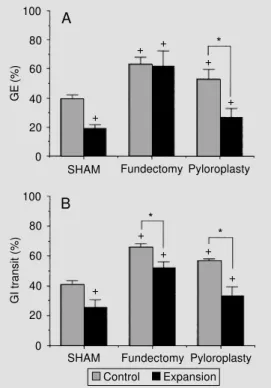
As can be seen in Figure 2A and B, fundec-tomy and pyloroplasty significantly increased GE of liquid rates by 37.9 and 25.4%, re-spectively (P<0.05). BV expansion decreased GE of liquid rates in SHAM laparotomized animals and in animals submitted to pyloro-plasty by 52.4 and 49.3%, respectively (P<0.05), but produced a minor and not sta-tistically significant GE decrease in animals previously submitted to fundectomy (2.3%, NS), as can be seen in Figure 2A.
Effect of BV expansion on GI transit of liquid in SHAM laparotomized animals and in animals previously submitted to fundectomy or pyloroplasty
Fundectomy and pyloroplasty significant-ly increased the rates of GI transit of liquid
GE (%)
100
80
60
40
20
0
Fundectomy Pyloroplasty
SHAM Fundectomy Pyloroplasty
GI transit (%)
100
80
60
40
20
0
Control Expansion +
*
+ +
+
+
+ +
+ +
+
* A
B
Figure 2 - A, Rates of gastric emptying (GE) of liquid in nor-movolemic (Control) SHAM laparotomized (N = 5) and in laparotomized expanded animals (N = 5), as well as in normovole-mic and expanded (Expansion) animals previously submitted to fundectomy (N = 6 and 5, re-spectively) or pyloroplasty (N = 5 and 5, respectively). B, Rates of gastrointestinal (GI) transit of liq-uid in the same sequence. Blood volume (BV) expansion was per-formed by intravenous infusion of Ringer-bicarbonate in a vol-ume up to 5% body weight, 1 ml/min. +P<0.05 vs SHAM
con-trols (Student-Newman-Keuls test); *P<0.05 vs intragroup con-trol (Student-Newman-Keuls test).
*
by 39.6 and 30.9% (P<0.05). BV expansion decreased the rates of GI transit of liquid in SHAM operated animals and in animals sub-mitted to fundectomy or pyloroplasty by 36.7, 21.1 and 42.3% (P<0.05). Thus, as can be seen in Figure 2B, neither fundectomy nor pyloroplasty prevented the effect of BV ex-pansion on GI transit of liquid.
Effect of vagotomy on GE and GI transit in SHAM laparotomized animals and in animals previously submitted to fundectomy or pyloroplasty
Subdiaphragmatic vagotomy prevented the effect of BV expansion on the rates of GE and GI transit of liquid in SHAM laparotomized animals. BV expansion also did not modify the rates of GE and GI transit of liquid in vagotomized animals previously submitted to fundectomy or pyloroplasty. Thus, vagotomy prevented 1) the effect of BV expansion on GE and GI transit in SHAM laparotomized animals, 2) the effect of BV expansion on GE and GI transit in animals submitted to pyloroplasty, and 3) the effect of BV expansion on GI transit in animals submitted to fundectomy, as can be seen in Figure 3.
Effect of BV expansion on MAP, CVP and hematocrit
MAP before BV expansion was 112.2 ± 3.2 mmHg. BV expansion did not change MAP either during expansion or after expan-sion was completed (117.8 ± 4.4 and 115.7 ± 7.1, respectively, N = 4, NS). However, after BV expansion, CVP levels increased from
2.4 ± 3.6 to 7.3 ± 2.1 cmH2O (P<0.05, N = 5)
and the mean hematocrit values decreased from 49.6 ± 1.6% (N = 5) to 34 ± 1.1% 10 min after expansion (P<0.05, N = 5).
Discussion
Acute blood volume expansion delays
the gastric emptying and gastrointestinal tran-sit of liquid in awake rats (9; Gondim F de-AA, Oliveira GR, Graça JRV da, Cavalcante DIM, Sousa MAN, Santos AA and Rola FH, unpublished results). In the present study we demonstrated that, as observed in intact con-trols (9), GE and GI transit rates were de-creased in SHAM laparotomized animals after BV expansion, as well as in animals previously submitted to pyloroplasty. How-ever, fundectomy prevented the effect of BV expansion on GE but not on GI transit.
The effect of acute BV expansion on GE and GI transit of liquid appears to be related to alterations in GI motility since BV expan-sion increases the gastroduodenal resistance offered to saline flow in anesthetized dogs and rats (7,8). In addition, we have previ-ously demonstrated that BV expansion also decreases GE rates in animals submitted to
ip administration of ranitidine (9; Gondim F
de-AA, Oliveira GR, Graça JRV da, Cavalcante DIM, Sousa MAN, Santos AA and Rola FH, unpublished results), which minimizes the possible interference of a high level of gastric secretion induced by BV
Figure 3 - Effect of subdiaphrag-matic vagotomy on the rates of gastric emptying (GE) (A) and gastrointestinal (GI) transit (B) in normovolemic animals (Va-gotomy) and in animals submit-ted to blood volume (BV) expan-sion (Vago + Exps) and to SHAM laparotomy, fundectomy or py-loroplasty (N = 5 for each exper-imental variation). BV expansion was performed by intravenous infusion of Ringer-bicarbonate in a volume up to 5% body weight, 1 ml/min. NS, Not significant.
GE (%)
100
80
60
40
20
0
Fundectomy Pyloroplasty
SHAM Fundectomy Pyloroplasty
GI transit (%)
100
80
60
40
20
0
Vagotomy Vago + Exps
A
B
NS
NS NS
NS NS
expansion with the evaluation of actual gas-tric emptying (13). Thus, the changes in the GE of liquid observed here did not appear to be related to secretory pattern modifica-tions.
The proximal stomach has an important reservoir function, receiving and storing in-gesta (15) and is important for GE control (10). Fundectomy decreases the gastric vol-ume capacity, increases postprandial intra-gastric pressure and accelerates GE, as could be observed in our experiments. The present findings indicate that the proximal stomach is an essential element for the development of the GE delay due to BV expansion, since fundectomy prevented it. This contrasts with our results obtained in anesthetized rats (16). This difference can be explained by the in-ability of the previous perfusion system (8,16) to separate fundic from antral function. An-other point to consider is the possible activa-tion of cardiopulmonary receptors (17) which can increase fundic relaxation and cannot be easily evidenced from our previous experi-mental protocols (8,16).
The role of the pylorus in GE control is still obscure. However, the pylorus has been proposed as an effective resistance to transpyloric flow of liquid by increased lo-calized pyloric contractions (18), and delays in GE due to changes in pyloric pressure waves, obstructing the flow through the py-lorus, have also been reported (19). Pyloric resistance was not essential to trigger the effect of BV expansion on GE, in contrast to previous results in anesthetized animals, which clearly demonstrated its participation (16). In fact, pyloroplasty increased GE and GI transit, but did not block the effect of BV expansion.
In our experimental protocol, changes in GI transit of liquid were influenced by GE as well as by the small intestine propulsion, since the test meal was intragastrically deliv-ered. Concerning the delay in GI transit of liquid, neither fundectomy (which prevented GE delay), nor pyloroplasty prevented it.
Thus, since GE rates were markedly de-creased by BV expansion, it would not be surprising to find a decrease in GI transit, which could be entirely related to the de-crease in GE. This statement is valid consid-ering the experiments in animals submitted to SHAM laparotomy or pyloroplasty, since both GE and GI transits were significantly decreased by BV expansion. However, in animals submitted to fundectomy, GE rates were not decreased by BV expansion (indi-cating that fundectomy prevented the effect of BV expansion on GE), while GI transit rates were significantly decreased. In this case, the delay in GI transit could not be explained only by the delay in GE, indicating that the GI transit delay due to BV expansion may occur without gastric participation, prob-ably due to small intestine motility activa-tion, which has been suggested previously in anesthetized animals (6). In addition, it has been demonstrated that changes in small intestine transit time could occur independ-ently of changes in GE (20) since the rate of GE can influence the transit of food down the initial intestinal portions, but has a more limited influence down the whole small in-testine (20) and other studies have also dem-onstrated that GE can be increased, but the final GI transit delayed (21).
A relationship between delayed GE and MAP changes was not found, since MAP levels did not change significantly after BV expansion. However, CVP levels were sig-nificantly increased after BV expansion. Hematocrit values decreased significantly 10 min after BV expansion. Thus, besides delayed GE, acute BV expansion also modi-fied hematocrit and CVP, but not MAP lev-els. The GE delay due to acute BV expansion also appears to be influenced by the infused volume but not necessarily by the composi-tion of the expanding solucomposi-tion (8).
animals and in animals submitted to pyloro-plasty, in agreement with our previous ob-servation (9) indicating that vagal pathways are involved in the phenomenon. In fact, BV expansion activates cardiopulmonary recep-tors which release their signals through va-gal pathways (17). However, despite these
findings, yohimbine, an α-2 blocker, has
also been reported to be effective in blocking
References
1. Michel AR (1986). The gut: the unobtru-sive regulator of sodium balance. Per-spectives in Biology and Medicine, 29: 203-213.
2. Sawchenko PE & Fridman MI (1979). Sen-sory functions of the liver. American Jour-nal of Physiology, 236: R5-R20.
3. Duffy PA, Granger DN & Taylor AE (1978). Intestinal secretion induced by volume expansion in the dog. Gastroenterology, 75: 413-418.
4. Levens NR (1985). Control of intestinal absorption by the renin-angiotensin sys-tem. American Journal of Physiology, 249: G3-G15.
5. Lee JS (1983). Relationship between in-testinal motility, tone, water absorption and lymph flow in the rat. American Jour-nal of Physiology, 345: 489-499. 6. Rola FH, dos-Santos AA, Xavier-Neto J,
Cristino-Filho G, Rocha CI, Santiago Jr AT, Gondim FAA, Pereira JM & Capelo LR (1989). Effects of acute volemic changes on jejunal compliance in dogs. Brazilian Journal of Medical and Biological Re-search, 22: 523-531.
7. Santos AA, Xavier-Neto J, Santiago-Jr AT, Souza MAN, Martins AS, Alzamora F & Rola FH (1991). Acute volaemic changes modify the gastroduodenal resistance to the flow of saline in anaesthetized dogs. Acta Physiologica Scandinavica, 143: 261-269.
8. Xavier-Neto J, dos Santos AA & Rola FH (1990). Acute hypervolemia increases the gastroduodenal resistance to the flow of saline in rats. Gut, 31: 1006-1010.
9. Gondim F de AA, Oliveira GR, Graça JRV da, Cavalcante DIM, Santiago Jr AT, Santos AA & Rola FH (1996). Neural mechanisms involved in gastric emptying delay due to acute expansion of the extra-cellular fluid volume. Neurogastroenterol-ogy and Motility, 1: 74 (Abstract). 10. Wilbur BG, Kelly KA & Code CF (1974).
Effect of gastric fundectomy on canine gastric electrical and motor activity. American Journal of Physiology, 226: 1445-1449.
11. Ormsbee III HS & Bass P (1976). Gastro-duodenal motor gradients in the dog after pyloroplasty. American Journal of Physiol-ogy, 230: 389-397.
12. Rego MCV, Graça JRV, Gondim F de AA, Gondim RBM, Dantas RP & Rola FH (1996). Role of proximal stomach and py-lorus on gastric emptying and gastrointes-tinal transit delays elicited by acute blood volume expansion in awake rats. Gut, 39 (Suppl 3): 214 (Abstract).
13. Scarpignato C, Capovilla T & Bertaccini G (1980). Action of caerulein on gastric emp-tying of the conscious rat. Archives Inter-nationales de Pharmacodynamie et de Therapie, 246: 286-293.
14. Green AF (1959). Comparative effects of analgesics on pain threshold, respiratory frequency and gastrointestinal propulsion. British Journal of Pharmacology, 14: 26-34.
15. Kelly KA (1980). Gastric emptying of liq-uids and solids: roles of proximal and dis-tal stomach. American Journal of Physiol-ogy, 239: G71-G76.
16. Graça JRV, Gondim F de-AA, Cavalcante DIM, Xavier-Neto J, Messias ELM, Marques JAP, Santos AA & Rola FH (1997). Gastroduodenal resistance and neural mechanisms involved in saline flow decrease elicited by acute blood volume expansion in anesthetized rats. Brazilian Journal of Medical and Biological Re-search, 30: 1257-1266.
17. Thames MD (1978). Contribution of cardi-opulmonary receptors to the control of the kidney. Federation Proceedings, 37: 1209-1213.
18. Treacy PJ, Jamieson GG & Dent J (1994). Pyloric motility and liquid gastric empty-ing durempty-ing barostatic control of gastric pressure in pigs. Journal of Physiology, 474: 361-366.
19. Houghton LA, Read NW, Hedge R, Horowitz M, Collins PJ, Chatterton B & Dent J (1988). Relationship of the motor activity of the antrum, pylorus, and duode-num to gastric emptying of a solid-liquid mixed meal. Gastroenterology, 94: 1285-1291.
20. Read NW, Cammack J, Edwards C, Holgate AM, Cann PA & Brown C (1982). Is the transit time of a meal through the small intestine related to the rate at which it leaves the stomach? Gut, 23: 824-828. 21. Stewart JJ, Battarbee HD & Betzing KW
(1992). Intestinal myoelectrical activity and transit time in chronic portal hyper-tension. American Journal of Physiology, 263: G474-G479.
the BV expansion effect on GE and GI tran-sit (9).