.
Hydrogen sulfi de: a new endogenous player in an old mechanism of
plant tolerance to high salinity
Cristiane J. da-Silva
1and Luzia V. Modolo
1*Received: June 10, 2017 Accepted: August 22, 2017
ABSTRACT
High salinity aff ects plants due to stimulation of osmotic stress. Cell signaling triggered by nitric oxide (NO) and hydrogen sulfi de (H2S) activates a cascade of biochemical events that culminate in plant tolerance to abiotic and biotic stresses. For instance, the NO/H2S-stimulated biochemical events that occur in plants during response to high salinity include the control of reactive oxygen species, activation of antioxidant system, accumulation of osmoprotectants in cytosol, induction of K+ uptake and Na+ cell extrusion or its vacuolar compartmentation among others. Th is review is a compilation of what we have learned in the last 10 years about NO participation during cell signaling in response to high salinity as well as the role of H2S, a new player in the mechanism of plant tolerance to salt stress. Th e main sources of NO and H2S in plant cells is also discussed together with the evidence of interplay between both signaling molecules during response to stress.
Keywords: abiotic stress, cell signaling, hydrogen sulfi de, nitric oxide, NO and H2S biosynthesis, salt stress
Introduction
It is estimated that over 800 million hectares of land throughout the world are overloaded with salt, which represents more than 6 % of the world’s total land area (Munns & Tester 2008). High salinity leads to osmotic stress, cell toxicity by ions excess and ultimately nutrition disorders and oxidative stress in plants (Munns & Tester 2008). A signaling cascade involving expression of specifi c genes and accumulation of certain metabolites is pivotal for plants successfully acclimating and tolerating high salinity (Gupta & Huang 2014). Nitric oxide (NO), and
more recently hydrogen sulfi de (H2S), were recognized as important players in cell signaling triggered during plant response to biotic and abiotic stresses (Delledonne et al.
1998; Durner et al. 1998; Zhang et al. 2008). Th e role of these signaling molecules in salt stress has been explored over the past few years.
Th e NO is a gaseous free radical widely produced in living organisms. Its production was fi rst reported in plants by Dr. Lowell Klepper by the end of the 1970s (Klepper 1979). Nevertheless, the advent of researches focusing on NO in plants took place 19 years later (Delledonne et al. 1998;
Durner et al. 1998) when Drs. Robert F. Furchgott, Louis J.
1 Departamento de Botânica, Instituto de Ciências Biológicas, Universidade Federal de Minas Gerais, Av. Pres. Antônio Carlos, 6627, Pampulha, 31270-901, Belo Horizonte, MG, Brazil
* Corresponding author: [email protected]
Ignarro and Ferid Murad were jointly laureate with the Nobel Prize for the disclosure of NO as an endothelium-derived relaxing factor in mammals. Since then, NO has been shown to influence plant response to salt stress by improving seed vigor and germination (Hayat et al. 2012) and controlling the cellular levels of reactive oxygen species (ROS) (Keyster
et al. 2012; Ahmad et al. 2016), nutrient (Kong et al. 2016; Liu et al. 2016) and osmoprotectants (Wu et al. 2011; Tian
et al. 2015). The NO biosynthesis in plant cells can occur by non-enzymatic and enzymatic means. Nitrite (NO2-) or nitrate (NO3-), in the presence of ascorbic acid (AsA), may be non-enzymatically converted to NO (Klepper 1990). The acidic condition of aleurone layers was also demonstrated to favor NO production in apoplast (Bethke et al. 2004). The light-dependent production of NO from nitrogen dioxide, assisted by carotenoids, has been reported (Cooney et al.
1994). On the other hand, the enzymatic mechanisms that drive NO production in plant cells are still under debate since a plethora of examples are reported in the literature. The H2S is a small and lipophilic molecule that was pointed out as a possible cellular signaling component in mammalians (Abe & Kimura 1996). Indeed, H2S is considered the third gas transmitter in addition to NO and carbon monoxide (Wang 2002). Its ability to induce seed germination and relief copper stress was demonstrated in the late 2000s (Zhang et al. 2008). However, earlier H2S was believed to be exclusively phytotoxic. Since then, H2S was implicated in the ROS control through the activation of antioxidant system (Yu et al. 2013; Shan et al. 2014; da-Silva et al. 2017), maintenance of high K+/Na+ ratio
(Lai et al. 2014; Deng et al. 2016) and accumulation of osmolytes (Shi et al. 2013) during plant response to high salt concentrations. Recent research demonstrates that H2S is primarily produced in plant tissues from L/D-cysteine or sulfide (Li 2015).
This review describes the main enzymatic sources of NO and H2S in plants and compiles what it is known from the past 10 years on the role of these signaling molecules during plants response to high salinity.
Biosynthesis of NO and H
2S in
plant cells
Enzymes involved in NO biosynthesis
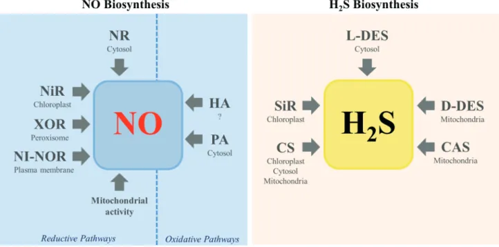
A body of evidence indicates that the production of NO in plant cells may come from both reductive and oxidative pathways (Fig. 1). Reductive mechanisms for NO synthesis include NO3- or NO
2- as the primary substrates for nitrate reductase (NR), a plasma membrane-bound nitrite:NO reductase (NI-NOR), a mitochondrial protein system or xanthine oxidoreductase (XOR). The oxidative pathway comprises the polyamines and hydroxylamines metabolisms by still unknown mechanisms (Fig. 1).
The NR, a cytosolic enzyme, is able to catalyze directly or indirectly the NO production from NO3- (Yamasaki & Sakihama 2000; Modolo et al. 2005). The genes Nia1 and
Nia2 encode for NR in Arabidopsis thaliana (Wilkinson
Figure 1. Schematic representation of the main pathways of NO and H2S production reported in plant cells. CAS, β-cyanoalanine
& Crawford 1993). This enzyme promotes the NAD(P) H-dependent reduction of NO3- to NO
2- and then to NO (Magalhaes et al. 2000; Yamasaki & Sakihama 2000). The direct production of NO from NR, however, represents only 1−2 % of the nitrate-reducing capacity of this enzyme (Rockel et al. 2002; Planchet et al. 2005). This is because relatively high amounts of NO2- (K
M= 100 µM) are required for NR reducing NO2- to NO, while the formation of NO is prevented in the presence of NO3- in amounts as low as 50 µM (Rockel et al. 2002). The NR can also indirectly
contribute to NO production by catalyzing the formation of the substrate (NO2-) for other enzymatic systems. For instance, in vitro experiments showed that a plasma membrane-bound protein (NI-NOR) of tobacco (Nicotiana tabacum) roots was able to reduce NO2- to NO (Stöhr et al. 2001) that, in turn, yielded higher rates of tobacco root colonization by the arbuscular mycorrhizal Glomus mosseae
(Moche et al. 2010). However, the gene that encodes for NI-NOR, the protein amino acid sequence and the electron donor that assists the NO2- reduction remain unknown. Mitochondrial activities were also determined to contribute to NO production from NO2- in Arabidopsis plants, tobacco cell suspensions and Oryza sativa (rice) (Modolo et al. 2005; Planchet et al. 2005; Stoimenova et al. 2007). The reduction
of NO2- to NO has also been reported to be catalyzed by the peroxisomal enzyme XOR. This enzyme mainly catalyzes the formation of uric acid and O2- from xanthine oxidation. However, formation of NO from NO2- was observed to be assisted by XOR in the presence of NADH or xanthine as reducing agents (Li et al. 2004). Production of O2- or NO was recorded in pea (Pisum sativum) and attributed
to XOR activity, depending on the cell redox state (Del Río et al. 2004) while white lupin (Lupinus albus) roots upon phosphate deficiency produced NO via XOR (Wang et al. 2010). The nitrite reductase (NiR) was also shown to be a possible source of NO in spinach (Spinacia oleracea) chloroplasts via reduction of NO2- assisted by ferredoxin. Then, NO would be a byproduct of the pathway that leads to NH4+ formation (Kuznetsova et al. 2004). The affinity of NO2- for mitochondrial NiR (mNiR) was determined to be very low, indicating that NO production via mNiR activity is
only relevant under conditions in which NO2- accumulates in the organelle, such as hypoxia (Gupta et al. 2011).
Metabolization of polyamines constitutes an example of oxidative process that might drive NO production in plant cells. Arabidopsis supplemented with exogenous polyamines exhibited increased NO production in cells (Tun et al. 2006). Similar results were found in
cadmium-stressed wheat (Triticum aestivum) (Groppa et al. 2008) and drought-stressed cucumber (Cucumis sativus) (Arasimowicz-Jelonek et al. 2009). The increment of arginase activity in tobacco leaves upon high salinity was recently determined to be accompanied of NO accumulation in cells (da-Silva et al. 2017). Arginase catalyzes the conversion of L-arginine to urea and L-ornithine, in which the latter may originate
polyamines (e.g. putrecine, spermidine and/or spermine). The mechanism by which polyamines are oxidized to NO still remains to be elucidated. Another potential oxidative pathway that leads to NO production includes hydroxylamine as substrate. Treatment of NR-deficient tobacco cell cultures with exogenous hydroxylamine resulted in cellular accumulation of large amounts of NO (Rümer
et al. 2009). The NO biosynthesis from the oxidation of hydroxylamine is believed to be involved in the regulation of ROS levels in plant cells, especially during the reoxygenation of anoxic tissues (Rümer et al. 2009). Nonetheless, both, the enzymatic system involved in the hydroxylamine-dependent NO formation and the site where such pathway takes place are still unknown. Many authors suggest that plant cells are also able to produce NO from L-arginine oxidation, with concomitant formation of L-citrulline, in a reaction catalyzed by a nitric oxide synthase (NOS)-like enzyme as it occurs in mammalian cells. Despite that, no gene or protein that encodes for an NOS-like enzyme has been so far isolated from plant cells. Likewise, the Nitric Oxide-Associated protein 1 (ATNOA1) of Arabidopsis (Guo et al.
2003) was initially believed to catalyze NO biosynthesis from L-arginine oxidation. Instead, evidence suggests that ATNOA1 somehow modulates NO accumulation in plant cells according to environmental conditions as ATNOA1 -defective mutant plants may present normal NO levels (Moreau et al. 2008).
Enzymes involved in H
2S biosynthesis
Five enzymatic systems have been reported to contribute to H2S biosynthesis in plant cells (Li 2015; Fig. 1). The majority of publications that deal with H2S production in plants usually focus on the activity of L-cysteine desulfhydrase (L-DES) (Romero et al. 2013), a cytoplasmic
enzyme that converts L-cysteine to pyruvate with release of H2S and NH4+ (Harrington & Smith 1980; Álvarez et al. 2010; Li 2015), using pyridoxal phosphate as a cofactor (Calderwood & Kopriva 2014). The L-DES was also shown to regulate the L-cysteine homeostasis in Arabidopsis (Álvarez et al. 2010). Under physiological conditions,
DES1 expression, which encodes for L-DES, was induced
by abscisic acid in Arabidopsis guard cells (Scuffi et al.
2014). Furthermore, the treatment of alfalfa (Medicago sativa) or tobacco with high NaCl concentrations enhanced
L-DES activity (Lai et al. 2014; da-Silva et al. 2017). The L-DES was also stimulated in heat-stressed maize (Zea mays) plants incubated with salicylic acid or H2O2 (Li et al.
each other and their physiological implications remain to be clarified (Calderwood & Kopriva 2014). The expression of DCD1 (a D-DES gene) increased in cadmium-stressed chinese cabbage (Brassica rapa), which resulted in H2S
accumulation in cells (Zhang et al. 2015). Increment of D-DES activity in a time-dependent manner was reported in salt-stressed alfalfa (Cui et al. 2014).
The mitochondrial enzymeβ-cyanoalanine synthase (CAS) catalyzes the condensation of L-cysteine to cyanide (CN-) to yield H2S (Akopyan et al. 1975; Hatzfeld et al. 2000; Li 2015). Its activity helps plant to control the cell levels of CN- during ethylene production as this anion is a potent inhibitor of mitochondrial respiratory chain. Cysteine synthase (CS), present in cytosol, mitochondria and chloroplasts, catalyzes the reversible reaction between L-cysteine and acetate to form O-acetyl-L-serine and H2S (Wirtz & Hell 2006; Li 2015). It is also documented that high concentrations of NaCl stimulated CAS and CS activities in tobacco and resulted in H2S accumulation in leaves (da-Silva et al. 2017). Besides these sources, plant cells are able to reduce SO32- to H
2S in the presence of ferredoxin and sulfite reductase (SiR), a chloroplast enzyme (Nakayama et al. 2000; Li 2015). The SO32- may originate from either SO42- (through sulphur nutrition) or SO
2 (uptaken from atmosphere). In this sense, SO42- is activated by ATP sulfurylase to form adenosine 5′-phosphosulfate (APS). The formed APS is further reduced to SO32-via APS reductase activity (Nakayama et al. 2000; Li 2015). Notably, salt-stressed tobacco plants presented decreased SiR activity explained by the occurrence of stomatal closure, a condition that prevented SO2 from entering into plant leaves (da-Silva
et al. 2017). Therefore, the enzymes L-DES, CAS and CS, but not SiR, contribute to H2S biosynthesis during tobacco response to high salinity.
Cell signaling in salt-stressed plants
mediated by NO and H
2S
Integrated plant cell signaling must be orchestrated to provide with metabolic and structural changes for individuals survival and tolerance to salt stress. The next three sections will focus on current knowledge about NO and H2S roles during plant responses to high salinity.
Nitric oxide
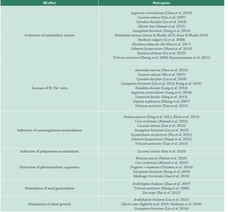
Important roles have been ascribed to NO in plants tolerance to abiotic stress, in which the increment of this signaling molecule in cells was associated to a variety of strategies used by plants to cope high salinity (Tab. 1). The
Atnoa1 Arabidopsis mutant plants, that exhibit impaired NO biosynthesis, were demonstrated to be highly sensitive to high salinity, being more vulnerable to oxidative stress and presenting lower germination and survival rates under such condition (Zhao et al. 2007).
Activation of the antioxidant system is one of the NO roles in plants under salt stress. Several findings indicate an improvement in the performance of enzymatic and non-enzymatic antioxidant systems in salt-stressed plants treated with NO donors. Cucumber seedlings hydroponically grown in medium containing 50 mM NaCl and 100 µM sodium nitroprusside (SNP; NO+ donor) showed higher activity of superoxide dismutase (SOD), catalase (CAT), peroxidase and ascorbate peroxidase (APX) when compared to cucumber solely treated with 50 mM NaCl (Fan et al.
2007). As a result, cells from these seedlings presented lower membrane permeability and decreased levels of O2-, H2O2 and lipid peroxides (Fan et al. 2007). The NO was found to have dual role on SOD by positively modulating the FeSOD expression and negatively affecting Cu/ZnSOD
one. With this, NO furnished a differential antioxidant protection to salt-stressed sunflower (Helianthus annuus)
seedlings (Arora & Bhatla 2015). Likewise, the challenge of tobacco roots with NaCl led to accumulation of endogenous NO in leaves that was accompanied by an increment of SOD and CAT activities (da-Silva et al. 2017). The activity of monodehydroascorbate reductase, dehydroascorbate reductase (DHAR), glutathione reductase (GR), glutathione
S-transferase, glutathione peroxidase (GPX), glyoxalase I and glyoxalase II (both related to methylglyoxylate detoxification) was also stimulated by SNP in wheat plants treated with 300 mM NaCl (Hasanuzzaman et al. 2011).
Induction of non-enzymatic antioxidant system [AsA and reduced glutathione (GSH)] was observed in wheat seedlings treated with SNP prior to NaCl exposure (Hasanuzzaman et al. 2011). The treatment with SNP decreased ferritin levels in barley (Hordeum vulgare) seedlings, which contributed to the attenuation of oxidative stress triggered by high salinity (Li et al. 2008). Likewise, exogenous NO alleviated
high-salinity-triggered oxidative stress in soybean (Glycine max;Simaei et al. 2012), mangrove (Aegiceras corniculatum; Chen et al. 2014), tomato (Solanum lycopersicum; Manai et al. 2014), cotton (Gossypium hirsutum; Dong et al. 2014), spinach (Du et al. 2015), sunflower (Kaur & Bhatla 2016) and bermudagrass (Cynodon dactylon; Liu et al. 2016).
The protective role of NO on plant photosynthetic apparatus is also documented. The SNP at 100 µM restored chloroplast pigments and maximum photochemical efficiency of photosystem II to normal levels in strawberries (Fragaria × ananassa cv. ‘Camarosa’) plants challenged with high salinity (Christou et al. 2014). Similar results were observed in salt-stressed cotton seedlings wherein application of 100 µM SNP to leaves improved plants photosynthetic performance (Liu et al. 2014). The treatment of chickpea (Cicer arietinum) plants with 100 mM NaCl and
50 µM of S-nitroso-N-acetylpenicillamine (SNAP; an NO donor) provided higher amounts of chlorophylls a and b
and carotenoids in plant leaves in comparison with those solely treated with NaCl (Ahmad et al. 2016). Aspersion of
Table 1: Roles of nitric oxide (NO) during plant response to salt stress.
NO effect Plant species
Activation of antioxidant system
Aegiceras corniculatum (Chen et al. 2014) Cucumis sativus (Fan et al. 2007) Cynodon dactylon (Liu et al. 2016)
Glycine max (Simaei et al. 2012) Gossypium hirsutum (Dong et al. 2014)
Helianthus annuus (Arora & Bhatla 2015; Kaur & Bhatla 2016) Hordeum vulgare (Li et al. 2008)
Nicotiana tabacum (da-Silva et al. 2017) Solanum lycopersicum (Manai et al. 2014)
Spinacia oleracea (Du et al. 2015)
Triticum aestivum (Zheng et al. 2008; Hasanuzzaman et al. 2011)
Increase of K+/Na+ ratio
Avicennia marina (Chen et al. 2010) Cucumis sativus (Shi et al. 2007) Cynodon dactylon (Liu et al. 2016) Gossypium hirsutum (Liu et al. 2013; Kong et al. 2016)
Kandelia obovata (Lang et al. 2014) Aegiceras corniculatum (Lang et al. 2014)
Limonium bicolor (Ding et al. 2013) Populus euphratica (Zhang et al. 2007)
Triticum aestivum (Tian et al. 2015)
Induction of osmoregulators accumulation
Brassica juncea (Zeng et al. 2011; Khan et al. 2012) Cicer arietinum (Ahmad et al. 2016)
Cucumis sativus (Fan et al. 2013) Gossypium hirsutum (Liu et al. 2013) Lycopersicom esculentum (Wu et al. 2011) Solanum lycopersicum (Hayat et al. 2012) Triticum aestivum (Tian et al. 2015)
Induction of polyamines accumulation Cucumis sativus (Fan et al. 2013)
Protection of photosynthetic apparatus
Brassica juncea (Fatma et al. 2016) Cicer arietinum (Ahmad et al. 2016) Fragaria × ananassa (Christou et al. 2014)
Gossypium hirsutum (Kong et al. 2016) Medicago truncatula (Jian et al. 2016)
Stimulation of seed germination
Arabidopisis thaliana (Zhao et al. 2007) Triticum aestivum (Zheng et al. 2008)
Zea mays (Bai et al. 2011)
Stimulation of plant growth
Arabidopisis thaliana (Liu et al. 2015) Glycine max (Egbichi et al. 2014; Vaishnav et al. 2016)
Gossypium hirsutum (Liu et al. 2014)
and increased chlorophylls content and photosynthetic rate (Kong et al. 2016). The NO and sulfur nutrition were found to prevent chloroplasts damage in salt-exposed mustard (B. juncea) plants (Fatma et al. 2016). The SNP stimulated the expression of AOX, a component of plant mitochondrial electron transport, in barrelclover (M. truncatula)under high salinity thus, alleviating oxidative stress and photosynthetic damages (Jian et al. 2016).
Inhibition of plasma membrane H+-ATPase and tonoplast H+-PPase caused by NaCl was prevented by 50 µM SNP in cucumber plants (Shi et al. 2007). Additionally, the gene expression of a plasma membrane H+-ATPase was stimulated by SNP in salt-stressed calluses of desert poplar (Populus euphratica), which in turn resulted in higher K+/Na+ ratio
(Zhang et al. 2007). Plasma membrane H+-ATPase and tonoplast Na+/H+ antiporter proteins were also induced by SNP in salt-stressed Avicennia marina and caused an increment of K+/Na+ ratio due to Na+ efflux from cells towards salt glands (Chen et al. 2010). Similar results were observed in cotton (Kong et al. 2016), Kandelia obovate and A. corniculatum
(Lang et al. 2014). Besides intense Na+ secretion from sea-lavender (Limonium bicolor) leaves under stress, SNP caused an increment in the number of Na+-loaded salt glands in salt-stressed plants (Ding 2013). In addition to increasing K+/Na+ ratio, SNP enhanced Ca2+ and Mg2+ uptake in salt-stressed plants (Liu et al. 2013; Tian et al. 2015; Liu et al. 2016).
incubated with SNP prior to salt stress exhibited higher relative water content (RWC) than salt-stressed plants devoid of NO treatment (Dinler et al. 2014). Exogenous NO also stimulated proline accumulation in several plant species (Wu
et al. 2011; Zeng et al. 2011; Hayat et al. 2012; Khan et al.
2012; Fan et al. 2013; Liu et al. 2013). The activity of pyrroline-5-carboxylate synthetase and proline dehydrogenase, enzymes involved in L-proline biosynthesis, and L-proline accumulation were boosted by SNP in cucumber seedlings under high salinity (Fan et al. 2013). Then, cellular turgor was maintained at normal levels and seedlings overcame NaCl stress. Mustard plants subjected to salt stress exhibited higher amounts of glycine betaine when treated with SNP (Khan et al. 2012), while wheat plants accumulated soluble carbohydrates in cells (Tian et al. 2015). The NO released from SNAP also induced accumulation of L-proline, L-glycine betaine, soluble proteins and carbohydrates in leaves of salt-stressed chickpea (Ahmad et al. 2016).
The combined treatment of cucumber seedlings with NaCl and SNP caused an increment of spermine levels and (spermidine + spermine)/putrescine ratio, which in turn helped plant cells to cope with the abiotic stress imposed (Fan et al. 2013). Polyamines, such as spermine and spermidine allows for protein, nucleic acid and cell membrane stabilization, besides being great osmolytes and inducers of plant growth and development (Fan et al. 2013). The SNP-induced germination of salt-stressed wheat seeds was attributed to the maintenance of K+/Na+ balance, increase of SOD and CAT activities and decrease of the lipid peroxides, H2O2 and O2- levels (Zheng et al. 2008). The SNAP, together with G-proteins, induced the protein accumulation,
the antioxidant enzymes activity, the proteins related to cell defense, the energy metabolism and the cell division in salt-treated maize seedlings (Bai et al. 2011).
The application of an NO donor on NaCl-treated soybean improved plants growth and biomass accumulation in shoot, root and nodules (Egbichi et al. 2014; Vaishnav et al. 2016). Indeed, the NaCl-triggered disruption of Pseudomonas simiae
(rhizobacteria) colonization in soybean was reverted by 100 µM SNP and allowed plant to tolerate salt stress (Vaishnav
et al. 2016). Conversely, increased levels of NO triggered the decrease of root meristems growth through auxin depletion in NaCl-treated Arabidopsis (Liu et al. 2015). In fact, removal of endogenous NO from roots rescued, in part,
PIN expression and destabilized IAA17 protein, involved in the repression of auxin signaling.
Hydrogen sulfide
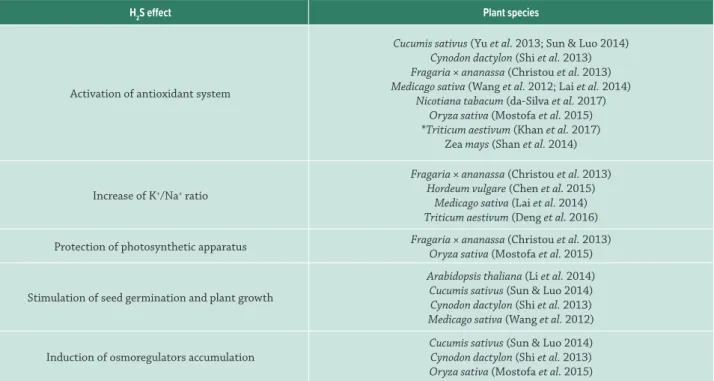
Many physiological processes were also found to be regulated by H2S in plants capable to tolerate different types of stress, including high salinity (Tab. 2).
Oxidative burst, an uncontrolled overproduction of ROS, is one of the first events elicited in plants cell upon salt stress, leading to intensification of electrolytes leakage, lipid peroxidation and protein oxidation. In fact, mitigation of oxidative stress in salt-stressed plants is one of the most studied roles of H2S. The activities of SOD, CAT, APX, GR, GPX and DHAR in stressed cucumber seedlings were increased by treatment with NaHS (an H2S-donor) while H2O2 and lipid peroxide levels decreased under the same experimental conditions (Yu et al. 2013). Undeniably,
Table 2: Roles of hydrogen sulfide (H2S) during plant response to salt stress.
H2S effect Plant species
Activation of antioxidant system
Cucumis sativus (Yu et al. 2013; Sun & Luo 2014) Cynodon dactylon (Shi et al. 2013) Fragaria × ananassa (Christou et al. 2013) Medicago sativa (Wang et al. 2012; Lai et al. 2014)
Nicotiana tabacum (da-Silva et al. 2017) Oryza sativa (Mostofa et al. 2015) *Triticum aestivum (Khan et al. 2017)
Zea mays (Shan et al. 2014)
Increase of K+/Na+ ratio
Fragaria × ananassa (Christou et al. 2013) Hordeum vulgare (Chen et al. 2015)
Medicago sativa (Lai et al. 2014) Triticum aestivum (Deng et al. 2016)
Protection of photosynthetic apparatus Fragaria × ananassa (Christou et al. 2013) Oryza sativa (Mostofa et al. 2015)
Stimulation of seed germination and plant growth
Arabidopsis thaliana (Li et al. 2014) Cucumis sativus (Sun & Luo 2014) Cynodon dactylon (Shi et al. 2013) Medicago sativa (Wang et al. 2012)
Induction of osmoregulators accumulation
Cucumis sativus (Sun & Luo 2014) Cynodon dactylon (Shi et al. 2013) Oryza sativa (Mostofa et al. 2015)
the suppression of endogenous H2S by infiltration of tobacco leaves with hypotaurine negatively affected the activity of SOD, CAT and APX in NaCl stress plants (da-Silva et al. 2017). The activity of enzymes involved in GSH (γ-glutamylcysteine synthetase) and AsA (L-galactono-1,4-lactone dehydrogenase) biosyntheses and further increment of GSH/oxidized glutathione and AsA/DHR ratios were stimulated by NaHS in leaves of salt-treated maize (Shan
et al. 2014). Similarly to the observed for NO donors, NaHS controlled methylglyoxylate levels in rice by increasing the activity of glyoxalase I and glyoxalase II (Mostofa et al.
2015). In addition, NaHS decreased in plants the activity of lipoxygenase, an enzyme implicated in the formation of lipid peroxides. Alleviation of NaCl-induced oxidative stress by H2S exogenous was also observed in alfalfa, bermudagrass, strawberry and cucumber (Wang et al. 2012; Christou et al. 2013; Shi et al. 2013; Lai et al. 2014; Sun & Luo 2014).
The maintenance of high K+/Na+ ratio in plant cells under salt-stress was also reported to be induced by H2S. Wheat seedlings treated with 50 µM NaHS, followed by 100 mM NaCl exposure exhibited increased K+/Na+ ratio with augment of selective transport of K+ over Na+ through nonselective cation channels and salt overly sensitive 1 (SOS1), a plasma membrane Na+/H+ antiporter (Deng et al. 2016). Induction of plasma membrane Na+/H+ antiporter genes (e.g.SOS2 -like, SOS3-like and SOS4) by NaHS was also described in strawberry plants under high salinity, indicating a role for H2S in K+ uptake (Christou et al. 2013). The K+/Na+ homeostasis in salt-treated alfalfa was shown to be maintained by NaHS through the prevention of K+ efflux likely triggered by lower expression of shaker-like K+ outward-rectifying channel genes (Lai et al. 2014). Similar results were shown in roots of salt-treated barley seedlings in the presence of NaHS (Chen et al.
2015). Remarkably, H2S maintained low Na+ levels in cells by increasing the transcription of genes that encode for plasma membrane H+-ATPase, H+-ATPase subunit β and vacuolar Na+/H+ antiporter and augmenting Na+ compartmentation in vacuoles (Chen et al. 2015).
Germination of alfalfa seeds under 100 mM NaCl was stimulated by 100 µM NaHS (Wang et al. 2012). The improvement of seed germination rate caused by H2S may be a result of the induction of starch break down in the endosperm as the activity of α-amylase and β-amylase increased in salt-stressed cucumber seeds upon treatment with NaHS and ultimately led to hypocotyl and radicle growth (Sun & Luo 2014). The inhibition of root growth in Arabidopsis under salt stress was abolished by NaHS (Li et al. 2014), while this H2S donor improved the survival rate of
salt-treated bermudagrass (Shi et al. 2013). The treatment of strawberry roots with NaHS prior to NaCl exposure resulted in increased photosynthetic rate, stomatal conductance and RWC in leaves in comparison with plants solely exposed to NaCl (Christou et al. 2013). Similarly, an H2S donor increased chlorophyll, carotenoid and total protein contents in rice under salinity (Mostofa et al. 2015).
Exogenous H2S also led to the accumulation of L-proline, sucrose and other soluble carbohydrates in NaCl-stressed bermudagrass cells (Shi et al. 2013). Soluble carbohydrates also accumulated in hypocotyl and radicle cells of cucumber plants stressed with sodium bicarbonate and treated with NaHS (Sun & Luo 2014).
Interplay between NO and H
2S during plant response
to salt stress
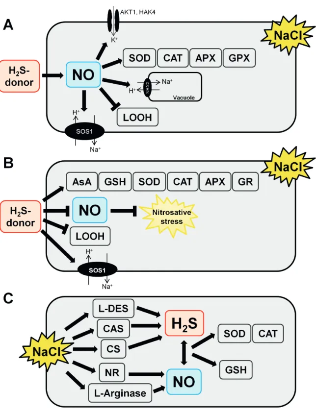
It is unquestionable that NO and H2S share roles in the signaling pathway that leads to plant tolerance to environmental stresses. Then, the extent of the cooperative function between these signaling molecules has received considerable attention recently.
Studies carried out with alfalfa seeds treated with NaCl for 24 h and barley seedlings challenged with NaCl for 48 h suggested that H2S might induce NO production during the response to the stress (Tab. 2; Wang et al. 2012; Chen
et al. 2015). The simultaneous treatment of alfalfa with 100 µM NaHS (H2S donor) and 100 mM NaCl increased NO levels in cells by 30 %. This increment was accompanied of an increase in K+/Na+ ratio and transcription levels of SOD, CAT, APX and guaiacol peroxidase genes and a decrease in lipid peroxides (Wang et al. 2012). The use of a specific
NO scavenger reversed the NaHS effects on alfalfa, clearly indicating the influence of exogenous H2S on NO endogenous levels. The NaHS (100 µM) also boosted NO production in barley seedlings by 30 % in comparison to control, detected
in situ using a fluorophore specific to NO (Chen et al. 2015). Meanwhile, NaHS maintained ionic homeostasis through the decrease of K+ cell efflux and increase of Na+ in vacuoles. The gene expression of an inward-rectifying potassium channel (HvAKT1) and a high-affinity K+ uptake (HvHAK4) protein system also increased in barley upon concomitant treatment with NaCl and NaHS. Up-regulation of transcriptional levels of vacuolar Na+/H+ antiporter (HvVNHX2), H+-ATPase subunit β (HvVHA-β) and protein expression of vacuolar Na+/H+ antiporter (NHE1) was also observed in barley under the experimental conditions tested (Chen et al. 2015). In contrast, the treatment of NaCl-exposed strawberries plants with NaHS (100 µM) decreased NO levels in plant cells by 1.7-fold (Christou et al. 2013; Tab. 2). The decrease in the NO levels in strawberry was attributed by the authors to a possible control of nitrosative stress. Likewise, the lipid peroxide levels decreased while increment of expression of genes encoding for antioxidant enzymes and biosynthesis of AsA, GSH and SOS was recorded. Another line of evidence shows that endogenous NO and H2S stimulate the production of one another in tobacco leaves after NaCl stress for 10 days, as hypotaurine (an H2S scavenger) compromised NO accumulation in ca. 1.3-fold and cPTIO (an NO scavenger) undermined H2S production in ca. 1.6-fold (da-Silva et al.
prevent water loss and drove to the control of oxidative stress assisted by GSH. Additionally, 200 µM S-nitrosoglutathione (an NO donor) enhanced the levels of L-cysteine by 10 % and the activity of L/D-DES and CS that, in turn, led to 20 % higher amounts of H2S in wheat seedlings under osmotic stress (Khan et al. 2017). The increase of H2S, provoked by exogenous NO, controlled oxidative stress by improving SOD, CAT, APX, GR, NR and peroxidase activities in plant cells and relieving H2O2 and O2- effects. Accumulation of L-proline and glycine betaine was also observed in osmotic-stressed wheat seedlings supplemented with exogenous NO (Khan et al. 2017).
Figure 2 summarizes the known interactions between NO and H2S, regardless of their origin (endogenous or not), determined during plant response to high salinity.
Concluding remarks
Both NO and H2S may originate in plants from several pathways in which NR seems to be indirectly the main source of NO while the majority of H2S produced comes from L-DES activity. The role of NO in the mitigation of oxidative burst in plants upon (a)biotic stress is known for roughly two decades and most recently, H2S has emerged as a new player in such signaling pathway, orchestrating biochemical events that lead plants tolerance to high salinity. Recent studies show that NO and H2S act together and influence the production of one another during plant response to relatively long periods of salt stress to improve plant antioxidant system, K+ uptake over Na+ and production of osmoprotective molecules. The extent of the interaction between these signaling molecules deserves more investigation, since there is still controversy with respect to which molecule triggers the cascade. Understanding this interplay will expand our knowledge on the complex biochemical cascade activated in plant cells with competence to cope with high salinity.
Acknowledgements
Part of the work cited in this review was financially supported by the Conselho Nacional de Desenvolvimento Científico e Tecnológico (CNPq), the Fundação de Amparo à Pesquisa do Estado de Minas Gerais (FAPEMIG) and the Coordenação de Aperfeiçoamento de Pessoal de Nível Superior (CAPES). LVM is supported by a CNPq research fellowship and CJS was granted with a CNPq studentship.
References
Abe K, Kimura H. 1996. The possible role of hydrogen sulfide as an endogenous neuromodulator. The Journal of Neuroscience 16: 1066-1071. Ahmad P, Latef AAA, Hashem A, Abd-Allah EF, Gucel S, Tran LSP. 2016.
Nitric oxide mitigates salt stress by regulating levels of osmolytes and antioxidant enzymes in chickpea. Frontiers in Plant Science 7: 1-11.
Akopyan TN, Braunstein AE, Goryachenkova EV. 1975. Beta-cyanoalanine synthase: purification and characterization. Proceedings of the National Academy of Sciences 72: 1617-1621.
Álvarez C, Calo L, Romero LC, García I, Gotor C. 2010. An O-acetylserine(thiol) lyase homolog with L-cysteine desulfhydrase activity regulates cysteine homeostasis in Arabidopsis. Plant Physiology 152: 656-669. Arasimowicz-Jelonek M, Floryszak-Wieczorek J, Kubiś J. 2009. Interaction
between polyamine and nitric oxide signaling in adaptive responses to drought in cucumber. Journal of Plant Growth Regulation 28: 177-186. Arora D, Bhatla SC. 2015. Nitric oxide triggers a concentration-dependent
differential modulation of superoxide dismutase (FeSOD and Cu/ ZnSOD) activity in sunflower seedling roots and cotyledons as an early and long distance signaling response to NaCl stress. Plant Signaling & Behavior 10: 1-10.
Bai X, Yang L, Yang Y, Ahmad P, Yang Y, Hu X. 2011. Deciphering the protective role of nitric oxide against salt stress at the physiological and proteomic levels in maize. Journal of Proteome Research 10: 4349-4364.
Bethke PC, Badger MR, Jones RL. 2004. Apoplastic synthesis of nitric oxide by plant tissues. The Plant Cell 16: 332-341.
Calderwood A, Kopriva S. 2014. Hydrogen sulfide in plants: from dissipation of excess sulfur to signaling molecule. Nitric Oxide 41: 72-78. Chen J, Wang WH, Wu FH, et al. 2015. Hydrogen sulfide enhances
salt tolerance through nitric oxide-mediated maintenance of ion homeostasis in barley seedling roots. Scientific Reports 5: 1-19. Chen J, Xiao Q, Wang C, et al. 2014. Nitric oxide alleviates oxidative stress
caused by salt in leaves of a mangrove species, Aegiceras corniculatum. Aquatic Botany 117: 41-47.
Chen J, Xiao Q, Wu F, et al. 2010. Nitric oxide enhances salt secretion and Na+ sequestration in a mangrove plant, Avicennia marina, through
increasing the expression of H+-ATPase and Na+/H+ antiporter under
high salinity. Tree Physiology 30: 1570-1585.
Christou A, Manganaris GA, Fotopoulos V. 2014. Systemic mitigation of salt stress by hydrogen peroxide and sodium nitroprusside in strawberry plants via transcriptional regulation of enzymatic and non-enzymatic antioxidants. Environmental and Experimental Botany 107: 46-54. Christou A, Manganaris GA, Papadopoulos I, Fotopoulos V. 2013. Hydrogen
sulfide induces systemic tolerance to salinity and non-ionic osmotic stress in strawberry plants through modification of reactive species biosynthesis and transcriptional regulation of multiple defence pathways. Journal of Experimental Botany 64: 1953-1966. Cooney RV, Harwood PJ, Custer LJ, Franke AA. 1994. Light-mediated
conversion of nitrogen dioxide to nitric oxide by carotenoids. Environmental Health Perspectives 102: 460-462.
Cui W, Chen H, Zhu K, et al. 2014. Cadmium-induced hydrogen sulfide synthesis is involved in cadmium tolerance in Medicago sativa by reestablishment of reduced (homo) glutathione and reactive oxygen species homeostases. PloS One 9: 1-12. https://doi.org/10.1371/ journal.pone.0109669.
da-Silva CJ, Fontes EPB, Modolo LV. 2017. Salinity-induced accumulation of endogenous H2S and NO is associated with modulation of the antioxidant and redox defense systems in Nicotiana tabacum L. cv. Havana. Plant Science 256: 148-159.
Del-Río LA, Corpas FJ, Barroso JB. 2004. Nitric oxide and nitric oxide synthase activity in plants. Phytochemistry 65: 783-792.
Delledonne M, Xia Y, Dixon RA, Lamb C. 1998. Nitric oxide functions as a signal in plant disease resistance. Nature 394: 585-588.
Deng YQ, Bao J, Yuan F, Liang X, Feng ZT, Wang BS. 2016. Exogenous hydrogen sulfide alleviates salt stress in wheat seedlings by decreasing Na+ content. Plant Growth Regulation 79: 391-399.
Ding F. 2013. Effects of salinity and nitric oxide donor sodium nitroprusside (SNP) on development and salt secretion of salt glands of Limonium bicolor. Acta Physiologiae Plantarum 35: 741-747.
Dinler BS, Antoniou C, Fotopoulos V. 2014. Interplay between GST and nitric oxide in the early response of soybean (Glycine max L.) plants to salinity stress. Journal of Plant Physiology 171: 1740-1747. Dong YJ, Jinc SS, Liu S, Xu LL, Kong J. 2014. Effects of exogenous nitric
Du ST, Liu Y, Zhang P, Liu HJ, Zhang XQ, Zhang RR. 2015. Atmospheric application of trace amounts of nitric oxide enhances tolerance to salt stress and improves nutritional quality in spinach (Spinacia oleracea L.). Food Chemistry 173: 905-911.
Durner J, Wendehenne D, Klessig DF. 1998. Defense gene induction in tobacco by nitric oxide, cyclic GMP, and cyclic ADP-ribose. Proceedings of the National Academy of Sciences 95: 10328-10333.
Egbichi I, Keyster M, Ludidi N. 2014. Effect of exogenous application of nitric oxide on salt stress responses of soybean. South African Journal of Botany 90: 131-136.
Fan HF, Du CX, Guo SR. 2013. Nitric oxide enhances salt tolerance in cucumber seedlings by regulating free polyamine content. Environmental and Experimental Botany 86: 52-59.
Fan H, Guo S, Jiao Y, Zhang R, Li J. 2007. Effects of exogenous nitric oxide on growth, active oxygen species metabolism, and photosynthetic characteristics in cucumber seedlings under NaCl stress. Frontiers of Agriculture in China 1: 308-314.
Fatma M, Masood A, Per TS, Khan NA. 2016. Nitric oxide alleviates salt stress inhibited photosynthetic performance by interacting with sulfur assimilation in mustard. Frontiers in Plant Science 7: 1-16. Groppa MD, Rosales EP, Iannone MF, Benavides MP. 2008. Nitric
oxide, polyamines and Cd-induced phytotoxicity in wheat roots. Phytochemistry 69: 2609-2615.
Guo FQ, Okamoto M, Crawford NM. 2003. Identification of a plant nitric oxide synthase gene involved in hormonal signaling. Science 302: 100-103.
Gupta B, Huang B. 2014. Mechanism of salinity tolerance in plants: physiological, biochemical, and molecular characterization. International Journal of Genomics 2014: 1-18.
Gupta KJ, Fernie AR, Kaiser WM, van-Dongen JT. 2011. On the origins of nitric oxide. Trends in Plant Science 16: 160-168.
Harrington HM, Smith IK. 1980. Cysteine metabolism in cultured tobacco cells. Plant Physiology 65: 151-155.
Hasanuzzaman M, Hossain MA, Fujita M. 2011. Nitric oxide modulates antioxidant defense and the methylglyoxal detoxification system and reduces salinity-induced damage of wheat seedlings. Plant Biotechnology Reports 5: 353-365.
Hatzfeld Y, Maruyama A, Schmidt A, Noji M, Ishizawa K, Saito K. 2000. β-Cyanoalanine synthase is a mitochondrial cysteine synthase-like protein in spinach and Arabidopsis. Plant Physiology 123: 1163-1172. Hayat S, Yadav S, Wani AS, Irfan M, Alyemini MN, Ahmad A. 2012. Impact of sodium nitroprusside on nitrate reductase, proline content, and antioxidant system in tomato under salinity stress. Horticulture, Environment and Biotechnology 53: 362-367.
Jian W, Zhang DW, Zhu F, et al. 2016. Alternative oxidase pathway is involved in the exogenous SNP-elevated tolerance of Medicago truncatula to salt stress. Journal of Plant Physiology 193: 79-87. Jin Z, Xue S, Luo Y, et al. 2013. Hydrogen sulfide interacting with abscisic
acid in stomatal regulation responses to drought stress in Arabidopsis. Plant Physiology and Biochemistry 62: 41-46.
Kaur H, Bhatla SC. 2016. Melatonin and nitric oxide modulate glutathione content and glutathione reductase activity in sunflower seedling cotyledons accompanying salt stress. Nitric Oxide 59: 42-53. Keyster M, Klein A, Ludidi N. 2012. Caspase-like enzymatic activity and
the ascorbate-glutathione cycle participate in salt stress tolerance of maize conferred by exogenously applied nitric oxide. Plant Signaling & Behavior 7: 349-360.
Khan MN, Mobin M, Abbas ZK, Siddiqui MH. 2017. Nitric oxide-induced synthesis of hydrogen sulfide alleviates osmotic stress in wheat seedlings through sustaining antioxidant enzymes, osmolyte accumulation and cysteine homeostasis. Nitric Oxide 68: 91-102. Khan MN, Siddiqui MH, Mohammad F, Naeem M. 2012. Interactive role
of nitric oxide and calcium chloride in enhancing tolerance to salt stress. Nitric Oxide 27: 210-218.
Klepper L. 1979. Nitric oxide (NO) and nitrogen dioxide (NO2) emissions from herbicide-treated soybean plants. Atmospheric Environment 13: 537-542.
Klepper L. 1990. Comparison between NOx evolution mechanisms of wild-type and nr1 mutant soybean leaves. Plant Physiology 93: 26-32. Kong X, Wang T, Li W, Tang W, Zhang D, Dong H. 2016. Exogenous nitric
oxide delays salt-induced leaf senescence in cotton (Gossypium hirsutum L.). Acta Physiologiae Plantarum 38: 1-9.
Kuznetsova S, Knaff DB, Hirasawa M, Lagoutte B, Sétif P. 2004. Mechanism of spinach chloroplast ferredoxin-dependent nitrite reductase: spectroscopic evidence for intermediate states. Biochemistry 43: 510-517.
Lai D, Mao Y, Zhou H, et al. 2014. Endogenous hydrogen sulfide enhances salt tolerance by coupling the reestablishment of redox homeostasis and preventing salt-induced K+ loss in seedlings of Medicago sativa.
Plant Science 225: 117-129.
Lang T, Sun H, Li N, et al. 2014. Multiple signaling networks of extracellular ATP, hydrogen peroxide, calcium, and nitric oxide in the mediation of root ion fluxes in secretor and non-secretor mangroves under salt stress. Aquatic Botany 119: 33-43.
Li H, Samouilov A, Liu X, Zweier JL. 2004. Characterization of the effects of oxygen on xanthine oxidase-mediated nitric oxide formation. Journal of Biological Chemistry 279: 16939-16946.
Li J, Jia H, Wang J, Cao Q, Wen Z. 2014. Hydrogen sulfide is involved in maintaining ion homeostasis via regulating plasma membrane Na+/
H+ antiporter system in the hydrogen peroxide-dependent manner
in salt-stress Arabidopsis thaliana root. Protoplasma 251: 899-912. Li QY, Niu HB, Yin J, et al. 2008. Protective role of exogenous nitric oxide
against oxidative-stress induced by salt stress in barley (Hordeum vulgare). Colloids and Surfaces B: Biointerfaces 65: 220-225. Li ZG. 2015. Chapter Thirteen: analysis of some enzymes activities of
hydrogen sulfide metabolism in plants. Methods in Enzymology 555: 253-269.
Li ZG, Xie LR, Li XJ. 2015. Hydrogen sulfide acts as a downstream signal molecule in salicylic acid-induced heat tolerance in maize (Zea mays L.) seedlings. Journal of Plant Physiology 177: 121-127.
Liu A, Fan J, Gitau MM, Chen L, Fu J. 2016. Nitric oxide involvement in bermudagrass response to salt stress. Journal of the American Society for Horticultural Science 141: 425-433.
Liu S, Dong Y, Xu L, Kong J. 2014. Effects of foliar applications of nitric oxide and salicylic acid on salt-induced changes in photosynthesis and antioxidative metabolism of cotton seedlings. Plant Growth Regulation 73: 67-78.
Liu S, Dong YJ, Xu LL, Kong J, Bai XY. 2013. Roles of exogenous nitric oxide in regulating ionic equilibrium and moderating oxidative stress in cotton seedlings during salt stress. Journal of Soil Science and Plant Nutrition 13: 929-941.
Liu W, Li RJ, Han TT, Cai W, Fu ZW, Lu YT. 2015. Salt stress reduces root meristem size by nitric oxide-mediated modulation of auxin accumulation and signaling in Arabidopsis. Plant Physiology 168: 343-356.
Magalhaes JR, Monte DC, Durzan D. 2000. Nitric oxide and ethylene emission in Arabidopsis thaliana. Physiology and Molecular Biology of Plants 6: 117-127.
Manai J, Gouia H, Corpas FJ. 2014. Redox and nitric oxide homeostasis are affected in tomato (Solanum lycopersicum) roots under salinity-induced oxidative stress. Journal of Plant Physiology 171: 1028-1035. Moche M, Stremlau S, Hecht L, Göbel C, Feussner I, Stöhr C. 2010. Effect
of nitrate supply and mycorrhizal inoculation on characteristics of tobacco root plasma membrane vesicles. Planta 231: 425-436. Modolo LV, Augusto O, Almeida IM, Magalhaes JR, Salgado I. 2005. Nitrite
as the major source of nitric oxide production by Arabidopsis thaliana in response to Pseudomonas syringae. FEBS Letters 579: 3814-3820. Moreau M, Lee GI, Wang Y, Crane BR, Klessig DF. 2008. AtNOS/AtNOA1 is a functional Arabidopsis thaliana cGTPase and not a nitric-oxide synthase. Journal of Biological Chemistry 283: 32957-32967. Mostofa MG, Saegusa D, Fujita M, Tran LSP. 2015. Hydrogen sulfide
regulates salt tolerance in rice by maintaining Na+/K+ balance, mineral
homeostasis and oxidative metabolism under excessive salt stress. Frontiers in Plant Science 6: 1-14.
Munns R, Tester M. 2008. Mechanisms of salinity tolerance. Annual Review of Plant Biology 59: 651-681.
Parihar P, Singh S, Singh R, Singh VP, Prasad SM. 2015. Effect of salinity stress on plants and its tolerance strategies: a review. Environmental Science and Pollution Research 22: 4056-4075.
Planchet E, Gupta KJ, Sonoda M, Kaiser WM. 2005. Nitric oxide emission from tobacco leaves and cell suspensions: rate limiting factors and evidence for the involvement of mitochondrial electron transport. The Plant Journal 41: 732-743.
Riemenschneider A, Riedel K, Hoefgen R, Papenbrock J, Hesse H. 2005. Impact of reduced O-acetylserine(thiol)lyase isoform contents on potato plant metabolism. Plant Physiology 137: 892-900.
Rockel P, Strube F, Rockel A, Wildt J, Kaiser WM. 2002. Regulation of nitric oxide (NO) production by plant nitrate reductase in vivo and in vitro. Journal of Experimental Botany 53: 103-110.
Romero LC, García I, Gotor C. 2013. L-cysteine desulfhydrase 1 modulates the generation of the signaling molecule sulfide in plant cytosol. Plant Signaling & Behavior 8: 4621-34.
Rümer S, Gupta KJ, Kaiser WM. 2009. Plant cells oxidize hydroxylamines to NO. Journal of Experimental Botany 60: 2065-2072.
Scuffi D, Álvarez C, Laspina N, Gotor C, Lamattina L, García-Mata C. 2014. Hydrogen sulfide generated by L-cysteine desulfhydrase acts upstream of nitric oxide to modulate abscisic acid-dependent stomatal closure. Plant Physiology 166: 2065-2076.
Shan C, Liu H, Zhao L, Wang X. 2014. Effects of exogenous hydrogen sulfide on the redox states of ascorbate and glutathione in maize leaves under salt stress. Biologia Plantarum 58: 169-173.
Shi H, Ye T, Chan Z. 2013. Exogenous application of hydrogen sulfide donor sodium hydrosulfide enhanced multiple abiotic stress tolerance in bermudagrass (Cynodon dactylon (L). Pers.). Plant Physiology and Biochemistry 71: 226-234.
Shi Q, Ding F, Wang X, Wei M. 2007. Exogenous nitric oxide protect cucumber roots against oxidative stress induced by salt stress. Plant Physiology and Biochemistry 45: 542-550.
Simaei M, Khavari-Nejad RA, Bernard F. 2012. Exogenous application of salicylic acid and nitric oxide on the ionic contents and enzymatic activities in NaCl-stressed soybean plants. American Journal of Plant Sciences 3: 1495-1503.
Stöhr C, Strube F, Marx G, Ullrich WR, Rockel P. 2001. A plasma membrane-bound enzyme of tobacco roots catalyses the formation of nitric oxide from nitrite. Planta 212: 835-841.
Stoimenova M, Igamberdiev AU, Gupta KJ, Hill RD. 2007. Nitrite-driven anaerobic ATP synthesis in barley and rice root mitochondria. Planta 226: 465-474.
Sun YD, Luo WR. 2014. Effects of exogenous hydrogen sulphide on seed germination and seedling growth of cucumber (Cucumis sativus) under sodium bicarbonate stress. Seed Science and Technology 42: 126-131. Tian X, He M, Wang Z, et al. 2015. Application of nitric oxide and calcium nitrate enhances tolerance of wheat seedlings to salt stress. Plant Growth Regulation 77: 343-356.
Tun NN, Santa-Catarina C, Begum T, et al. 2006. Polyamines induce rapid biosynthesis of nitric oxide (NO) in Arabidopsis thaliana seedlings. Plant and Cell Physiology 47: 346-354.
Vaishnav A, Kumari S, Jain S, Varma A, Tuteja N, Choudhary DK. 2016. PGPR‐mediated expression of salt tolerance gene in soybean through volatiles under sodium nitroprusside. Journal of Basic Microbiology 56: 1274-1288.
Wang BL, Tang XY, Cheng LY, et al. 2010. Nitric oxide is involved in phosphorus deficiency‐induced cluster‐root development and citrate exudation in white lupin. New Phytologist 187: 1112-1123. Wang RUI. 2002. Two’s company, three’s a crowd: can H2S be the third
endogenous gaseous transmitter?. The FASEB Journal 16: 1792-1798. Wang Y, Li L, Cui W, Xu S, Shen W, Wang R. 2012. Hydrogen sulfide
enhances alfalfa (Medicago sativa) tolerance against salinity during seed germination by nitric oxide pathway. Plant and Soil 351: 107-119. Wilkinson JQ, Crawford NM. 1993. Identification and characterization
of a chlorate-resistant mutant of Arabidopsis thaliana with mutations in both nitrate reductase structural genes NIA1 and NIA2. Molecular and General Genetics MGG 239: 289-297.
Wirtz M, Hell R. 2006. Functional analysis of the cysteine synthase protein complex from plants: structural, biochemical and regulatory properties. Journal of Plant Physiology 163: 273-286.
Wu X, Zhu W, Zhang H, Ding H, Zhang HJ. 2011. Exogenous nitric oxide protects against salt-induced oxidative stress in the leaves from two genotypes of tomato (Lycopersicom esculentum Mill.). Acta Physiologiae Plantarum 33: 1199-1209.
Yamasaki H, Sakihama Y. 2000. Simultaneous production of nitric oxide and peroxynitrite by plant nitrate reductase: in vitro evidence for the NR‐dependent formation of active nitrogen species. FEBS Letters 468: 89-92.
Yu LX, Zhang CJ, Shang HQ, et al. 2013. Exogenous hydrogen sulfide enhanced antioxidant capacity, amylase activities and salt tolerance of cucumber hypocotyls and radicles. Journal of Integrative Agriculture 12: 445-456.
Zeng CL, Liu L, Wang BR, Wu XM, Zhou Y. 2011. Physiological effects of exogenous nitric oxide on Brassica juncea seedlings under NaCl stress. Biologia Plantarum 55: 345-348.
Zhang F, Wang Y, Yang Y, Wu HAO, Wang DI, Liu J. 2007. Involvement of hydrogen peroxide and nitric oxide in salt resistance in the calluses from Populus euphratica. Plant, Cell & Environment 30: 775-785. Zhang H, Hu LY, Hu KD, He YD, Wang SH, Luo JP. 2008. Hydrogen sulfide
promotes wheat seed germination and alleviates oxidative damage against copper stress. Journal of Integrative Plant Biology 50: 1518-1529. Zhang L, Pei Y, Wang H, et al. 2015. Hydrogen sulfide alleviates
cadmium-induced cell death through restraining ROS accumulation in roots of Brassica rapa L. ssp. pekinensis. Oxidative Medicine and Cellular Longevity 1-11.
Zhao MG, Tian QY, Zhang WH. 2007. Nitric oxide synthase-dependent nitric oxide production is associated with salt tolerance in Arabidopsis. Plant Physiology 144: 206-217.