Mating type loci analysis indicates that
Villosiclava virens
, the
casual agent of false smut disease of rice, is a homothallic
fungus
Rongtao Fu, Chuanchun Yin, Qiming Deng, Ping Li & Aiping Zheng
Rice Research Institute, Sichuan Agricultural University, 211 Huiming Road, Wenjiang District, Chengdu City, Sichuan Province, China
Author for correspondence: Aiping Zheng, e-mail: aipingzh@163.com
ABSTRACT
Villosiclava virens (anamorph: Ustilaginoidea virens) was isolated from infected rice plants, where it caused false smut. Similar to other ascomycetes, the sexual compatibility of V. virens is controlled by the mating type (MAT) locus. In this study, we applied a PCR-based approach to screen mating type genes in the single asexual spore isolate. Our results showed that V. virens possesses the two master genes required for mating (MAT1-1-1 and MAT1-2-1), suggesting that this fungus is likely to be homothallic. Mating type primer pairs were developed to genotype the single ascospore isolates from different places in China. These analyses provided further evidence that V. virens is a homothallic species and suggest novel genetic mechanisms of sexual reproduction in V. virens.
Key words:Ustilaginoidea virens, asexual spores, homothallic, MAT, multiplex-PCR.
INTRODUCTION
Villosiclava virens (Nakata) E. Tanaka & C. Tanaka
is the causative agent of the important false smut of rice (Oryza sativa L.) and maize (Zea mays L.). The anamorphic
state of the causal fungus was called Ustilaginoidea virens
(Cooke) Takah. (Takahashi, 1896). The teleomorphic state of this fungus is allied with the family Clavicipitaceae and named Claviceps virens M. Sakurai ex Nakata (Sakurai,
1934). However, phylogenetic analyses have demonstrated that Ustilaginoideae species are not allied with the genus Claviceps (White et al., 2000; Bischoff et al., 2004).
Therefore, a new genus Villosiclava was proposed and V. virens has been used as the name for the rice false smut
fungus (Tanaka et al., 2008). The pathogen can infect roots at the young seedling stage and rice coleoptiles and staminal filaments at the earlier bolting stage (Tang et al., 2013). The typical symptom of rice false smut is the formation of greenish spore balls in spikelets, which are covered by an abundance of chlamydospores. The spore balls are initially yellow and turn greenish black with maturity, which is when they contaminate the rice grains. Ustiloxins are an additional concern for humans and livestock (Koiso et al., 1994). Villosiclavavirens can
form conidia and ascospores (Fu et al., 2012), which are often the source of infection. The sclerotia of V. virens,
which often form on the surface of mature spore balls, may germinate and produce sexually reproductive stages of the fungus (Singh & Dubey, 1984). Sexually reproduction
The sexual cycle of the pathogen plays an important role in disease epidemiology (Hayden et al., 2003). Mating type genes are the master loci that govern sexual reproduction and development in fungi (Metin et al., 2010; Butler, 2010), whereas sexual identity in plants and animals is known to be governed by genes located on the sex chromosomes (Fraser & Heitman, 2005; Marais & Galtier, 2003). Fungi can reproduce by selfing or outcrossing. In heterothallic ascomycetes such as Tuber melanosporum Vittad. (Rubini
et al., 2011), mating requires a partner of the opposite mating type. In different strains, the genes of opposite mating types are dissimilar sequences: one encodes an alpha-box-domain protein (MAT1-1), and the other encodes
a high mobility group (HMG) protein (MAT1-2) (Turgeon
& Yoder, 2000). In contrast to heterothallic species, in most homothallic ascomycetes a single strain contains the genes for both transcription factors, MAT1-1 and MAT1-2. This
genetic organization confers self-fertile mating ability (Debuchy & Turgeon, 2006; Yunet al., 2000). These genes in homothallic fungi are usually linked, but in some case they are not, such as in
Aspergillus nidulans (Eidam) G. Winter (Paoletti et al., 2007).
Similar to other ascomycetes, sexual reproduction in
V. virens is controlled by the Mat loci. In previous studies,
Yokoyama et al. (2006) suggested that V. virens (Claviceps oryzae-sativae in his work) was an heterothallic fungus
using a PCR-based assay. Here, we reexamined the V. virens
mating type genes in single asexual-spore (chlamydospore) isolates and ascospores isolates collected from China and determined the mating type genotype using multiplex PCR.
the distribution of mating types (Dyer et al., 2001). In addition, we analyzed the distribution of mating type genes in several isolates from the natural population.
MATERIAL AND METHODS
Fungal isolates and culture conditions
Isolates derived from conidia and ascospores used in this study are listed in Tables 1 and 2, respectively.
Isolation from conidia
These isolates were collected in southwestern and eastern China in 2011 and were obtained from naturally infected rice spikelets showing typical false smut symptoms. The yellowish rice kernels, which were covered with a mass of V. virens chlamydospores, were surface sterilized with
75% ethanol for 2 min and subsequently rinsed three times with sterile water. The treated specimens were resuspended into a chlamydospore suspension and diluted to 103 ml-1. An
aliquot (150 μl) of the spore suspension was poured onto potato dextrose agar (PDA) solid medium containing 100 μg/ml chloramphenicol. The plate was incubated at 28ºC in the dark. When visible colonies appeared, the colonies were transferred individually onto fresh PDA plates and incubated at 28ºC in the dark. Each isolate was individually maintained at 4°C on a separate 9-cm Petri dishes containing PDA solid medium.
Isolation from ascospores
Sclerotia were collected in the rice field and were buried in moist sterile sand at 25°C under light. The single ascospore was isolated as previously described (Singh & Dubey, 1984). The isolates were maintained at 4°C as described above.
DNA isolation and ITS-PCR
Villosiclava virens mycelia were inoculated into 50
ml potato dextrose liquid and kept on an incubator-shaker at 150 rpm. The mycelia were harvested by filtration and ground to a powder in the presence of liquid nitrogen. DNA was isolated as reported by Murray and Thompson (1980), resuspended in deionized water, and stored at -20°C.
Specific internal transcribed spacer (ITS) primers were used to verify the identity of V. virens isolates. The
polymerase chain reaction (PCR) mix (50 μl) contained 2 U polymerase (Takara), 5μl of 10× Taq buffer, 400 mM of dNTP’s, 2 μmol of DNA template and primers US1-5 and US3-3 (Table 3) (Zhou et al., 2003). PCR was performed using the following conditions: 96°C for 1 min, followed by 30 cycles of denaturing at 96°C for 20 s, annealing at 58°C for 30 s and extension 72°C for 30 s, followed by a final extension for 7 min at 72°C. PCR-amplified products were resolved by standard agarose gel electrophoresis and detected by Gelview (BioTeke) staining.
Isolates Locality Longitude Latitude Strain number a
MAT1 -1-1 MAT1 -2-1
Uvsf 104.16 31.19 8 + +
Uvmy 104.42 31.30 10 + +
Uvdz 107.29 31.14 6 + +
Uvmz 104.19 31.32 12 + +
Uvnj 105.02 29.36 9 + +
Uvzg 104.46 29.23 7 + +
Uvfa 119.39 27.06 6 + +
Uvlz 105.24 28.54 12 + +
Uvsn 105.33 30.31 9 + +
Uvkm 102.42 25.04 14 + +
Uvcd 104.04 30.40 26 + +
Uvhf 117.17 31.52 12 + +
Uvnc 106.04 30.49 10 + +
Uvfz 119.18 26.05 16 + +
Uvya 102.59 29.59 10 + +
Uvql 103.34 30.26 7 + +
Uvhz 120.10 30.16 10 + +
Uvxc 102.16 27.54 14 + +
Uvcz 103.40 30.39 8 + +
NBRC31672
Shifang
Mianyang
Dazhou
mianzhu
Neijiang
Zigong
Fuan
Luzhou
Shuining
Kunming
Chengdou
Hefei
Nanchong
Fuzhou
Yaan
Qionglai
Hangzhou
Xichang
Chongzhou
Japan 1 + +
TABLE 1 - Isolates of conidia used in this study.
aA total of 206 isolates collected in 19 Chinese regions. +Detected.
PCR amplification of mating type genes and DNA
sequencing
The primers for the MAT1 genes were designed based
on the high similarity to amino acid sequences of the alpha box and HMG box in Clavicipitaceae. We constructed two
sets of primers (MAT1-F1/MAT1-R1, MAT2-F1/MAT2-R1; Table 3) to amplify the relatively conserved MAT1-1
and MAT1-2 genes in V. virens.
PCR reactions were carried out in a 25 μl volume containing 25 ng DNA template, 20 mM Tris-HCl pH 8.4, 1.5 Mm MgCl2, 200 mM dNTP’s, 25 pmol of each
primer, and 1 U Taq polymerase (Takara). Amplification conditions were 95°C for 1 min, followed by 35 cycles of denaturing at 95°C for 30 s, annealing at 56°C for 1 min and extension at 72°C for 40 s, followed by a final extension for 7 min at 72°C. The PCR amplicons were resolved, and their sizes were determined on a 1.2 % agarose gel run in 0.5× TBE.
The amplicons of the MAT1-1 and MAT1-2 genes
were excised from the gel and purified using a Gel Extraction kit (Axygen). The purified fragments were cloned into the pEASY-T1 vector (TransGene) according to the manufacturer’s instructions. Sequences were obtained using Illumina GA technology, and analysis was carried out using the National Center for Biotechnology Information (NCBI) BLAST
Analysis of mating type gene distribution in natural populations of Villosiclava virens in China
We designed two sets of specific primers (MAT1-F2/MAT1-R2, MAT2-F2/MAT2-R2; Table 3) to amplify the MAT genes in V. virens isolated from naturally
infected rice spikelets showing typical false smut symptom by multiplex PCR. The PCR reaction mixture (20 μl) contained 10× Taq buffer, 4 mM MgCl2, 0.4 mM
each dNTP, 0.2 μM of each of the four primers and 1 U of Taq DNA polymerase (Takara). PCR products were amplified according to the following conditions: initial denaturation at 95°C for 1 min, follow by 40 cycles of denaturing at 95°C for 30 s, annealing at 56°C for 40 s and extension at 72°C for 40 s, and a final elongation step for 7 min at 72°C.
RESULTS
Isolation and characterization of Villosiclava virens
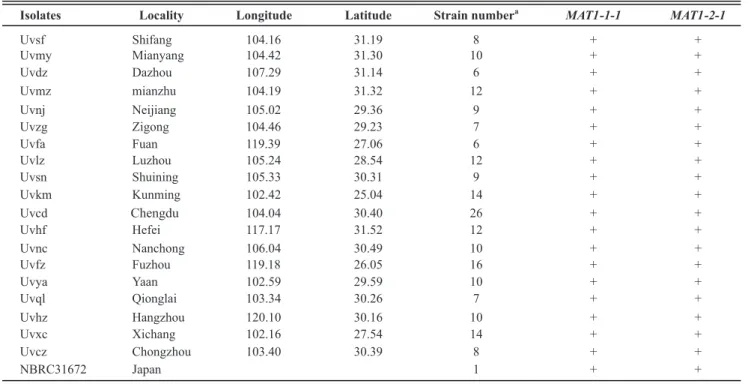
Chlamydospores of V. virens germinated easily on
PDA medium. Colonies appeared as white dots at 5 days after inoculation. As the fungus grew, the colony gradually resembled white bread (Figure 1). A single colony of V. virens was transferred and maintained in a fresh culture.
In this study, we obtained 206 conidia isolates from southwestern and eastern China (Table 1) and 26 ascospore
Genes Primers Sequences
ITS
MAT1 -1-1
MAT1 -2-1
US1-5
US3-3
MAT1 F1
MAT1 R1
MAT1 F2
MAT1 R2
MAT2 F1
MAT2 R1
MAT2 F2
MAT2 R2
5’-CCG GAG GAT ACA ACC AAA AAA ACT CT-3’
5’-GCT CCA AGT GCG AGG ATA ACT GAA T-3’
5’-GAA ACT CYA ACT CAA ACR AAG TCG-3’
5’-GKA AAC YTT GGC TAT CAR CGC CC-3’
5’-GAA ACT CCA ACT CAA ACG AAG TCG-3’
5’-GTA AAC TTT GGC TAT CAA CGC CC-3’
5’-GGA GCR ACA TAA TAC CGT YAA AGA-3’
5’-GGR GTG TTT TWC TAA GAR GGC CT-3’
5’-GGA GCG ACA TAA TAC CGT CAA AGA-3’
5’-GGG GTG TTT TTC TAA GAG GGC CT-3’
TABLE 3 - Primers used in this study.
Isolates Locality Longitude Latitude Strain number a
MAT1 -1-1 MAT1 -2-1
Uvascd Chengdou 104.04 30.40 8 + +
Uvasql Qionglai 103.34 30.26 6 + +
Uvasya Yaan 102.59 29.59 5 + +
Uvnc Nanchong 106.04 30.49 7 + +
TABLE 2 - Analysis of the mating type in single ascospores isolates.
aA total of 26 isolates. +Detected.
FIGURE 2 - Gelview-stained agarose gel showing representative results of PCR amplification of the ITS regions of V. virens isolates.
Lane 1, DNA marker (Trans2K, TransGen); lane 2, negative control; lane 3, positive control; lanes 4-22, isolates Uvsf2, Uvmy5, Uvdz1, Uvmz10, Uvnj9, Uvzg7, Uvfa1, Uvlz9, Uvsn1, Uvkm1, Uvcd1, Uvhf10, Uvnc1, Uvfz12, Uvya10, Uvql6, Uvhz9, Uvxc12, Uvcz8.
comparison of KC920892 with AB124632 (which was already deposited in GenBank) indicated 89% identity. The length of the mating type genes MAT1-1-1 and MAT1-2-1 were 256 bp and 222 bp, respectively (Figure
3).
Mating type gene distribution in natural populations The presence and distribution of mating type genes collected from natural populations of the fungus were estimated using a multiplex PCR method. As shown in Table 1, V. virens isolated from each local population contained
both MAT1-1 and MAT1-2 genes.
V. virens resulting from selfing
To confirm our hypothesis that V. virens is
derived from selfing and that it is a homothallic fungus, we performed multiplex PCR using two sets specific primers (MAT1-F2/MAT1-R2 and MAT2-F2/MAT2-R2) to screen mating type genes in ascospore isolates. The results showed that the ascospore isolates exhibited both
MAT1-1-1 and MAT1-2-1 genes (Figure 4). These results
suggested that V. virens is self-fertile and does not need
the participation of a partner.
DISCUSSION
In this study, the ITS from all isolates tested, including the reference strain for V. virens, showed 99.9 to
100% sequence similarity, confirming that these isolates are genetically closely-related.
Fungal mating type genes play an important role in regulating sexuality, virulence and survival. Sun et al. (2013) have reported that the genetic diversity of U. virens
was high in some areas of China. In this study, we used asexual spores to amplify the mating type genes of V. virens and found that a single asexual spore isolate
carries both MAT1-1-1 and MAT1-2-1 genes. This
FIGURE 1 - Colony morphology of asexual spores
(chlamydospores) germination (5 days).
The identity of the pathogen was further verified by ITS-PCR. The specific primers of V. virens amplified a
380-bp product (Figure 2) from all the isolates specific to the false smut pathogen.
Identification of MAT1-1 and MAT1-2
After DNA extraction from asexual single spore isolates, their mating type genes were identified by PCR amplification. The results showed that V. virens possesses
two types of mating type loci, MAT1-1-1 and MAT1-2-1.
The genomic sequences of the MAT1 genes have been
FIGURE 3 - Goldview-stained
agarose gel showing representative results of PCR amplification of mating-type gene fragments in V. virens isolates. A. Fragment of
MAT1-1-1 gene (256 bp) (accession number KC920891); B. Fragment of MAT1-2-1 (222 bp) (accession number KC920892). Lane 1, DNA marker (Trans2K, TransGen); lane 2, negative control; lane 3 positive control; lanes 4-22, isolates Uvsf2, Uvmy5, Uvdz1, Uvmz10, Uvnj9, Uvzg7, Uvfa1, Uvlz9, Uvsn1, Uvkm1, Uvcd1, Uvhf10, Uvnc1, Uvfz12, Uvya10, Uvql6, Uvhz9, Uvxc12, Uvcz8.
A
B
FIGURE 4 - Multiplex PCR
amplification of mating-type genes from representative ascospore isolates. Lane 1, DNA marker (Trans2K, TransGen); lane 2, negative control; lanes 3-18, isolates Uvascd1, Uvascd2, Uvascd6, Uvascd8, Uvasql2, Uvasql3, Uvasql5, Uvasql6, Uvasya1, Uvasya2, Uvasya3, Uvasya5, Uvasnc2, Uvasnc3, Uvasnc5, Uvasnc6.
result differs from those reported by Yokoyama et al. (2006), who found only MAT1-2 gene in U. virens.
This difference may be due to primer design and PCR conditions. Mating type genes have previously been described in several heterothallic and homothallic filamentous fungi as master regulators of sexual compatibility and sexual reproduction (Casselton, 2002; Kronstad 2007; Paoletti et al., 2007). These genes in homothallic fungi are usually linked. An exception is self-compatible Neurospora africana L.H.
Huang & Backus, which contains only MAT-A-1 present in the genome (Glass & Smith, 1994). Homothallic fungi such as A.nidulans are also cross-fertile (Pontecorvo
et al.,1953). Pontecorvo et al. (1953) propose the term “relative heterothallism” to explain the ability of a
suggest that sexual identity in homothallic systems might be regulated by differential expression of mating type genes or selective epigenetic silencing one of two
MAT genes (Raju & Perkins, 2000; Pöggeler, 2002;
Scazzocchio, 2006). Further research is necessary to determine whether or not the MAT genes in the genome
of V. virens are linked.
In this study, we developed a simple and reliable multiplex PCR-based marker for determining the distribution of mating type genes in V. virens, and our results showed that all
isolates displays both MAT genes. Furthermore, the multiplex
PCR codominant marker was used to screen mating type genes in ascospore isolates, and the results showed that the ascospore isolates exhibited both MAT1-1-1 and MAT1-2-1 genes. The
multiplex PCR was also used to ascertain the heterothallism in the ascomycete Tuber melanosporum (Rubini et al., 2011).
Our studies suggest a novel reproductive mechanism of V. virens. Determination of the mating type of field isolates
may provide important insights into the genetic basis and molecular mechanism of reproduction in V. virens.
ACKNOWLEDGEMENTS
This work was financially supported by the Ministry of Agriculture of China (No. 2008ZX08009003).
REFERENCES
Bulter G (2010) Fungal sex and pathogenesis. Clinical Microbiology 23:140-159.
Bischoff JF, Sullivan RF, Kjer KM, White JF Jr (2004) Phylogenetic placement of the anamorphic tribe Ustilaginoideae (Hypocreales, Ascomycota). Mycologia 96:1088-1094. Casselton LA (2002) Mate recognition in fungi. Heredity 88:142-147.
Dyer PS, Furneau X, Douhan G, Murray D (2001) A multiplex PCR test for determination of mating type applied to the plant pathogens Tapesia yallundae and Tapesia acuformis. Fungal Genetics and Biology 33:173-180.
Debuchy R, Turgeon BG (2006) Mating-type structure, evolution and function in Euascomycetes. In: Kües U, Fischer R (Eds.) The Mycota Vol. I. Growth, Differentiation and Sexuality. Amsterdam, The Netherlands. Springer. pp. 293-323.
Fraser JA, Heitman J (2005) Chromosomal sex-determining regions in animals, plants and fungi. Current Opinion in Genetics and Development 15:645-651.
Fu RT, Ding L, Zhu J, Li P, Zheng AP (2012) Morphological structure of propagules and electrophoretic karyotype analysis of false smut Villosiclava virens in rice. Journal of Microbiology 50:263-269.
Glass NL, Smith ML (1994) Structure and function of a mating-type gene from the homothallic species Neurospora africana. Molecular and General Genetics 244:401-409.
Hayden HL, Carlier J, Aitken EAB (2003) Genetic structure of Mycosphaerella fijiensis populations from Australia, Papua New Guinea and the Pacific Islands. Plant Pathology 52:703-712.
Kronstad JW (2007) Self-fertility: The genetics of sex in lonely fungi. Current Biology 17:R843-R845.
Koiso HC, Li Y, Iwasaki S, Hanaoka K, Kobayashi T, Sonoda R, Fujita Y, Yaegashi H, Sato Y (1994) Ustiloxin, antimitotic cyclic peptides from false smut balls on rice panicles caused by Ustilaginoidea virens. Journal of Antibiotics 47:765-773. Murray MG, Thompson WF (1980) Rapid isolation of high molecular weight plant DNA. Nucleic Acids Research 8:4321-4326.
Metin B, Findley K, Heitman J (2010) The mating type locus (MAT) and sexual reproduction of Cryptococcus heveanensis: Insights into the evolution of sex and sex-determining
chromosomal regions in fungi. PLoS Genetics 6:1-19.
Marais G, Galtier N (2003) Sex chromosomes: How XY recombination stops. Current Biology 13:R641-R643.
Pöggeler S (2002) Genomic evidence for mating abilities in the asexual pathogen Aspergillus fumigates. Current Genetics 42:153-160.
Paoletti M, Seymour FA, Alcocer MJC, Kaur N, Calvo AM, Archer DB, Dyer PS (2007) Mating type and the genetic basis of self-fertility in the model fungus Aspergillus nidulans. Current Biology 17:1384-1389.
Pontecorvo G, Roper JA, Hemmons LM, MacDonald KD, Bufton AW (1953) The genetics of Aspergillus nidulans. Advances in Genetics 5:141-238.
Rubini A, Belfiori B, Riccioni C, Tisserant E, Arcioni S, Martin F, Paolocci F (2011) Isolation and characterization of MAT genes in the symbiotic ascomycete Tuber melanosporum. New Phytologist 189:710-722.
Raju NB, Perkins DD (2000) Programmed ascospore death in the homothallic ascomycete Coniochaeta tetraspora. Fungal Genetics and Biology 30:213-221.
Sakurai M (1934) On the causal fungus of rice false smut. Annals of the Phytopathological Society of Japan 3:70-71. Scazzocchio C (2006) Aspergillus genomes: Secret sex and the secrets of sex. Trends in Genetics 22:521-525.
Singh RA, Dubey KS (1984) Sclerotial germination and ascospores formation of claviceps oryzae- sativae in India. Indian Phytopathology 37:168-170.
Sun XY, Kang S, Zhang YJ, Tan XQ, Yu YF, He HY, Zhang YF, Wang S, Sun WX, Cai L, Li SJ (2013) Genetic diversity and population structure of rice pathogen Ustilaginoidea virens in china. PLoS One 8:1-11.
Takahashi Y (1896) On Ustilago virens Cooke and a new species of Tilletia parasitica on rice plant. Botanical Magazine Tokyo 10:16-20.
Tanaka T, Ashizawa T, Sonoda R, Tanaka C (2008) Villosiclava virens gen. nov., com. nov., teleomorph of Ustilaginoidea virens, the causal agent of rice false smut. Mycotaxon 106:491-501.
Tang YX, Jin J, Hu DW, Yong LM, Xu Y, He LP (2013) Elucidation of the infection process of Ustilaginoidea virens (teleomorph: Villosiclava virens) in rice spikelets. Plant Pathology 62:1-8.
Turgeon BG, Yoder OC (2000) Proposed nomenclature for mating type genes of filamentous ascomycetes. Fungal Genetics and Biology 31:1-5.
White JF Jr, Sullivan R, Moy M, Patel R, Duncan R (2000) An overview of problems in the classification of plant-parasitic Clavicipitaceae. Studies in Mycology 45:95-105.
Yun SH, Arie T, Kaneko I, Yoder OC, Turgeon BG (2000) Molecular organization of mating type loci in heterothallic, homothallic, and asexual Gibberella/Fusarium species. Fungal Genetics and Biology 31:7-20.
TPP-2013-0180 Submitted: 26 October 2013 Revisions requested: 26 November 2013 Accepted: 30 December 2013 Section Editor: Meike Piepenbring Zhou YL, Izumitsu K, Sonoda R, Nakazaki T, Tanaka T, Tsuda
M, Tanaka C (2003) PCR-based detection of Ustilaginoidea