Márcia Cristina David Palma
Mestre em Microbiologia Aplicada
Elucidating the mating system of
Phaffia
rhodozyma
, an astaxanthin
-
producing
yeast with biotechnological potential
Dissertação para obtenção do Grau de Doutor em
Biologia
Orientador: Doutora Paula Gonçalves, Profª. Auxiliar, FCT/UNL
Co-orientador: José Paulo Sampaio, Prof. Associado, FCT/UNL
Júri:
Presidente: Profª. Doutora Maria Adelaide de Almeida Pedro de Jesus Arguentes: Profª. Doutora Ursula Kües
Doutora Sara Mayer Branco
Vogais: Profª. Doutora Paula Maria Theriaga Mendes Bernardo Gonçalves Prof. Doutor Fernando José Santos Rodrigues
i
Márcia Cristina David Palma
Elucidating the mating system of
Phaffia rhodozyma
, an astaxanthin-producing
yeast with biotechnological potential
“
c
opyright”
by Márcia David-Palma, FCT/UNL
iii
Acknowledgments
I want to express my gratitude to my thesis advisors, Paula Gonçalves and José Paulo Sampaio for their encouragement, guidance and trust along the years. To Diego Libking I express my gratitude for the many years of scientific collaboration and cooperation, and above all, for providing the crucial genomic information of P. rhodozyma that made possible my PhD project.
I also want to thank the members of the thesis committee, Fernando Rodrigues and Isabel Gordo, for their interest and suggestions throughout the development of my work. I would like to thank Gerhard Sandmann and Victor Cifuentes for providing plasmids that helped me get started and the assistance of Luís Jaime Mota for providing plasmids and strains for the two-hybrid systems used in this work.
I am indebted to all my lab colleagues (past and present), for their friendship and collaboration which provided for a great lab atmosphere. I would like to express my gratitude to Lia Godinho and Mário Ferreira for sharing with me their knowledge of molecular genetics, their friendship (and some of their restriction enzymes as well). I’m also thankful for all the help provided by our lab technician Nicole Soares.
I’m especially grateful to Carla Gonçalves that along the years has shared with me “the good, the bad and the ugly” of the scientific reality. She has been my traveling companion on the “scientific road” since the beginning and along the way became my treasured friend.
The support of my family has been essential throughout all my life. For the ones that are still beside
me, I’m extremely grateful for their patience, love and support through the trying times that marked the years in which this work was developed. To the ones that have already transformed into stardust, my deepest gratitude and love. Your force will always be with me. Special thanks go to my fellow scientist, best friend and husband for never failing faith on me and for being the light in the darkest of days. Thank you.
v
Resumo
Nos fungos basidiomicetes a identidade sexual é geralmente determinada por duas regiões genómicas designadas locus PR e locus HD (ou coletivamente denominadas de loci MAT) que habitualmente estão presentes em cromossomas distintos. O primeiro locus pode ser constituído por vários genes que codificam receptores de feromona (Ste3) e precursores de feromonas (Mfa) enquanto o segundo inclui, na sua versão mais simples, um par de genes, HD1 e HD2, transcritos em direcções opostas, que codificam factores de transcrição com um domínio de ligação ao DNA. A maioria dos basidiomicetes apresentam um comportamento heterotálico, que requer a existência de compatibilidade entre ambos os lociMAT, sendo para tal necessário que cada indivíduo apresente diferentes alelos dos genes MAT. Existem, no entanto, fungos cujo comportamento sexual dispensa o cruzamento entre dois indivíduos geneticamente compatíveis, designados fungos homotálicos. Espécies homotálicas possuem a capacidade de completar o seu ciclo de vida em monocultura, dando origem a progenia meiótica. Enquanto os determinantes genéticos que regulam o comportamento heterotálico em basidiomicetes são já conhecidos em detalhe em várias espécies modelo, muito pouco se sabe sobre os genes e mecanismos moleculares que se encontram na base do homotalismo. O principal objectivo do trabalho apresentado nesta tese, foi preencher tanto quanto possível essa lacuna, estudando os genes e mecanismos moleculares que governam o ciclo de vida homotálico da levedura Phaffia rhodozyma. Foram encontrados seis genes MAT em P. rhodozyma, organizados em dois loci distintos, PR e HD, provavelmente localizados em cromossomas distintos. O locusPR encontrado apresenta dois conjuntos de genes, separados por aproximadamente 5 kb, onde cada conjunto é constituído por um gene STE3 e um gene MFA. O locus HD é composto por um par de genes, HD1 e HD2, transcritos em direcções opostas e apresentando o característico domínio de ligação ao DNA. A construção de mutantes com deleções de vários genes MAT permitiu propor um mecanismo molecular para o comportamento homotálico de P. rhodozyma. No modelo molecular proposto (i) a feromona Mfa1 activa o receptor Ste3-2 enquanto a feromona Mfa2 activa o receptor Ste3-1, existindo, portanto, redundância do sistema receptor/feromona enquanto (ii) os dois fatores de transcrição, Hd1 e Hd2, são ambos necessários para a ocorrência de reprodução sexuada. Procedeu-se igualmente à comparação das regiões MAT de P. rhodozyma com as regiões MAT de duas espécies novas entretanto incluídas no género Phaffia, (i.e. P. novazelandica e P. tasmanica) assim como com outras espécies pertencentes à ordem Cystofilobasidiales. Este trabalho permitiu pela primeira vez a elucidação dos mecanismos molecular básicos responsáveis pelo ciclo de vida homotálico de um basidiomiceta. Simultaneamente, a manipulação genética de P. rhodozyma, uma espécie com potencial biotecnológico para produção de astaxantina, permitiu gerar mutantes com determinantes sexuais
“compatíveis” que poderão ser utilizados para melhoramento de estirpes industriais através de cruzamentos selectivos.
vii
Abstract
In fungi belonging to the phylum Basidiomycota, sexual identity is usually determined by two genetically unlinked MAT loci, one named PR locus, which encodes one or more pheromone receptors (Ste3) and pheromone precursors (Mfa), and the other, named HD locus, that comprehends at least one pair of divergently transcribed genes encoding homeodomain transcription factors (HD1 and HD2). The two MAT loci work as two distinct mating compatibility check points. Most basidiomycete species are heterothallic, meaning that sexual reproduction requires mating between two sexually compatible individuals harboring different alleles at both MAT loci. However, some species are known to be homothallic, one individual can complete the sexual cycle without mating with a genetically distinct partner. While the molecular underpinnings of the heterothallic life cycles of several basidiomycete model species have been dissected in detail, much less is known concerning the molecular basis for homothallism. The general aim of this research was to study the molecular mechanisms of sexual reproduction in fungi, specifically those governing the homothallic life cycle of P. rhodozyma.
Six MAT genes were found in P. rhodozyma, organized in two MAT loci, most likely located on different chromosomes. The PR locus was shown to be composed of two clusters, at approximately 5 kb from one another, each encoding one STE3 gene and one MFA gene, while the HD locus encompassed two divergently transcribed homeodomain transcription factors genes, HD1 and HD2. Functional genetic analysis was performed by targeted gene deletion of the MAT elements found in P. rhodozyma and the results allowed the proposal of a molecular model controlling the homothallic sexual behavior of P. rhodozyma. In this model (i) each pheromone interacts with the pheromone receptor of the other cluster (Mfa1 activates Ste3-2 while Mfa2 activates Ste3-1); since neither pheromone receptor is required per se for sporulation they seem to be functionally redundant; and (ii) both homeodomain proteins appear to work together to regulate genes required for sexual development. Comparison of the MAT regions of additional Phaffia species and of other representatives within the order Cystofilobasidiales, indicate that transitions to homothallism probably occurred several times independently. Furthermore, it revealed a particularly dynamic pattern of MAT gene evolution, with the generation of new receptors within each genus and exceptionally large numbers of mature pheromones encoded in the genomes of some of the species.
In conclusion, this work allowed for the first time the elucidation of the basic molecular mechanisms governing the homothallic life cycle of a basidiomycete. At the same time the genetic manipulation of the MAT genes of P. rhodozyma also allowed the generation of preferably outcrossing strains, which may be potentially useful for further improvement of this yeast as an industrial organism by way of selective breeding.
ix
Abbreviations
AIC Akaike information criterion
bp Base pair
D1/D2 Domains 1 and 2 of the 26S rDNA
DNA Deoxyribonucleic acid
GDF Gene deletion fragment
HD Homeodomain
HMG High-mobility group
HPLC-PAD High-performance liquid chromatography with pulsed amperometric detector
ITS Internal transcribed spacer JGI Joint Genome Institute
kb Kilobase pair
LSU Large subunit (ribosome)
MAP Mitogen activated protein
MAPK Mitogen activated protein kinase
MAT Mating type
Mb Megabase pair
min Minute(s)
ML Maximum likelihood
NCBI National Center for Biotechnology Information
ORF Open reading frame
rDNA ribosomal DNA
s Second(s)
PFGE Pulsed-field gel electrophoresis
PCR Polymerase chain reaction
Abbreviations of relevant culture collections
CBS Centraalbureau voor Schimmelcultures, Utrecht, The Netherlands
JCM Japan Collection of Microorganisms, Riken Bioresource Center, Saitama, Japan
PYCC Portuguese Yeast Culture Collection, CREM, Faculdade de Ciências e Tecnologia,
x
Abbreviated species names
C. albicans Candida albicans C. cinerea Coprinopsis cinerea
C. deneoformans Cryptococcus deneoformans S. cerevisiae Saccharomyces cerevisiae
S. commune Schizophyllum commune
S. pombe Schizosaccharomyces pombe
S. reilianum Sporisorium reilianum C. bisporidii Cystofilobasidium bisporidii C. ferigula Cystofilobasidium ferigula C. capitatum Cystofilobasidium capitatum C. macerans Cystofilobasidium macerans P. rhodozyma Phaffia rhodozyma
P. novazelandica Phaffia novazelandica P. tasmanica Phaffia tasmanica
K. huempii Krasilnikovozyma huempii M. blollopis Mrakia blollopis
M. frigida Mrakia frigida
M. aquatica Mrakia aquatica
M. perniciosa Moniliophthora perniciosa T. pamirica Tausonia pamirica T. pullulans Tausonia pullulans
xi
Index
Chapter 1
General Introduction
1.1.
Sexual reproduction in fungi
... 21.1.1. Sexual and asexual reproduction ... 2
1.1.2. Sexual identity in fungi ... 3
1.1.3. Mating-type (MAT) genes ... 3
1.1.3.1. MAT genes in Ascomycota ... 3
1.1.3.2. MAT genes in Basidiomycota ... 4
1.1.4. Mating behavior in fungi ... 6
1.1.4.1. Heterothallic mating ... 7
1.1.4.2. Homothallic mating ... 7
1.1.4.2.1. Primary homothallism ... 8
1.1.4.2.2. Secondary homothallism - Pseudohomothallism ... 9
1.1.4.2.3. Secondary homothallism - Mating-type switching ... 9
1.1.4.2.4. Unisexual mating ... 10
1.1.5. Homothallic basidiomycetes ... 11
1.1.5.1. Moniliophthora permiciosa ... 11
1.1.5.2. Filobasidiela depauperata ... 12
1.1.6. Molecular mechanisms enabling transitions between mating systems and mating behaviors ... 13
1.2.
Phaffia rhodozyma
... 141.2.1. Discovery and taxonomic classification ... 14
1.2.2. Biogeography and ecological associations of Phaffia rhodozyma ... 15
1.2.3. Life cycle of Phaffia rhodozyma ... 16
1.2.4. Biotechnological relevance of Phaffia rhodozyma ... 18
xii
Chapter 2
Identification of
MAT
and meiosis genes in
P. rhodozyma
genomes
2.1. Introduction
... 222.2.
Materials and methods
... 232.2.1. Search for putative MAT and meiosis genes in P. rhodozyma genomes and their characterization .. 23
2.2.2. Comparison of MAT gene sequences between the three available P. rhodozyma genomes ... 24
2.2.3. Phylogenetic analysis of the putative MAT genes in P. rhodozyma strains representing distinct populations ... 24
2.2.4. Phylogenetic analysis of the pheromone receptor proteins of P. rhodozyma and other Tremellomycetes ... 25
2.3.
Results
... 262.3.1. MAT and mating related genes in P. rhodozyma ... 26
2.3.2. Meiosis genes in P. rhodozyma ... 28
2.3.3. MAT gene variability within P. rhodozyma ... 28
2.3.4. Phylogenies of MAT genes within P. rhodozyma ... 29
2.3.5. Phylogenetic relationship of pheromone receptors from P. rhodozyma with other basidiomycetes ... 30
2.4.
Discussion
... 322.4.1. MAT genes are present in P. rhodozyma ... 32
2.4.2. Mating related genes and meiotic genes are present in P. rhodozyma ... 33
2.5.
Conclusion
... 34Chapter 3
Genetic dissection of the homothallic sexual reproduction of
P. rhodozyma
3.1.
Introduction
... 363.2.
Materials and methods
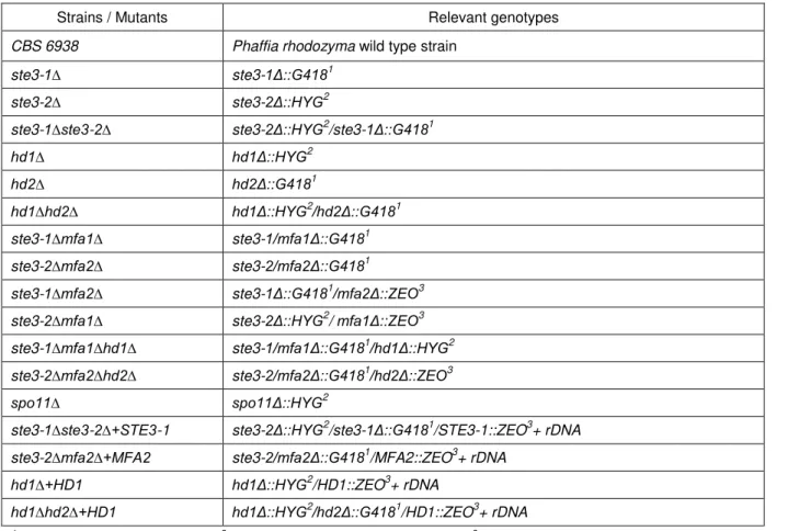
... 373.2.1. Strains and culture conditions ... 37
3.2.2. Construction of gene deletion fragments and complementation plasmids ... 37
3.2.3. Transformation of P. rhodozyma ... 38
3.2.4. Sporulation assays ... 38
3.2.5. Crosses between deletion mutant strains ... 39
3.2.6. Bacterial two-hybrid assays ... 39
xiii
3.2.8. Search for potential alternative homeodomain transcription factor proteins ... 40
3.3.
Results
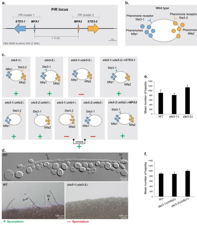
... 413.3.1. Pheromone receptors are required for sexual reproduction ... 41
3.3.2. Evidence for recipocal compatibility between receptors and pheromones encoded by the two PR gene clusters ... 43
3.3.3. Genes HD1 and HD2 are required for sexual development in P. rhodozyma ... 43
3.3.4. Genetic evidence for the involvement of meiosis in the sexual cycle of P. rhodozyma ... 45
3.3.5. Construction of heterothallic strains ... 46
3.4.
Discussion
... 46Chapter 4
Two new species in the genus
Phaffia
-
Phaffia novazelandica
sp. nov. and
Phaffia tasmanica
sp. nov.
4.1.
Introduction
... 524.2.
Materials and methods
... 544.2.1. Phenotypical characterization ... 54
4.2.2. Genome sequencing ... 54
4.2.3. Genome assembly, prediction of coding regions and search for relevant genes ... 54
4.2.4. Phylogenetic analysis ... 56
4.3.
Results
... 574.3.1. Morphological and physiological characterization of P. novazelandica and P. tasmanica ... 57
4.3.2. The divergent Australasian lineages belong to the genus Phaffia ... 60
4.3.3. Astaxanthin production by P. tasmanica and P. novazelandica ... 61
4.3.4. Organization and comparative analysis of Phaffia MAT loci ... 62
4.4.
Discussion
... 644.4.1. Phylogenetic assignment of P. tasmanica and P. novazelandica in the genus Phaffia and its placement within the Cystofilobasidiales ... 64
4.4.2. Phenotypic and MAT genomic traits of Phaffia ... 64
4.4.3. Association of P. tasmanica and P. novazelandica with Nothofagus ... 65
xiv
Chapter 5
MAT
loci evolution in
Phaffia
and in other Cystofilobasidiales
5.1.
Introduction
... 685.2.
Materials and methods
... 695.2.1. Genome sequencing, identification of MAT genes and synteny analysis ... 69
5.2.2. MAT gene content and comparison across Cystofilobasidiales ... 70
5.2.3. Phylogenetic analyses ... 70
5.3.
Results
... 715.3.1. Identification of MAT genes in Cystofilobasidiales ... 71
5.3.2. Pheromone receptors in Cystofilobasidiales ... 72
5.3.3. Pheromones in Cystofilobasidiales ... 74
5.3.4. Homeodomain transcription factors in homothallic and heterothallic Cystofilobasidiales ... 77
5.3.5. The PR locus in Cystofilobasidiales ... 79
5.3.6. The HD locus in Cystofilobasidiales... 84
5.4.
Discussion
... 875.4.1. MAT loci in Phaffia ... 87
5.4.2. MAT loci in Mrakia ... 88
5.4.3. MAT loci in Cystofilobasidium ... 90
5.4.4. MAT loci in other members of the Cystofilobasidiales ... 91
5.5.
Conclusion
... 92Chapter 6
Concluding remarks and future perspectives
6.1.
Concluding remarks and future perspectives
... 96xv
Appendix I
Additional information pertaining to Chapter 2 ...
114Appendix II
Additional information pertaining to Chapter 3 ...
122Appendix III
Additional information pertaining to Chapter 4 ...
136III.1. Physiological characteristics of P. novazelandica and P. tasmanica ... 136
III.2. Additional information pertaining to the species phylogeny and to the MAT genes found in P. novazelandica and P. tasmanica ... 137
III.3. Additional information pertaining to the MAT and astaxanthin related genes found in P. novazelandica and P. tasmanica ... 138
Appendix IV
Additional information pertaining to Chapter 5 ...
146IV.1. Quality statistics for the draft genome assemblies ... 146
IV.2. Additional information pertaining to the MAT genes found in Cystofilobasidiales ... 146
xvii
Figure Index
Chapter 1
Figure 1.1. Summary of the modes of sexual reproduction in fungi ...6
Figure 1.2. Distinc habitats associated with P. rhodozyma ... 15
Figure 1.3. Distribution, phylogeny and host tree associations of P. rhodozyma ... 16
Figure 1.4. Life cycle of P. rhodozyma ... 17
Figure 1.5. Sexual state of P. rhodozyma ... 18
Chapter 2
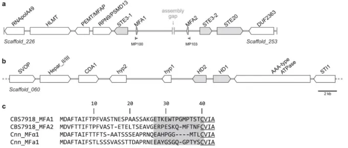
Figure 2.1. Gene content and organization around the putative mating type genes of P. rhodozyma ... 26Figure 2.2. Features of Hd1 and Hd2 proteins of P. rhodozyma ... 27
Figure 2.3. Alignments of the predicted proteins for Hd1 and Hd2 of distinct P. rhodozyma strains ... 28
Figure 2.4. Phylogeny of MAT genes ... 29
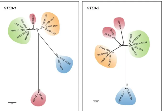
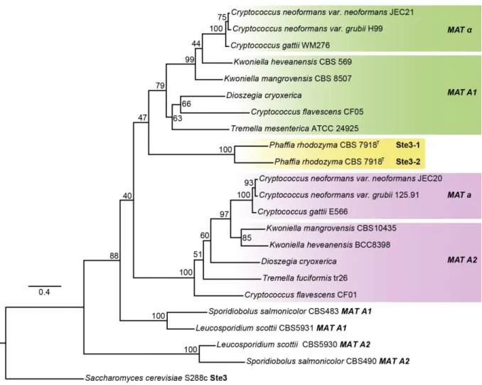
Figure 2.5. Maximum likelihood phylogenetic tree of the pheromone receptors (Ste3) from Tremellomycetes ... 31
Chapter 3
Figure 3.1. MAT loci in P. rhodozyma ... 36Figure 3.2. Deletion mutants in the PR locus ... 42
Figure 3.3. Deletion mutants in the HD locus ... 44
Figure 3.4. SPO11 deletion mutant ... 45
xviii
Chapter 4
Figure 4.1. ITS phylogeny inferred with maximum likelihood for Phaffia and
Cystofilobasidium species ... 53
Figure 4.2. Detail of the Tremellomycetes phylogenetic tree based on a seven gene dataset ... 53
Figure 4.3. Australasia and the collection sites of Phaffia spp. isolates ... 57
Figure 4.4. Vegetative cells of P. novazelandica and P. tasmanica ... 58
Figure 4.5. Sexual structures of P. novazelandica and P. tasmanica ... 59
Figure 4.6. Phylogeny of the species of the order Cystofilobasidiales ... 60
Figure 4.7. Phaffia and astaxanthin production ... 61
Figure 4.8. Scaffolds encompassing PR and HD genes in the different Phaffia species ... 62
Figure 4.9. Phylogeny of Phaffia pheromone receptors ... 63
Figure 4.10. Pheromone precursor proteins from all Phaffia species ... 63
Chapter 5
Figure 5.1. Phylogeny of pheromone receptors of the Cystofilobasidiales ... 72Figure 5.2. Protein sequence alignment of the pheromone precursors of members of the Cystofilobasidiales ... 75
Figure 5.3. Genomic regions of strains of M. blollopis depicting the MFA genes found and their similarity ... 76
Figure 5.4. Alignments of Hd1 and Hd2 predicted proteins ... 78
Figure 5.5. PR regions in members of the Cystofilobasidiales ... 81
Figure 5.6. Repeat regions present in the PR scaffolds of M. blollopis and M. frigida ... 83
xix
Appendix I
Figure I.1. MAT protein alignments ... 116
Figure I.2. Predicted pheromone receptor proteins Ste3-1 and Ste3-2 of strain CBS 7918T ... 119
Figure I.3. Predicted Hd1 and Hd2 proteins of strain CBS 7918T ... 119
Appendix II
Figure II.1. Plasmids used and constructed in this work ... 129Figure II.2. Southern blot analysis of selected deletion mutants ... 130
Figure II.3. Synthetic coding sequences of the HD1 and HD2 genes ... 131
Figure II.4. Bacterial two-hybrid assays... 132
Figure II.5. Yeast two-hybrid assays ... 133
Appendix III
Figure III.1. Alignment of the proteins from the biosynthetic route of astaxanthin of the three Phaffia species ... 138Figure III.2. Predicted proteins of the pheromone receptor genes of P. novazelandica and P. tasmanica ... 143
Figure III.3. Predicted proteins of the homeodomain transcription factor genes of P. novazelandica and P. tasmanica ... 144
Appendix IV
Figure IV.1. Phylogenetic inferences illustrating the likelihood of strain Nwmf-AP1 belonging to Mrakia blollopis species instead of Mrakia frigida ... 147Figure IV.2. Pheromone receptor phylogeny ... 148
xxi
Table Index
Chapter 2
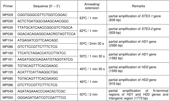
Table 2.1. List of strains used in the phylogenetic study... 24
Table 2.2. List of primers and PCR conditions used to amplify MAT genes in P.
rhodozyma strains ... 25
Table 2.3. List of MAT genes found in P. rhodozyma genomes ... 26
Chapter 3
Table 3.1. List of P. rhodozyma strains used and corresponding genetically manipulated
derivatives generated in this study ... 41
Chapter 4
Table 4.1.
List of strains selected for whole genome sequencing...
54Table 4.2.
List of draft genomes from species of Cystofilobasidiales...
55Table 4.3.
List of strains used in phenotypic tests...
58Table 4.4.
Maximum growth temperatures of P. novazelandica and P. tasmanica...
58Table 4.5.
Location of genes of the biosynthetic route of astaxanthin in the draft genomesof P. novazelandica and P. tasmanica
...
61Chapter 5
Table 5.1.
List of all draft genome assemblies from distinct Cystofilobasidiales speciesused in this chapter
...
69Table 5.2.
List of MAT genes found in the analyzed Cystofilobasidiales genomes...
71Table 5.3.
Percentage of amino acid identity between different Ste3 proteins in severalspecies of Cystofilobasidiales
...
73Table 5.4.
MFA genes from Cystofilobasidiales, their pheromone precursor proteins andxxii
Appendix I
Table I.1.
Complete list of MAT genes and MAT related genes searched in the draftgenome assembly of CBS 7918T
...
114Table I.2.
Complete list of meiotic genes searched in the draft genome assembly of CBS7918T
...
114Table I.3.
Amino acid identity (%) between different alleles of pheromone receptors fromselected basidiomycetes
...
114Table I.4.
Accession numbers and information for all sequences obtained and used inchapter 2
...
114Appendix II
Table II.1.
List of strains used in bacterial and yeast two-hybrid assays...
122Table II.2.
Primers and plasmids used for the construction of all gene deletion fragmentsexcept for gene STE3-2.
...
123Table II.3.
Primers and plasmid used for the construction of the STE3-2 deletionfragment by overlap extension PCR
...
124Table II.4.
Primers and plasmid used for the construction of the zeocin resistancecassette
...
125Table II.5.
Primers and plasmid used for the construction of the complementationplasmids
...
125Table II.6.
Sporulation data pertaining to plot (e) from Figure 3.2, plot (c) from Figure 3.3and plot (a) from Figure 3.4
...
126Table II.7.
Sporulation data pertaining to plot (f) from Figure 3.2...
126Table II.8.
Data from crosses of double and triple mutant strains...
126Table II.9.
List of plasmids used in the Bacterial Adenylate Cyclase Two-Hybrid System...
127Table II.10.
List of primers used in the Bacterial Adenylate Cyclase Two-Hybrid System...
127Table II.11.
List of plasmids used in the Matchmaker Gold Yeast Two-Hybrid System...
128Table II.12.
List of primers used in the Matchmaker Gold Yeast Two-Hybrid System...
128xxiii
Appendix III
Table III.1.
Physiological tests for three strains of each of the new Phaffia species...
136Table III.2.
Statistics for the genomes assembled for this work (Chapter 4)...
137Appendix IV
CHAPTER 1
___________________________________________________________________
Chapter | 1
2
1.1. Sexual reproduction in fungi
The study of sexual reproduction in fungi has demonstrated that these organisms are an exceptional model for understanding the evolution of sex determination in eukaryotes (Fraser et al. 2004), including in metazoans with which fungi are evolutionarily closely related (Baldauf & Palmer 1993; Wainright et al. 1993; Idnurm 2011a). Of the several lineages that compose the fungal kingdom, the majority of the described fungal species are part of the Dikarya clade (James et al. 2006), composed of the phyla Ascomycota and Basidiomycota, which have been the object of most of the studies about sexual reproduction (Idnurm 2011a).
1.1.1. Sexual and asexual reproduction
Chapter | 1
3
1.1.2. Sexual identity in fungi
Sexual identity in fungi is determined in the haploid stage (Billiard et al. 2011) by one or two specialized genomic regions named mating-type loci (MAT), which in addition to defining the cell mating-type, are also responsible for orchestrating the sexual cycle (Herskowitz et al. 1992; Casselton 2002; Stanton & Hull 2007). The fungal MAT loci have been shown to vary greatly, both in length and gene content throughout both ascomycetes and basidiomycetes (Casselton 2002; Stanton & Hull 2007). Depending on whether the mating type-determining genes are genetically linked in one single MAT locus or genetically unlinked in two distinct MAT loci, the mating-system of a fungal species may be respectively, bipolar or tetrapolar (Casselton & Olesnicky 1998; Fraser & Heitman 2003). In species with a bipolar mating-system, cells of two distinct (and compatible) mating-types (e.g. a and α) exist in the population, where cell identity is governed by a single MAT locus that encodes one of two possible idiomorphs in Ascomycota (Butler 2007) or alleles in Basidiomycota (Fraser & Heitman 2003; Fraser et al. 2007). In contrast, species with a tetrapolar mating-system have two different and unlinked MAT loci defining the mating-type of each individual. This means that, in order to successfully mate, individuals must differ from one another at both MAT loci (Fraser & Heitman 2003; Fraser et al. 2007). If each of the two MAT loci harbor only two alleles (biallelic), four distinct mating-types will exist in the population. However, most tetrapolar species are multiallelic in at least one of the MAT loci, giving rise to a large number of different mating-types (Casselton & Olesnicky 1998; Fraser & Heitman 2003; Kües 2015). A more recently described intermediate system, named pseudo-bipolar, was found in basidiomycetes, which involves a configuration in which the PR and HD loci are partially linked enabling limited recombination (Coelho et al. 2010; Gioti et al. 2013a; Wu et al. 2015). In the Ascomycota the bipolar mating-system is the norm while in the Basidiomycota the prevailing system is the tetrapolar one, albeit also encompassing bipolar and pseudo-bipolar species (Fraser & Heitman 2003; Fraser et al. 2007; Morrow & Fraser 2009; Coelho et al. 2010). Given that tetrapolarity is absent in the ascomycetes and has been found in all the major lineages of the basidiomycetes (Ustilaginomycotina, Pucciniomycotina and Agaricomycotina), the tetrapolar system is considered to be the ancestral mating-system of this phylum (Fraser et al. 2007; Kües et al. 2011; Maia et al. 2015).
1.1.3. Mating-type (
MAT
) genes
1.1.3.1. MAT genes in Ascomycota
Chapter | 1
4
ascomycetes display different sets of transcription factors present at their MAT loci (Butler 2007; Stanton & Hull 2007; Bennett & Turgeon 2016). Nevertheless, in general the MATα idiomorphs (also designated as MAT1-1) are defined by the consistent presence of a gene encoding a protein with a α -domain motif, whereas the MATa idiomorphs (also designated as MAT1-2) are usually characterized by the presence of a gene encoding a transcription factor with a HMG domain (Butler et al. 2004; Butler 2007; Bennett & Turgeon 2016). In some species, however, MATα may harbor more than one gene, like in Saccharomyces cerevisiae in which MATα encodes a protein with an α domain (MATα1) and another with a homeodomain (MATα2) (Herskowitz et al. 1992; Butler 2007). In ascomycetes, alternate versions of the MAT locus are named idiomorphs rather than alleles (Butler 2007) because, although they typically occupy the same chromosomal location in different haploid genomes, they are completely dissimilar and encode unrelated proteins. The transcription factors encoded at the MAT loci regulate an array of genes essential for mate recognition, meiosis and sexual development, like pheromones (MFa and MFα) and pheromone receptors (STE2 and STE3) genes belonging to the G-protein-coupled receptor family, all of which are encoded outside of the MAT loci (Kronstad & Staben 1997; Chen & Thorner 2007; Tsong et al. 2007; Bennett & Turgeon 2016).
1.1.3.2. MAT genes in Basidiomycota
As previously mentioned, the most frequent mating-system in the Basidiomycota is the tetrapolar system, which means that sexual identity (mating-type) is generally determined by two genetically unlinked MAT loci (Casselton & Olesnicky 1998). One distinct feature of the tetrapolar mating-system and consequently of mating in the Basidiomycota is that the genes encoding pheromones and pheromone receptors have acquired a mating-type defining role, in opposition to what occurs in the Ascomycetes (Fraser et al. 2007). The MAT regions of Basidiomycetes that have been characterized thus far show an enormous level of diversity, nevertheless they all appear to derive from a common component set: a locus encoding pheromones and pheromone receptor genes, usually named PR, and a locus encoding homeodomain transcription factor genes referred to as HD (Fraser et al. 2007; Kües 2015).
The archetype of the PR locus encodes pheromone receptor genes homologous to the STE3 receptor gene form S. cerevisiae, a G-protein-coupled receptor with seven transmembrane domains, and pheromone precursor genes, which are homologous to the lipid-modified MFa pheromone gene of S. cerevisiae (Bölker et al. 1992; Casselton & Olesnicky 1998; Raudaskoski & Kothe 2010; Kües 2015). Basidiomycete pheromones are first translated into pheromone precursor proteins which subsequently undergo posttranslational processing at both N and C-terminal regions. A general feature of pheromone precursor proteins is the presence of a “CaaX” motif at the carboxyl terminus of the
C-Chapter | 1
5
terminal proteolytic cleavage of the motif “aaX”, followed by carboxylmethylation of the cysteine residue and proteolytic cleavage at the N-terminus, originating mature lipopetide pheromones ranging from 9 to 15 amino acids (Caldwell et al. 1995; Kües et al. 2011; Kües 2015; Freihorst et al. 2016). The site for N-terminal cleavage is conserved in the pheromone precursor proteins of some species, like Coprinopsis cinerea were the protease recognition site is a pair of charged amino acids such as glutamate-arginine, or one single charged residue, as in some Schizophyllum commune precursors (Riquelme et al. 2005; Freihorst et al. 2016). Alignments of the predicted pheromone precursor proteins in some cases allow inference of the N-terminal cleavage site due to the levels of amino acid similarity on either side of the region that composes the mature pheromone (Fowler et al. 2004; Riquelme et al. 2005; Freihorst et al. 2016).
Regarding the HD locus, its most simple configurations contains two divergently transcribed genes encoding homeodomain transcription factors of different classes, HD1 and HD2 (Kües & Casselton 1992). The Hd1 homeodomain transcription factors are considered to have an atypical homeodomain, with three additional amino acids between helices one and two of the three helices comprising the homeodomain motif. Conversely, the Hd2 homeodomain transcription factors have a canonical homeodomain, with 60 residues forming a three helical structure and a highly conserved DNA-binding region (Kües & Casselton 1992; Kües et al. 2011; Freihorst et al. 2016). In order for these transcription factors to be functional in mating, it is necessary for them to heterodimerize with each other, however this can only occur after cell fusion, because dimerization is restricted to subunits (Hd1 and Hd2) encoded by different alleles of the HD loci, that originate from distinct mating partners (Banham et al. 1995; Kämper et al. 1995). It was shown that the discrimination between interactions of self and non-self-subunits is due to the N-terminal regions of both Hd1 and Hd2, which confer mating-type specificity and also mediate dimerization (Yee & Kronstad 1993; Kües et al. 1994; Kämper et al. 1995; Yue et al. 1997; Freihorst et al. 2016). Because the sequences from the N-terminal regions of different alleles of HD1 and also HD2 are usually highly divergent when compared to the homeodomain and
C-terminal regions, it was hypothesized that these genes have evolved under a pattern of “divergence
Chapter | 1
6
1.1.4. Mating behavior in Fungi
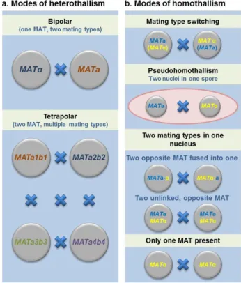
Sexual reproduction in fungi may occur by two distinct mating behaviors: heterothallism or homothallism (Figure 1.1) (Blakeslee 1904). Heterothallic species (self-sterile) require fusion of two compatible partners of different mating types in order for successful mating to occur, while in homothallic species, each individual cell has the ability to undergo sexual reproduction by itself and produce sexual progeny (Blakeslee 1904; Lin & Heitman 2007; Casselton 2008; Wilson et al. 2015b). Both mating behaviors conserve the key features of sexual reproduction (ploidy changes, meiosis and production of recombinant progeny), but they differ in the fact that homothallic species do not require a compatible partner (Lin & Heitman 2007; Ni et al. 2011). Homothallism is considered to be common in
the ascomycetes and relatively rare in basidiomycetes, where only about ≈10% of the accessed species (Whitehouse 1949) present this mating behavior (Lin & Heitman 2007; Kües et al. 2011). Many homothallic species are not hampered from mating with genetically distinct strains, therefore being able of both inbreeding and outcrossing (Lin & Heitman 2007; Kües et al. 2011). Additionally, some species have the ability to reproduce via both homothallic and heterothallic behavior, and are sometimes referred to as amphithallic (Ene & Bennett 2014; Roach et al. 2014). Finally, some species that are mostly heterothallic may present homothallic life cycles depending on the environmental conditions (Lin & Heitman 2007). Homothallism may in fact confer an adaptive advantage by removing the need to find a compatible partner (Billiard et al. 2011; Billiard et al. 2012) hence diminishing the cost associated with sexual reproduction (Roach et al. 2014).
Chapter | 1
7
Two strains need to possess opposite alleles at both MAT loci in order to successfully mate. (b) Modes of homothallism: (i) mating-type switching in which an α daughter cell mates with an a mother cell; (ii)
pseudohomothallism in which two nuclei of opposite mating types are packaged into one spore; (iii) two MAT loci in one nucleus in which the two opposite MAT loci are either fused or unlinked in the genome; (iv) there is only one MAT idiomorph present and cells reproduce via unisexual mating. Adapted from Ni et al. (2011).
Compared to heterothallism, homothallism has been less studied and consequently less well understood within basidiomycetes. Therefore, most of the molecular determinants and mechanisms underpinning the different types of homothallism are most often illustrated in ascomycetes models (Lin & Heitman 2007; Roach et al. 2014; Wilson et al. 2015b).
1.1.4.1. Heterothallic mating
In basidiomycetes, mating between two homokaryotic hyphae (or two haploid yeast cells) usually originates a dikaryotic filamentous stage in which the two parental nuclei are replicated in a coordinated fashion. Fusion of the two nuclei (karyogamy) of distinct mating-types only takes place in specialized structures, either basidia or teliospores, which allow for the occurrence of meiosis and the formation of basidiospores which are the meiotic progeny (Hibbett et al. 2007). Molecularly, heterothallic mating between two compatible haploid partners, encoding different alleles of the MAT genes at each of their MAT loci, is initiated when mature pheromones are exported from the cell via a specific transporter, Ste6 (Hsueh & Shen 2005) and perceived by the other mating partner leading to cell fusion (Kües et al. 2011). Binding of the pheromone to its appropriate receptor causes conformational changes at the C-terminus of the receptor (Ste3) located at the plasma membrane, which in turn interacts with an heterotrimeric G protein (Hsueh et al. 2007; Li et al. 2007; Xue et al. 2008). The pheromone signal is subsequently transmitted via the pheromone-activated signal transduction cascade (Regenfelder et al. 1997; Davidson et al. 2003; Raudaskoski & Kothe 2010; Roach et al. 2014), which then is interconnected with the regulatory pathways activated by the heterodimer formed by the subunits Hd1/Hd2 encoded by the distinct mating-type alleles (Kües & Casselton 1992; Kües et al. 2011). This ultimately puts in place the transcriptional program that allows for the completion of the sexual cycle (Kües et al. 2011). Following comparable steps, heterothallic sexual reproduction in ascomycetes is orchestrated by the transcription factors encoded at their MAT loci that regulated the expression of genes, like those encoding pheromones and receptors. When sexually compatible cells interact successfully, cells fuse and karyogamy occurs forming a diploid cell, which in adequate environmental conditions undergoes meiosis generating haploid meiotic progeny (Stanton & Hull 2007).
1.1.4.2. Homothallic mating
Chapter | 1
8
al. 2011). However, this classification does not distinguish the different molecular and cellular mechanisms employed by each organism to achieve the completion of the life cycle and the production of sexual offspring (Wilson et al. 2015b). The advent of molecular genetics followed by the exploration of the wealth of genomic data generated, namely by comparative genomics (Scazzocchio 2014), has unraveled profound differences between the molecular mechanisms underpinning homothallism (Lin & Heitman 2007; Wilson et al. 2015b). Different types of homothallism have been defined according to their morphological behavior and/or molecular bases: (i) primary homothallism, (ii) secondary homothallism encompassing pseudohomothallism and mating-type switching and finally (iii) unisexual reproduction (Lin & Heitman 2007; Wilson et al. 2015b; Bennett & Turgeon 2016).
1.1.4.2.1. Primary homothallism
Chapter | 1
9
1.1.4.2.2. Secondary homothallism
–
Pseudohomothallism
Some fungi exhibit a particular system where post-meiotic nuclei of opposite mating types are packaged into one single spore that is then able to complete the sexual cycle on its own; hence the restitution of the diploid condition can always occur without the need to find a compatible partner (Whitehouse 1949; Billiard et al. 2011; Billiard et al. 2012). This behavior has been termed pseudohomothallism (Whitehouse 1949; Lin & Heitman 2007) and although morphologically it abides to the definition of homothallism, it actually corresponds to a heterothallic compatibility system in which automixis is strongly favored (Idnurm et al. 2015; Wilson et al. 2015b). The ascomycete Neurospora tetrasperma is one of the most studied species that exhibits this mating behavior. In this species, after sexual reproduction specialized structures called asci are formed, containing four ascospores, each of which possess two independent nuclei with opposite mating-types that allow for the completion of the sexual cycle (one MAT1-2 and the other MAT1-2) (Dodge 1931; Raju 1992; Raju & Perkins 1994) (Wilson et al. 2015b). A similar example can be found in the basidiomycete species Agaricus bisporus var. bisporus (Kerrigan et al. 1993).
1.1.4.2.3. Secondary homothallism
–
Mating-type switching
Chapter | 1
10
whereas the other is silenced or expressed only at a reduced level. The mechanism of mating-type switching occurs by recombination between inverted-repeat sequences flanking the MAT loci which results in expression of the opposite mating type (Hanson et al. 2014; Maekawa & Kaneko 2014). Because the mechanisms of mating-type switching mentioned in the previous examples are reversible processes they are called bidirectional, however there is also another form of mating-type switching that is irreversible and hence termed unidirectional (Wilson et al. 2015b). The self-fertile isolates of the ascomycetes Ceratocystidaceae fimbriata and C. albifundus encode at their MAT locus the genes encoding both compatible idiomorphs (MAT1-1 and MAT1-2). Ascospores produced during sexual reproduction segregate into fertile and sterile isolates. Genetically, the switch from a self-fertile to a self-sterile is the result of the complete deletion of the genetic information of MAT1-2 idiomorph and is, therefore, irreversible (Wilken et al. 2014; Wilson et al. 2015b). In the basidiomycetes there is only one species Agrocybe aegerita reported to undergo mating-type switching, although the molecular mechanisms remains to be explored (Labarere & Noel 1992; Nieuwenhuis & Immler 2016).
1.1.4.2.4. Unisexual mating
Chapter | 1
11
1.1.5. Homothallic basidiomycetes
Few strictly homothallic basidiomycete species have been thoroughly studied regarding the molecular determinants responsible for their homothallic life cycle. The homothallic species for which the most is known regarding their homothallic behavior and molecular determinants are Moniliophthora perniciosa (Griffith & Hedger 1994a) and Filobasidiella depauperata (Kwon-Chung 1975).
1.1.5.1. Moniliophthora perniciosa
Chapter | 1
12
1.1.5.2.
Filobasidiella depauperata
Filobasidiella depauperata is a homothallic, non-pathogenic, basidiomycete species that grows exclusively as hyphae and is found in association with decaying insects (Rodriguez-Carres et al. 2010). Spores of F. depauperata are uninucleated and each germinated spore establishes a monokaryotic mycelium without clamp connections that produces basidia and basidiospores (Kwon-Chung 1975; Rodriguez-Carres et al. 2010). This species belongs to the class Tremellomycetes and is closely related to the human pathogens Cryptococcus deneoformans and Cryptococcus gattii (Findley et al. 2009). While most basidiomycetes have a heterothallic tetrapolar mating-system, C. deneoformans has a heterothallic bipolar mating-systemin which a single MAT locus (MATa or MATα) contains the receptor and the pheromone precursor genes linked to one of the genes encoding the homeodomain proteins: MATα encodes the HD1 (SXI1) gene and MATa encodes the HD2 (SXI2) gene (Hull & Heitman 2002; Hull et al. 2005). This is also in contrast with most basidiomycetes that usually encode a pair of divergently transcribed HD1 and HD2 genes in the HD locus that characterizes each of the mating-types (Kües et al. 2011). Alongside the MAT genes, the MAT locus of C. deneoformans encode some 25 genes, spanning more than 100 kb, which corresponds to about 6% of the chromosome where it is located (Lengeler et al. 2002; Fraser et al. 2004; Fraser & Heitman 2005; Fraser et al. 2007). As previously mentioned, C. deneoformans is also capable of unisexual mating that allows an isolate of a specific mating type (either α or a) to complete the life cycle and
Chapter | 1
13
et al. 2014). It is likely that sequencing of the complete genome of F. depauperata will provide further insights into its sexual behavior.
1.1.6. Molecular mechanisms enabling transitions between mating systems and
mating behaviors
Transitions between heterothallic and homothallic mating behaviors are common in fungi (Lin & Heitman 2007; Ni et al. 2011; Roach et al. 2014), and frequently both modes of sexual reproduction can be observed in different species of the same genus (e.g. Neurospora) (Wik et al. 2008), or even within distinct strains of the same species (e.g. Cystofilobasidium macerans) (Libkind et al. 2009). Additionally, some heterothallic fungi display the ability to undergo homothallic mating under specific environmental conditions (Lin & Heitman 2007; Ni et al. 2011). These transitions between heterothallism and homothallism have been viewed as a choice between outcrossing and inbreeding and depending on the environmental conditions one may be favored over the other (Ni et al. 2011). It is still a matter of debate whether homothallism evolved from heterothallism or if it was the other way around, as there is evidence to support both hypotheses (Ni et al. 2011). Some authors, studying the ascomycete genus Neurospora postulated that the transitions from heterothallism to homothallism occur in a single direction, suggesting that homothallism is an evolutionary dead end (Gioti et al. 2012). Contrastingly, others highlight the great plasticity of sexual determination and modes of reproduction in fungi and hypothesize that these transitions have occurred frequently throughout the evolution of fungal lineages and in both directions (Raper 1966; Yun et al. 1999; Galagan et al. 2005; Lin & Heitman 2007; Idnurm 2011b). In fact, evidence was found for transitions from a homothallic ancestor giving rise to a heterothallic species, namely the analysis of the genomes of Sclerotinia sclerotiorum (homothallic) and Botrytis cinerea (heterothallic) where thecomparison of the MAT loci in both species, showed that the configuration of the B. cinerea MAT loci can be explained by the occurrence of two simple inversion and deletion events from a S. sclerotiorum-like homothallic ancestor (Amselem et al. 2011).
Chapter | 1
14
As previously mentioned, in most basidiomycetes the PR locus and the HD locus are in different chromosomes, constituting a tetrapolar mating system, however, in some basidiomycetes (e.g. C. deneoformans or Ustilago hordei), these two loci have become fused giving rise to the bipolar mating system (Fraser et al. 2007; Idnurm et al. 2015). Among the possible mechanisms that lead to that genomic configuration is ectopic recombination or non-homologous end joining (Sun & Heitman 2016). These same mechanisms have been proposed for the appearance of homothallic species from heterothallic ones (Fraser et al. 2007; Lin & Heitman 2007; Sun & Heitman 2016). Non-homologous recombination events bringing together compatible mating type alleles in the same haploid genome can lead to the formation of a self-fertile organism (Idnurm 2011b). Additionally to recombination, mutation is also a molecular mechanism that can be at the origin of transitions between mating systems as well as mating behaviors (Fraser et al. 2007). Mutations at the PR locus giving rise to pheromone-receptor self-activation, or constitutive pheromone receptor activation are known for some basidiomycetes, like Schizophyllum commune and Coprinopsis cinerea (Olesnicky et al. 2000; Fowler et al. 2001; Kües et al. 2011). Similarly, mutations at the HD locus may lead to self-compatible HD1/HD2 allele pairs (Kües et al. 1994; Kämper et al. 1995). It has been observed that homothallism evolved independently several times, indicating that many different evolutionary events may lead to the different types of genetic arrangement that translate into an homothallic life cycle (Reeve 2014; Roach et al. 2014).
1.2.
Phaffia rhodozyma
1.2.1. Discovery and taxonomic classification
Chapter | 1
15
1.2.2. Biogeography and ecological associations of
Phaffia rhodozyma
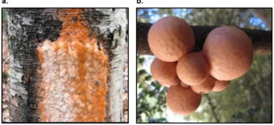
Since it was first isolated in 1967 by Herman Phaff (Phaff et al. 1972), several other isolates were obtained from exudates (also known as spring saps) of deciduous trees, belonging to genera Betula, Alnus, Fagus and Cornus throughout the Northern Hemisphere, e.g. Germany, Finland and Russia (Golubev et al. 1977; Weber et al. 2006). Due to the geographical distribution of the trees (Chen et al. 1999; Chen & Li 2004; Manchester et al. 2007) from which P. rhodozyma isolates were obtained, it was assumed that the species was only circumscribed to the Northern Hemisphere (Golubev 1995; Fell & Blatt 1999). However, in the beginning of the 21th century P. rhodozyma strains were isolated in the Southern Hemisphere (South America, Australia and New Zealand) from leafs of Nothofagus trees (David-Palma et al. 2014), as well as in association with the fruiting bodies of Cyttaria (Libkind et al. 2007; Libkind et al. 2011b), a biotrophic ascomycete restricted to Nothofagus (Peterson et al. 2010). The mycelium of Cyttaria develops inside the tree forming in its branches persistent tumors, from which fruiting bodies rich in simple sugars are annually produced, and from which isolates of P. rhodozyma are consistently isolated (Libkind et al. 2007; Libkind et al. 2008; Libkind et al. 2011b; David-Palma et al. 2014).
Figure 1.2. Distinct habitats associated with P. rhodozyma. (a) Exudate from a Betula tree trunk, Canada. (Photo courtesy of Beverly White); (b) branch of Nothofagus cunninghamii with Cyttaria gunnii fruiting bodies, Tasmania. (Photo courtesy of Diego Libkind)
Chapter | 1
16
Figure 1.3. Distribution, phylogeny and host tree association of P. rhodozyma. (a) World map showing the distribution of populations defined by the phylogenetic inference, which are represented by capital letters and distinct colors (b) Unrooted maximum likelihood phylogenies of Phaffia (left) based on a concatenated alignment of partial sequences of seven genes, and of the host trees (right) based on internal transcribed spacer sequences. Bootstrap values (1000 replicates) higher than 50% are indicated. The geographical origin of the isolates is indicated after strain number (A, Alaska; AUS, Australia; J, Japan; NA, North America; NE, Northern Europe; NZ, New Zealand; SA, South America; TAS, Tasmania). (Adapted from David-Palma et al., 2014).
1.2.3. Life cycle of
Phaffia rhodozyma
Chapter | 1
17
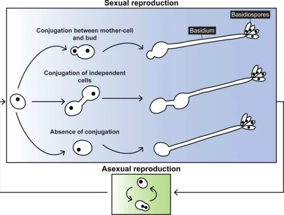
DNA of individual spores resulting from the crosses, revealed evidence of karyogamy, meiosis and recombination. However, segregation of the markers was not as expected from a normal meiosis, leading the authors to suggest the possible existence of aneuploids among the studied spores (Kucsera et al. 1998). The sexual stage of P. rhodozyma (Figure 1.5) represents an unusual departure from what is commonly observed in the sexual stages of most basidiomycetes, because it does not involve a transition between a unicellular/yeast stage to a filamentous/hyphal one (Fraser et al. 2007).
Chapter | 1
18
Figure 1.5. Sexual state of P. rhodozyma. (a-d) Photos from scanning electron microscopy showing a sporulating colony of P. rhodozyma cells. Yellow arrows point to two conjugated cells; orange arrows indicate basidia and blue arrows point to basidiospores. (Adapted from the site of the American Society for Microbiology, http://202.195.144.50/ASM/106-Introduce.htm. Photos by Carlos Echavarri-Erasun and Eric Johnson). (e) Micrograph of a colony with aerial basidiospores of strain CBS 6938. (Adapted from Kucsera et al., 2000).
1.2.4. Biotechnological relevance of
Phaffia rhodozyma
Chapter | 1
19
Dominguez-Bocanegra & Torres-Munoz 2004; Wozniak et al. 2011). All these studies spurred the development of different genetic tools including different yeast transformation protocols (Adrio & Veiga 1995; Rubinstein et al. 1996; Martinez et al. 1998; Wery et al. 1998; Visser et al. 2005) and mutant selection approaches, as well as, strategies for the construction of targeted knockout mutants and gene overexpression (Lin et al. 2012; Niklitschek et al. 2012; Hara et al. 2014a; Hara et al. 2014b).
1.3. Objectives and outline
The main objective of the work presented in this thesis was the elucidation of the molecular determinants and their underlying mechanisms governing the homothallic sexual cycle of P. rhodozyma. This work also aimed to provide the first insights regarding primary homothallism in basidiomycetes, which remain greatly understudied at the molecular level. Chapter 1 consists of a
general introduction highlighting the importance of sexual reproduction in fungi, summarizing its different modes of reproduction and indicating the most relevant molecular mechanisms that allow for the great diversity of sexual behaviors and mating type-determining systems observed in this group of organisms. Additionally, it describes the most significant aspects of the biology of the yeast Phaffia rhodozyma. Chapter 2 describes the identification, in available draft genomes of P. rhodozyma, of genes pertinent to mating determination in basidiomycetous fungi, as well as, key genes involved in the meiotic process. Identified MAT genes were further characterized and also amplified in a group of P. rhodozyma strains encompassing the known population diversity of this species, allowing for phylogenetic analyses. Chapter 3 describes the genetic manipulation of MAT genes in the haploid P. rhodozyma strain CBS 6938 by the construction of knockout mutants and their phenotypic characterization regarding their ability to undergo sexual reproduction. The obtained results allowed the proposition of a molecular model for the homothallic life cycle of P. rhodozyma. Chapter 4
CHAPTER 2
Identification of
MAT
and meiosis related genes in
Phaffia
rhodozyma genomes
Parts of the work presented in this chapter were published in:
Bellora N, Moliné M, David-Palma M, et al. (2016) Comparative genomics provides new insights into
the diversity, physiology, and sexuality of the only industrially exploited tremellomycete: Phaffia rhodozyma. BMC Genomics 17, 901.doi: 10.1186/s12864-016-3244-7
David-Palma M, Sampaio JP, Gonçalves P (2016) Genetic Dissection of Sexual Reproduction in a
Primary Homothallic Basidiomycete. PLOS Genetics 12(6): e1006110.
Chapter | 2
22
2.1. Introduction
Chapter | 2
23
2.2. Materials and Methods
2.2.1. Search for putative
MAT
and meiosis genes in
P. rhodozyma
genomes
and their characterization
The genomic regions containing MAT genes, namely the homeodomain transcription factors (HD1/HD2) and the mating pheromones (MFA) and receptors (STE3) were searched in the draft genome assemblies of P. rhodozyma CBS 7918T and CRUB 1149 (Bellora et al. 2016), or in local databases of proteins resulting from annotation of those draft genomes, by reciprocal BLASTP or TBLASTN, respectively. As queries, MAT proteins from C. deneoformans were used (Sxi1, Sxi2, Mfa1, and Ste3) (Table 2.3. and Table I.1, Appendix I). Pheromone precursor genes failing detection by BLAST due to their short length and highly variable sequences were identified manually upon inspection of the genomic regions near pheromone receptor genes, by searching for the existence of
ORFs whose deduced protein sequences contained a conserved “CaaX” motif at the C-terminus. To ascertain the contiguity of the scaffolds harboring the two sets of pheromone and receptor genes, a pair of primers (MP100 5′-TCCATCCTCAACTGATTGC-3′ and MP103 5′
-TTCATCTTGTCAGACAGC-3′) were used to amplify and partially sequence the region between both pheromone precursor genes.
Standard PCR and cycling conditions were used with Phusion® High-Fidelity DNA Polymerase using an annealing temperature of 51 °C and extension for 90 s. Protein sequences of genes involved in the pheromone signaling cascade in C. deneoformans (Roach et al. 2014), were used to identify the corresponding putative orthologs in P. rhodozyma by the same approach detailed above (Table I.1, Appendix I). Similarly, protein sequences of 61 genes known to be involved in meiosis in Saccharomyces cerevisiae, 41 of which have inferred orthology in C. deneoformans and other basidiomycetes (Schurko & Logsdon 2008; Halary et al. 2011; Gioti et al. 2013a), were retrieved from GenBank, and the corresponding orthologs in P. rhodozyma were identified (Table I.2, Appendix I). Core meiotic genes and meiosis-specific genes were categorized according to previously published sources (Ramesh et al. 2005; Malik et al. 2008; Halary et al. 2011).
The transmembrane regions in the pheromone receptor proteins were predicted by HMMTOP software (Tusnady & Simon 2001). For the deduced Hd1 and Hd2 proteins, homeodomain regions were determined using InterPro server (Mitchell et al. 2015) while nuclear localization signals (NLS) were predicted using SeqNLS server (Lin & Hu 2013) with a 0.5 cutoff. Potential alpha-helixes were predicted by Jpred4 (Drozdetskiy et al. 2015) and searches for coiled-coil dimerization motifs were