Low variation in ribosomal DNA and
internal transcribed spacers of the
symbiotic fungi of leaf-cutting ants
(Attini: Formicidae)
1Centro de Estudos de Insetos Sociais, and Departamentos de 2Bioquímica e Microbiologia, and 3Biologia, Instituto de Biociências,
Universidade Estadual Paulista, Rio Claro, SP, Brasil
4Departamento de Fonoaudiologia, Universidade Estadual Paulista, Marília, SP, Brasil 5Josephine Bay Paul Center for Comparative Molecular Biology and Evolution,
Marine Biological Laboratory, Woods Hole, MA, USA A.C.O. Silva-Pinhati1,
M. Bacci Jr.1,2,
G. Hinkle5,
M.L. Sogin5,
F.C. Pagnocca1,2,
V.G. Martins1,4,
O.C. Bueno1,3 and
M.J.A. Hebling1
Abstract
Leaf-cutting ants of the genera Atta and Acromyrmex (tribe Attini) are symbiotic with basidiomycete fungi of the genus Leucoagaricus (tribe Leucocoprineae), which they cultivate on vegetable matter inside their nests. We determined the variation of the 28S, 18S, and 5.8S ribosomal DNA (rDNA) gene loci and the rapidly evolving internal transcribed spacers 1 and 2 (ITS1 and ITS2) of 15 sympat-ric and allopatsympat-ric fungi associated with colonies of 11 species of leafcutter ants living up to 2,600 km apart in Brazil. We found that the fungal rDNA and ITS sequences from different species of ants were identical (or nearly identical) to each other, whereas 10 GenBank Leucoagaricus species showed higher ITS variation. Our findings suggest that Atta and Acromyrmex leafcutters living in geographic sites that are very distant from each other cultivate a single fungal species made up of closely related lineages of Leucoagaricus gongylophorus. We discuss the strikingly high similarity in the ITS1 and ITS2 regions of the Atta and Acromyrmex symbiotic L. gongylophorus studied by us, in contrast to the lower similarity displayed by their non-symbiotic counterparts. We suggest that the similarity of our L. gongylophorus isolates is an indication of the recent association of the fungus with these ants, and propose that both the intense lateral transmission of fungal material within leafcutter nests and the selection of more adapted fungal strains are involved in the homogenization of the symbiotic fungal stock. Correspondence
M. Bacci Jr. Centro de Estudos de Insetos Sociais, UNESP Av. 24A, 1515 13506-900 Rio Claro, SP Brasil
Fax: +55-19-3534-8523 E-mail: [email protected]
Research supported by FAPESP (No. 00/12538-5) and CAPES-University of Texas in Austin International Cooperation Program (CAPES-UT 005). M. Bacci Jr. and A.C.O. Silva-Pinhati are recipients of FAPESP fellowships.
Received October 20, 2003 Accepted June 22, 2004
Key words
•Atta
•Acromyrmex
•Internal transcribed spacer •Leaf-cutting ants
•Leucoagaricus
•Leucoagaricus gongylophorus
Introduction
The tribe Attini consists of approximately 200 species of ants living in symbiosis with three distinct groups of microorganisms, in what has been called ‘one of the most complex symbiotic association discovered in nature’
(1). These microorganisms include symbiotic basidiomycete fungi, which the ants utilize as food source, a parasite fungus of the genus
There are 13 genera of attines (2,3), the most derived being the leafcutter ants of the genera Atta and Acromyrmex, which are the dominant herbivores in the Neotropical re-gion between latitude 12º North and 33º South (4,5). This dominance results from the metabolic integration between the symbionts (6-9), which has been established during 50 million years of co-evolution (10,11).
To understand the processes involved in the association between leaf-cutting ants and symbiotic fungi, the diversity within each member of the symbiont pair needs to be characterized, but although the taxonomy of ants is well advanced (2), the taxonomy, and hence the diversity, of their symbiotic fungi has not yet been completely elucidated. Ba-sidiocarps collected from nests of Atta spe-cies have been named Leucocoprinus gon-gylophorus (12), Leucoagaricus gongylo-phorus (13) and Leucoagaricus weberi (14), but paired tests with mycelia suggest that some Atta and Acromyrmex ants cultivate a single species of fungus (15).
The multiple fungal species versus single species theories have more than taxonomic implications, being also related to the modes of fungal transmission within leafcutter
colo-nies. Vertical transmission occurs when queen ants leave their parent nest carrying the symbiotic fungus, which is then cultured by the newly founded colony (16). It has been proposed that vertical transmission has selected host-specific lineages of symbiotic fungi in leafcutter nests during the past 23 million years (10), so that the exclusively vertical transmission of fungal cultures within leafcutters is consistent with the existence of multiple symbiotic fungal species. On the other hand, the culturing of the same fungal species by Atta and Acromyrmex ants may indicate lateral transmission of fungal lin-eages. In laboratory nests, lateral transmis-sion of symbiotic fungal strains has been induced between and within species of basal attines of the genus Cyphomyrmex (17) as well as two Acromyrmex species (18) and also seems occasionally to occur in nature (18-21).
In the present paper, we provide new information on the systematics of Leuco-agaricus symbiotic fungi, which we obtained by sequencing the 28S, 18S and 5.8S rDNA loci and internal transcribed spacers 1 and 2 (ITS1 and ITS2) of two basidiocarp and thirteen mycelial fungal isolates collected from the nests of attine ants located in sev-eral geographic regions of Brazil between latitudes 1.52° and 23.43° South. Our results suggest that all the isolates studied belong to the species Leucoagaricus gongylophorus
and that lateral transmission was a common event through which selected lineages of the symbiotic fungus quickly proliferated in nests of many of the higher attine ants living in the tropical Americas.
Material and Methods
Fungal isolates and culture conditions
The Leucoagaricus isolates (Table 1) were collected as basidiocarps or as mycelial states from the nests of Atta and Acromyrmex
ants from July 1985 to December 2001 from
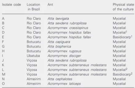
Table 1. Leucoagaricus isolates obtained from the nests of leaf-cutting ants.
Isolate code Location Ant Physical state
in Brazil of the culture
A Rio Claro Atta laevigata Mycelial
B Rio Claro Atta sexdens rubropilosa Mycelial
C Rio Claro Acromyrmex crassispinus Mycelial
D Rio Claro Acromyrmex hispidus fallax Mycelial1
E Rio Claro Acromyrmex hispidus fallax Basidiocarp1
F Botucatu Atta capiguara Mycelial
G Botucatu Atta bispherica Mycelial
H Botucatu Acromyrmex rugosus Mycelial
I Ubatuba Acromyrmex disciger Mycelial
J Viçosa Atta sexdens rubropilosa Mycelial
K Viçosa Acromyrmex subterraneus molestans Mycelial
L Viçosa Acromyrmex subterraneus molestans Mycelial2
M Viçosa Acromyrmex subterraneus molestans Basidiocarp2
N Almeirim Atta cephalotes Mycelial
O Almeirim Acromyrmex laticeps Mycelial
the geographic sites shown in Figure 1. The basidiocarp collected from Acromyrmex hispidus fallax was identified as Leuco-agaricus gongylophorus Heim (22). For the isolation of the mycelial form of the fungus from the fungus garden we used yeast nitro-gen base glucose chloramphenicol (YNBGC) agar, which increases the efficiency of isola-tion of this fungus (Silva-Pinhati ACO, Bacci M, Siqueira CG, Silva A, Pagnocca FC, Bueno OC and Hebling MJA, unpublished results) and consists of yeast nitrogen base medium (YNB, Difco 0392-15-9, Detroit, MI, USA) supplemented with 5 g/l glucose (product number 108342, Merck, Darmstadt, Ger-many), 17 g/l agar (Merck, product number 1.01614), 0.1 g/l chloramphenicol (product number C-0378, Sigma, St. Louis, MO, USA), and sufficient 2 M NaOH to adjust the pH to 6.0. For maintenance we used YNB-glucose agar (i.e., YNBG without chloramphenicol). The fungus garden material, collected from the underground nests of different ant spe-cies, was incubated in the dark at 25ºC and 80% humidity for 10 to 20 days on Petri dishes containing worker ants, which cleaned the garden material by removing soil frag-ments to a different part of the dish. When white mycelial spots of the fungal symbiont appeared on the leaf material some of the mycelium was collected using an aseptic technique and transferred to YNBGC agar on Petri dishes where it was incubated at 25ºC in the dark for 30 days. Alternatively, the fungus garden material from underground nests was collected and immediately plated onto YNBGC agar and incubated at 25ºC in the dark for 30 days. In both cases, after 30 days of cultivation on YNBGC agar the fun-gal isolates were subcultured to YNBG agar and cultured for a further 60 days, with the hyphae showing swollen tips (gongylidium), which are a distinctive characteristic of the symbiotic fungi of higher attine ants (5). The experiments described in this paper were carried out after two months to ten years of storage. When needed for DNA extraction,
mycelium was transferred to fresh YNBG agar and grown for 30 days, after which approximately 100 mg mycelium was pro-cessed as described below.
DNA extraction
Fungal mycelium was disrupted by placing 100 mg mycelium in a 1.5-ml microcentrifuge tube, freezing the tube and contents in liquid nitrogen, adding 0.5 ml ice-cold TE buffer, pH 8.6, containing 0.1% (w/v) SDS and homog-enizing the mycelia with a plastic pestle that fitted inside the tube. The nucleic acid was purified using buffered phenol, followed by a buffered phenol:chloroform mixture (1:1, v/v) and finally pure chloroform. Total nucleic acids were precipitated with ethanol and so-dium acetate, washed with ethanol and dis-solved in 100 µl TE buffer.
Amplification of ribosomal DNA and internal transcribed spacer regions
PCR amplification was carried out in a 100-µl reaction mixture using 100 ng fungal
Latitude, South
0º Atlantic Ocean
Almeirim
Rio Claro
Botucatu 3 2 1
Viçosa
Ubatuba
0 150 300 450 600 750 mi
0 250 500 750 1000 km
50º 45º 40º 35º 30º
Longitude, West
5º
10º
15º
20º
25º
30º
genomic DNA and 6 pmol each of primers F-5.8S (5'-GGATCACTCGGCTCRTGNRTC GATGAAG-3') and R-635 (5'-GGTCCGT GTTTCAAGACGG-3'). The amplification protocol consisted of initial denaturation at 94ºC for 3 min, followed by 30 PCR ampli-fication cycles of 94ºC for 10 s, 37ºC for 1 min and 72ºC for 3 min. The amplification produced fragments of approximately 1 kb comprising the 5.8S gene, the ITS2 region and 0.6 kb of the 28S gene. The same proce-dure and protocol were used to amplify the 18S rRNA gene and the ITS1-5.8S-ITS2 region, except that the primers for the 18S rRNA gene were the eukaryotic universal primers (23) A (5'-CCGAATTCGTCGACA ACCTGGTTGATCCTGCCAGT-3') and B (5'-CCCGGGATCCAAGCTTGATCCTTC TGCAGGTTCACCTAC-3') while the ITS4 and ITS5 primers (24) were used to amplify the ITS1-5.8S-ITS2 region. Primers were purchased from Invitrogen/Life Technolo-gies (São Paulo, SP, Brazil).
Cloning
PCR products were cloned in the pGEM-T vector (Promega, Madison, WI, USA), and transformed into competent XL1-BLU E. coli cells and recombinant clones selected and purified by a Miniprep procedure (25).
Sequencing
Both strands of recombinant plasmids or PCR products were individually sequenced with the SequiTherm kit (Epicentre Tech-nologies, Madison, WI, USA) according to manufacturer instructions. Each reaction contained 800 ng of purified DNA and 0.30-0.35 pmol infrared dye-labeled primer (Epicentre Technologies). The 5.8S-ITS2-28S fragments were sequenced using the vector primers M13F (5'-CACGACGTTG TAAAACGAC-3') and M13R (5'-GGATA ACAATTTCACACAGG-3') and the inter-nal primers F-63 (5'-TTCCTCCGCTTAT
TGATA-TGC-3') and R-63 (5'-TTCCTCC GCTTATTGATATGC-3') (Invitrogen/Life Technologies). Reaction conditions included denaturation at 94ºC for 3 min followed by 30 amplification cycles of 94º, 50º, and 70ºC for 30 s each. The same procedure was used for sequencing the 18S gene, using the M13F and M13R primers as well as the internal primers 514F (5'-TCTGGTGCCAGCASC CGCGG-3'), 536R (5'-TGGWATTACCGC GGSTGCTG-3'), 1055F (5'-GTGGTGGTGC ATGGCCG-3'), and 1055R (5'-AAGAAC GGCCATGCACCAC-3') (26). Reaction products were sequenced on a Li-Cor 4000L automated sequencing system (Li-Cor Bio-sciences, Lincoln, NE, USA). The ITS1-5.8S-ITS2 amplicons were sequenced using a 10-µl reaction mixture containing 6 pmol of the same primers as for their amplifica-tion, 100 ng template, 2.5 µl Big Dye reac-tant (product number 4303153, PE Applied Biosystems, Foster City, CA, USA), 2 µl 100 mM Tris and 2.5 mM MgCl2, pH 9.0. Reac-tion condiReac-tions included denaturaReac-tion at 96ºC for 1.5 min followed by 25 amplification cycles of 96ºC for 12 s, 50ºC for 8 s and 60ºC for 4 min. Reaction products were sequenced on an ABI 377 automated sequence system (Applied Biosystems).
Sequence analysis
network connecting the sequences was con-structed using the program Network 3.1.1.1 (available at www.fluxus-engineering.com) and the median joining algorithm (33). De-fault settings were chosen (r = 2 and ε = 0).
Results
In order to assess heterogeneity within and between isolates we sequenced indi-vidual clones containing the 5.8S-ITS2-28S amplicon. At least seven clones were se-quenced for each fungal isolate. We were especially interested in the ITS regions, which are known to incorporate changes at rela-tively high rates (see, e.g., 34,35). The 18S rDNA, which evolves comparatively slowly, was sequenced using pooled clones of each isolate. The 18S and 5.8S-ITS2-28S
se-quences generated are deposited in GenBank under accession numbers AF076380 to AF076430 and the ITS1-5.8S-ITS2 se-quences are deposited under accession num-bers AY642807 to AY642816.
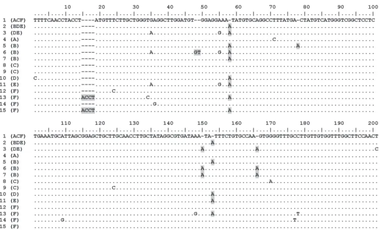
Few differences were found between iso-lates A to F, which were collected from two geographic sites (Table 1) and had identical 1760-bp 18S sequences, the 121-bp 5.8S and 690-bp 28S sequences showing more than 99% similarity. Variation in the ITS2 region (212 to 218 bp) was higher, with the 45 sequenced clones containing 15 distinct ITS2 haplotypes which had 25 variable posi-tions corresponding to 7 micro-satellites (1, 2 or 4 base repeats) and 14 transitions (8 C/ T and 6 A/G; Figure 2). Nucleotide diversity in the 45 ITS2 sequenced clones (π = 0.00589 ± 0.00110) was too low to allow population
Figure 2. Polymorphic sites of the 15 ITS2 haplotypes of six symbiotic fungal isolates. Fungal isolates were collected from nests of Atta laevigata (A),
Atta sexdens rubropilosa (B), Acromyrmex crassispinus (C), Acromyrmex hispidus fallax (D = mycelium, E = basidiocarp), and Atta capiguara (F). The
or to support a population study of these isolates, it was possible to group them by considering the three ITS2 haplotypes which were shared by several isolates (Table 2). The distribution of the shared haplotypes suggests that leafcutter fungal isolates A to F belong to two genetic groups, one group being composed of isolates A (from Atta laevigata), C (from Acromyrmex crassispi-nus) and F (from Atta capiguara), all con-taining ITS2 haplotype 1 but not haplotype 2, and the other group containing isolate B (from Atta sexdens rubropilosa) and isolates D and E (both from Acromyrmex hispidus fallax), all containing ITS2 haplotype 2 but not haplotype 1. Fungal isolates D and E also shared ITS2 haplotype 3. A phylogenetic network derived by the median-joining method showed that haplotypes 1 and 2 are likely centers of radiation/convergence of the ACF and BDE groups, respectively, so that some haplotypes are more closely re-lated within the ACF (haplotypes 1, 4, 8, 9, 12, 14) or BDE (haplotypes 2, 5, 10, 11) groups than between groups, although hap-lotypes 3, 6, 7, 13, and 15 were equally related to both ACF and BDE groups (Figure 3). These results suggest that the ACF and BDE genetic groups are in fact two distinct lineages of symbiotic fungi.
To extend our observations to a wider geographic region and other ant species, we investigated fungal isolates G to O (Table 1), collected from the nests of seven leafcutter species living in five different geographic sites. Both ITS1 and ITS2 regions were ana-lyzed by sequencing the amplified ITS1-5.8S-ITS2 fragments, but few polymorphic positions were found in the first clones ob-tained from isolates K and N. Because of this we decided to directly sequence the PCR products of isolates E and G to O and found them to be identical or to differ by only a maximum distance of 1.5%, in contrast to the 6.7 to 26% distance found between the 10 most closely related sequences of the GenBank Leucoagaricus species (see
spe-Figure 3. Phylogenetic relation-ship between the 15 ITS2 haplo-types of six symbiotic fungal iso-lates. Network nodes (circles) indicate haplotypes, which are numbered in bold and propor-tionally sized to the number of sequences they represent, i.e., 1, 2, 3, 5, 10, and 14 sequences. The areas of the colored parts of the circles are proportional to the number of sequence copies from each of the fungal isolates A to F (see also Table 2). Note that haplotypes from fungal iso-lates A, C and F are, in general, more closely related to each other than to haplotypes from isolates B, D and E.
studies and, similarly, the 2 parsimony-in-formative characters present in the 14 haplo-types (haplotype 11 was not considered in the parsimony analysis, since isolated D and E are from the same source nest) resulted in a highly polytomic tree (data not shown).
Although the information available was insufficient to reveal detailed relationships between our isolates in a phylogenetic tree
Table 2. Distribution and number of hits of each of the 15 ITS2 haplotypes found in the 45 sequenced clones of six sympatric or allopatric fungal isolates from Atta and
Acromyrmex leafcutter ants.
Isolate1 ITS2 haplotype Total
1 2 3 4 5 6 7 8 9 10 11 12 13 14 15
A 7 - - 1 - - - 8
B - 2 - - 3 1 1 - - - 7
C 5 - - - 1 2 - - - 8
D - 4 3 - - - 1 - - - 8
E - 4 2 - - - 1 - - - - 7
F 2 - - - 2 1 1 1 7
Total 14 10 5 1 3 1 1 1 1 1 1 2 1 1 1 45
1Fungus symbiotic with Atta laevigata (A), Atta sexdens rubropilosa (B), Acromyrmex
crassispinus (C), Acromyrmex hispidus fallax (D = mycelium, E = basidiocarp), and Atta
capiguara (F). Shaded numbers indicate the number of hits for haplotypes that were
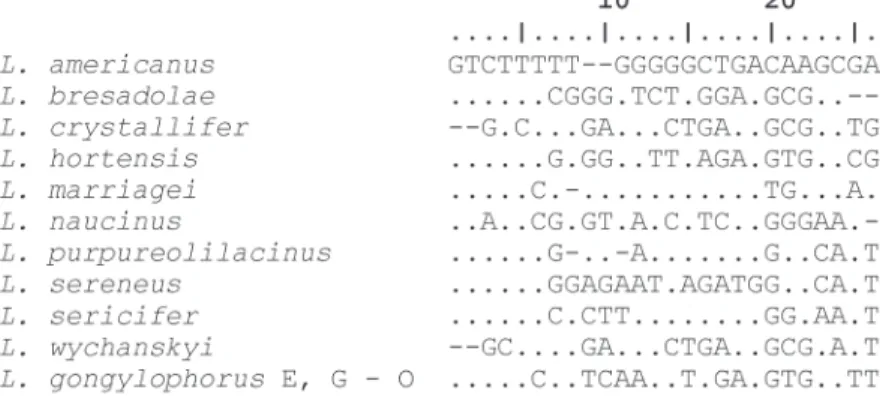
cies names in Figure 4).
Similarly, the distance between the ITS2 regions of isolates A to O was zero to 2.5%, while the distance between the ITS2 regions of the 10 most closely related GenBank Leu-coagaricus isolates was 9.4 to 26%. In addi-tion, the ITS1 locus presented a highly vari-able portion that had a base composition and sequence length characteristic for each of the GenBank 10 Leucoagaricus species in-vestigated. Conversely, this region was iden-tical for our leafcutter isolates E and G to O (Figure 4). These results indicate that the leafcutters studied by us cultivate very closely related fungal material, probably the same species, over a wide geographic region.
Discussion
In the past it has been difficult to demon-strate that a basidiocarp found in Atta or
Acromyrmex nests is that of the ant’s symbi-otic fungus and not one of the many contami-nant fungi existing in the nests (36). This situation has led to uncertainties in the iden-tification of ant fungi, which have received over a dozen names (4,37,38).
In our investigation we found that the
Leucoagaricus gongylophorus basidiocarp (E in Table 1) and the mycelial isolate (D in Table 1), both collected from the same
Acromyrmex hispidus fallax nest, had two identical (and some nearly identical) ITS2 sequences (Table 2). Since ITS regions have been used to distinguish species of fungi (e.g., 39) our results suggest that this Leu-coagaricus gongylophorus basidiocarp rep-resents the sexual stage of the symbiotic fungus of Acromyrmex hispidus fallax, sup-porting our previous results showing identi-cal RAPD fingerprints for these two isolates (22).
The Leucoagaricus gongylophorus ba-sidiocarp was also found to have ITS2 se-quences which were identical, or nearly iden-tical, to those of mycelial isolates from the nests of Atta laevigata (isolate A), Atta
sexdens rubropilosa (isolate B) and Acro-myrmex crassispinus (isolate C), all sympa-tric leafcutter species collected near the town of Rio Claro, SP, as well as to fungal isolate F collected from an Atta capiguara nest near the town of Botucatu, SP, 105 km from Rio Claro, and other fungal symbionts (isolates G to O) from sites near the towns of Botu-catu, Ubatuba, Viçosa, and Almeirim, which are up to 2,600 km apart from each other. The variations found in these ITS2 sequences were very low compared to those of GenBank ITS2 sequences belonging to 10 distinct Leu-coagaricus species. In addition, the highly variable portion of ITS1, which contains a characteristic signature for each Leucoagari-cus species, was identical in 10 of our fungal symbionts (isolates E and G to O) living in each of the investigated sites.
Taken together, these results indicate that the sympatric and allopatric leafcutter spe-cies studied in the present investigation are associated with closely similar fungal mate-rial, which may represent a single Leuco-agaricus species, suggesting that leaf-cut-ting ants cultivate the same fungal species over a wide geographic region of South America and indicating that the ‘single spe-cies theory’ regarding fungal culture by
leafcutters is more likely than the ‘multiple species theory’. Therefore, it is likely that the Leucoagaricus gongylophorus basidio-carp (E in Table 1) represents the sexual stage of all the mycelial state isolates studied in the present investigation.
The degree of polymorphism that we found in the ITS sequences was too low to reveal detailed relationships between our isolates in a phylogenetic tree. However, distribution of ITS sequences within our fun-gal isolates collected from the Rio Claro or Botucatu areas suggests that these isolates belong to distinct genetic groups character-ized by a specific ITS2 haplotype (Table 2), haplotype 1 only being found in isolates A, C and F (14 hits in 23 sequenced clones) and haplotype 2 only in isolates B, D and E (10 hits in 22 sequenced clones). These results, as well as the phylogenetic relationship be-tween haplotypes derived by median-joining analysis (Figure 3), suggest that these two genetic groups are in fact two distinct fungal lineages. Thus, it seems that the fungal strains cultured by Atta laevigata, Acromyrmex cras-sispinus and Atta capiguara (belonging to the ACF lineage) are more similar to each other than they are to the fungal strains cul-tured by Atta sexdens rubropilosa and Acro-myrmex hispidus fallax (BDE lineage). Chapela et al. (10) hypothesized that there has been restricted vertical transmission of symbiotic fungi in the higher attines for the last 23 million years, which suggests that the symbiotic fungi of Atta ants should be more similar to each other than they are to the symbiotic fungi of Acromyrmex ants. How-ever, our findings do not support the exclu-sively vertical transmission of symbiotic fungi in higher attines, suggesting instead that lat-eral transmission may occur under certain circumstances.
Our findings are supported in part by those of Bot et al. (18), who found that, in Panama, lateral transmission of fungal mate-rial may have occurred between the sympat-ric species Acromyrmex octospinosus and
Acromyrmex echinatior. However, these in-vestigators also mention that lateral transfer of symbiotic fungi between Atta and Acro-myrmex ants does not occur, although our results show that some Atta and Acromyr-mex species do indeed cultivate the same lineage of Leucoagaricus gongylophorus and hence have probably recently shared their symbiotic fungi.
from one nest to another or, conversely, this transmission may involve an intermediary free-living fungal stage so that many clones of the original symbiotic fungus could have been spread over the period of time during which these ants have been living in Ame-rica. Thus, it is possible that free-living close relatives (or even free-living forms) of Leu-coagaricus gongylophorus exist.
In addition to intensive lateral transmis-sion, some features of symbiotic fungal lin-eages may have shaped the population struc-ture of Leucoagaricus gongylophorus, such as resistance to antibiotics produced either by the ants (40) or the actinomycete sym-biont or to the parasite Escovopsis, so that more adapted fungal strains may have been
selected. Thus, the complete elucidation of the factors that have led to the low variation in Leucoagaricus gongylophorus ITS se-quences requires an intensive sampling and characterization of the two groups of symbi-otic microorganisms of many leafcutters from distinct geographic sites in America.
Acknowledgments
Dr. Fábio O. Freitas (Embrapa-Cenargem, Brasília, DF, Brazil) is acknowledged for his comments on median-joining analysis and Dr. Ulrich G. Mueller (The University of Texas, Austin, TX, USA) is also acknowl-edged for his comments on earlier versions of this paper.
References
1. Currie CR, Wong B, Stuart AE, Schultz TR, Rehner SA, Mueller UG, Sung GH, Spatafora JW & Strauss NA (2003). Ancient tripartite coevolution in the attine ant-microbe symbiosis. Science, 299: 386-388.
2. Bolton B (1995). A New General Catalogue of the Ants of the World. Harvard University Press, Cambridge, MA, USA.
3. Brandão CFR & Nunes AM (2001). A new fungus-growing ant genus
Mycetagroicus gen. n., with the description of three new species
and comments on the monophyly of the Attini (Hymenoptera: For-micidae). Sociobiology, 38: 639-665.
4. Weber NA (1979). Fungus culturing by ants. In: Batra LR (Editor),
Insect-Fungus Symbiosis, Mutualism and Commensalism. 20th
In-ternational Mycological Congress, Tampa, FL, USA. Halsted Press,
New York, 77-115.
5. Hölldobler B & Wilson EO (1990). The Ants. The Belknap Press of Harward University Press, Cambridge, MA, USA.
6. Martin MM (1970). The biochemical basis of the fungus-attine ant symbiosis. Science, 169: 16-20.
7. Bass M & Cherrett JM (1995). Fungal hyphae as a source of nutri-ents for the leaf-cutting ants Atta sexdens. Physiological Entomol-ogy, 20: 1-6.
8. North RD, Jackson CW & Howse PE (1997). Evolutionary aspects of ant-fungus interactions in the leaf-cutting ants. Trends in Ecology
and Evolution, 12: 386-389.
9. Siqueira CG, Bacci M, Pagnocca FC, Bueno OC & Hebling MJA (1998). Metabolism of plant polysaccharides by Leucoagaricus
gongylophorus, the symbiotic fungus of the leaf-cutting ant Atta
sexdens L. Applied and Environmental Microbiology, 64: 4820-4822.
10. Chapela IH, Rehner SA, Schultz TR & Muller UG (1994). Evolutionary history of the symbiosis between fungus-growing ants and their fungi. Science, 266: 1691-1694.
11. Hinkle G, Wetterer JK, Schultz TR & Sogin ML (1994). Phylogeny of the attine fungi based on analysis of small subunit ribosomal RNA gene sequences. Science, 226: 1695-1697.
12. Bononi VLR, Autuori M & Rocha MB (1981). Leucocoprinus
gongy-lophorus (Möller) Heim, o fungo do formigueiro de Atta sexdens
rubropilosa Forel. Rickia, 9: 93-97.
13. Fisher PJ, Stradling DJ & Pegler DN (1994). Leaf cutting ants, their fungus gardens and the formation of basidiomata of Leucoagaricus
gongylophorus. Mycologist, 8: 884-888.
14. Muchovej JJ, Della Lucia TM & Muchovej RM (1991).
Leucoagari-cus weberi sp nov. from a live nest of leaf-cutting ants. Mycological
Research, 95: 1308-1311.
15. Stradling DJ & Powel RJ (1986). The cloning of more highly produc-tive fungal strains: a factor in the speciation of fungus growing ants.
Experientia, 42: 962-964.
16. Weber NA (1972). Gardening ants, the attines. Memoirs of the
American Philosophical Society (Philadelphia, PA, USA), 92: 1-146.
17. Adams RMM, Mueller UG, Holloway AK, Green AM & Narozniak J (2000). Garden sharing and garden stealing in fungus-growing ants.
Naturwissenschaften, 87: 491-493.
18. Bot ANM, Rehner SA & Boomsma JJ (2001). Partial incompatibility between ants and symbiotic fungi in two sympatric species of
Acromyrmex leaf-cutting ants. Evolution, 55: 1980-2001.
19. Mueller UG, Lipari SE & Milgroom MG (1996). Amplified fragment length polymorphism (AFLP) fingerprinting of symbiotic fungi cul-tured by the fungus-growing ant Cyphomyrmex minutus. Molecular
Ecology, 5: 119-122.
20. Mueller UG, Rehner SA & Shultz TR (1998). The evolution of agricul-ture in ants. Science, 281: 2034-2038.
21. Green AM, Adams RMM & Mueller UG (2002). Extensive exchange of fungal cultivars between sympatric species of fungus-growing ants. Molecular Ecology, 11: 191-195.
22. Pagnocca FC, Bacci M, Fungaro MH, Bueno OC, Hebling MJA, Sant’anna A & Cappellari M (2001). RAPD analysis in basidiomata found in a nest of the leaf-cutting ant Acromyrmex hispidus fallax, Santschi. Mycological Research, 105: 173-176.
enzymatically amplified eucaryotic 16S-like rRNA coding regions.
Gene, 71: 491-499.
24. White TJ, Bruns T, Lee S & Taylor J (1990). Amplification and direct sequencing of fungal ribosomal RNA genes for phylogenetics. In: Innis MA, Gelfand DH, Sninsky JJ & White TJ (Editors), PCR
Proto-cols: A Guide to Methods and Applications. Academic Press, San
Diego, CA, USA, 315-322.
25. Sambrook J & Russel DW (2001). Molecular Cloning: A Laboratory
Manual. 3rd edn. Cold Spring Harbor Laboratory Press, Cold Spring
Harbor, New York.
26. Elwood HJ, Olsen GH & Sogin ML (1985). The small-subunit riboso-mal RNA gene sequences from the hypotrichous ciliates Oxytricha
nova and Stylonychia pustulata. Molecular Biology and Evolution, 2: 399-410.
27. Thompson JD, Higgins DG & Gibson TJ (1994). CLUSTAL W: im-proving the sensitivity of progressive multiple sequence alignment through sequence weighting, position specific gap penalties and weight matrix choice. Nucleic Acids Research, 22: 4673-4680. 28. Jukes TH & Cantor CR (1969). Evolution of protein molecules. In:
Munro HN (Editor), Mammalian Protein Metabolism III. Academic Press, New York.
29. Felsenstein J (1989). PHYLIP - Phylogeny Inference Package (Ver-sion 3.2). Cladistics, 5: 164-166.
30. Rozas J & Rozas R (1997). DnaSP version 2.0: a novel software package for extensive molecular populational genetic data analysis.
Computer Applications in the Biosciences, 13: 307-311.
31. Nei M (1988). Molecular Evolutionary Genetics. Columbia Univer-sity Press, New York.
32. Swofford DL (2000). Phylogenetic Analysis Using Parsimony,
Ver-sion 4.0b4a. Illinois Natural History Survey, Champaign, IL, USA.
33. Bandelt HJ, Forster P & Röhl A (1999). Median-joining networks for inferring intraspecific phylogenies. Molecular Biology and Evolution,
16: 37-48.
34. O’Donnel K (1992). Ribosomal DNA internal transcribed spacers are highly divergent in the phytopathogenic ascomycete Fusarium
sambucinum (Giberella pulicaris). Current Genetics, 22: 213-220.
35. Campbell AJD, Gasser RB & Chilton NB (1995). Differences in a ribosomal DNA sequence of Strongylus species allow identification of single eggs. International Journal of Parasitology, 25: 359-365. 36. Fisher PJ, Stradling DJ, Sutton BC & Petrini LE (1996). Microfungi in
the fungus gardens of the leaf-cutting ant Atta cephalotes: a prelimi-nary study. Mycological Reserch, 100: 541-546.
37. Wetterer JK (1994). Nourishment and evolution in fungus-growing ants and their fungi. In: Hunt JH & Nalepa CA (Editors),
Nourish-ment and Evolution in Insects Societies. Westview Press, Bolder,
CO, USA.
38. Kermarrec A, Decharme M & Febvay G (1986). Leaf-cutting ant symbiotic fungi: a synthesis of recent research. In: Lofgren CS & Van der Meer RK (Editors), Fire Ants and Leaf-Cutting Ants. Westview, Bolder, CO, USA and London, UK.
39. Sreenivasaprasad S, Mills PR, Meeham BM & Brown AE (1996). Phylogeny and systematics of 18 Colletotrichum species based on ribosomal RNA spacer sequence. Genome, 39: 499-512.