Vol.53, n. 1: pp. 141-152, January-February 2010
ISSN 1516-8913 Printed in Brazil BRAZILIAN ARCHIVES OF
BIOLOGY AND TECHNOLOGY
A N I N T E R N A T I O N A L J O U R N A L
Plant or Fungal Sequences? An Alternative Optimized PCR
Protocol to Avoid ITS (nrDNA) Misamplification
Vitor Fernandes Oliveira de Miranda
1, Vanderlei Geraldo Martins
2,3, Antonio Furlan
4and
Maurício Bacci Jr.
2*1Laboratório de Sistemática Vegetal e Herbarium Mogiense; Universidade de Mogi das Cruzes; 08780-911; Mogi
das Cruzes - SP - Brasil. 2Centro de Estudos de Insetos Sociais; Universidade Estadual Paulista; 13506-900; Rio Claro - SP - Brasil. 3Departamento de Fonoaudiologia; Universidade Estadual Paulista; Marília - SP - Brasil. 4Departamento de Botânica; Universidade Estadual Paulista; 13506-900; Rio Claro - SP - Brasil
ABSTRACT
The nuclear ribosomal DNA internal transcribed spacers (ITS1 and ITS2) from leaves of Drosera (Droseraceae) were amplified using “universal” primers. The analysis of the products demonstrated most samples were a molecular mixture as a result of unsuccessful and non-specific amplifications. Among the obtained sequences, two were from Basidiomycota fungi. Homologous sequences of Basidiomycota were obtained from GenBank database and added to a data set with sequences from Drosera leaves. Parsimony analysis demonstrated that one sequence was amplified from an Ustilaginomycetes fungus, and another from a Heterobasidiomycetes. Possibly these fungi were associated to leaves of Drosera, and not because of samples contamination. In order to provide optimization and a better specificity of PCR (polymerase chain reaction), a very successful method was demonstrated using dimethyl sulfoxide (DMSO) and bovine serum albumin (BSA) in reactions.
Key words: polymerase, DNA, Drosera, fungi, phylogeny
*Author for correspondence: mbacci@rc.unesp.br
INTRODUCTION
Many problems previously found were related to cladistics, such as the difficulties to survey the morphological homologous characters, as many of them were intractable and not able to be compared among all living organisms, therefore, they could only be transposed with the advent of molecular analyses. On the other hand, the considerable facility to obtain molecular data, as well as the growing knowledge of molecular biology as consequence of improvement in techniques has afforded much information for systematics. With the discovery of polymerase chain reaction (PCR),
the DNA sequencing became a alternative low cost and easy way in order to approach the phylogeny relations of living organisms of all taxonomical degrees (Baldwin et al., 1995), allowing the survey of homologous characters with more precision than provided by previous molecular techniques, such as the restriction maps. For this purpose, the ribosomal DNA has been demonstrated to be an important tool to provide a better comprehension of the living organisms’ history (Hillis and Dixon, 1991).
constituted by highly variable regions, which can be used for studies of taxonomical groups with recent diversification or even among the populations. On the other hand, the ribosomal DNA possesses highly conserved regions, which can be applied for organisms comparing with ancient diversification. The same conserved regions can be very useful for designing so called “universal primers” (White et al., 1990; Hillis and Dixon, 1991), in order to amplify the alternate variable regions.
Thousands of tandemly repeated copies of transcribed units and non-transcribed spacers (Long and Dawid, 1980; Hillis and Dixon, 1991) typically constitute the nuclear ribosomal DNA (nrDNA) of a eukaryote nuclear genome. The great amount of copies usually facilitates the amplification of nrDNA.
Nevertheless, one must regard the possibility of amplifying paralogous copies, and non-ortologous ones, of different taxa, providing erroneous phylogenetic inferences as a result of comparisons among non-homologous sequences. Another point is the possible existence of highly divergent paralogous copies (Dvorák, 1990; Suh et al., 1993; Dubcovsky and Dvorák, 1995; Buckler and Holtsford, 1996; Buckler et al., 1997), some of them possibly pseudogenes (Buckler et al., 1997). Thereby, special attention should be devoted to the PCR reactions in attempt to select the copies to be amplified.
The increasing sensibility of PCR reactions that allowed promoting amplifications from very small quantities of template DNA has been a point of extreme importance to the molecular studies, enabling phylogeny studies from tiny amounts of tissue or even from isolated cells (Lee and Taylor, 1990). However, when contaminant sequences are present in target DNA, the increasing sensibility of the PCR can be a problem and has been the reason of preoccupation in many studies (Sarkar and Sommer, 1990; Bobola et al., 1992; Smith and Klein, 1994, 1996; Liston and Alvarez-Buylla, 1995; Liston et al., 1996; Zhang et al., 1997; Chiang et al., 2001).
The carnivorous and cosmopolitan family Droseraceae comprises about 150 species, most of them grouped in the genus Drosera (Diels, 1906). In attempts to achieve a better comprehension of phylogenetic relations of Droseraceae family, studies have been developed with the survey of
morphological characters as well as sequencing of ITS region from nuclear ribosomal DNA (V. F. O. Miranda, A. Furlan, M. Bacci Jr. and V. G. Martins, unpublished data). Rivadavia et al. (2003) investigated the phylogenetic relations of Droseraceae using the analysis of rbcL sequences from chloroplast DNA. Although the analysis demonstrated very suitable phylogenetic relations among species from different continents, rbcL sequences provided unclear information about American and African species of Drosera species, which was understandable for the substitution rate for this gene (Wolfe et al., 1987). Regarding the lack of information and the little comprehension about American and African species of Drosera, ITS region was chosen since internal transcribed spacers (ITS1 and ITS2) could bring enough divergences to the phylogeny inferences (Hillis and Dixon, 1991; Baldwin et al., 1995), as studies have demonstrated (V. F. O. Miranda et al., unpublished data).
Initially, the amplifications of ITS regions of 15 Drosera species were carried out in order to sequenciate the amplicons directly. As the results showed high polymorphism to almost all samples, the improvement of PCR was intended, optimizing the amplification protocols (for example increasing the annealing temperature), as well as adding adjuvants reagents to the reactions, as dimethyl sulfoxide (DMSO) and bovine serum albumin (BSA). Surprisingly, two sequences obtained before the PCR optimization were from fungi, probably present in the plant tissue used for DNA extractions. This work had as its goal to identify the two “strange sequences” amplified as well as to demonstrate an alternative and successful way to optimize the ITS amplifications from nuclear ribosomal DNA.
MATERIALS AND METHODS
Plant tissues and sequences
Table 1 - Voucher numbers and GenBank accession numbers for the studied species.
Taxon Voucher numbers 1 GenBank accession numbers
Plantae
Capsella rubella AJ232913
Gymnocarpos mahranus AJ310970
Oryza sativa M16845
Sassafras tzumu AF272336
Vicia montbretti AF228075
Drosera anglica VM185 EU178843
Drosera brevifolia VM186 EU178844
Drosera burmannii VM187 EU178845
Drosera capensis VM188 EU178846
Drosera madagascariensis VM189 EU178847
Drosera nidiformis VM195 EU178848
Drosera ordensis VM198 EU178849
Drosera villosa VM205 EU178850
Fungi Ascomycota
Candida albicans AB049122
Ceratocystis fimbriata AF264904
Claviceps sorghi AJ242869
Metarhizium anisopliae AB071714
Neurospora tetrasperma AF388929
Basidiomycota
Agaricus bisporus AF188035
Amanita gemmata AF335440
Auricularia delicate AF291269
Auricularia fuscosuccinea AF291270
Auricularia mesenterica AF291271
Ceratobasidium bicorne AF200514
Ceratobasidium oryzae-sativae isolate IMI062599 AJ000192
Ceratobasidium oryzae-sativae isolate IMI375133 AJ000194
Exidia truncata AF291279
Exidiopsis calcea AF291280
Heterochaete sp. USJ 55639 AF291285
Ingoldiomyces hyalosporus strain S053 AF399891
Pseudozyma antarctica strain CBS 516.83 AF294698
Pseudozyma aphidis strain CBS 517.83 AF294699
Pseudozyma prolifica strain CBS 319.87 AF294700
Pseudozyma rugulosa strain CBS 170.88 AF294697
Puccinia miscanthi AJ406072
Rhizoctonia cerealis isolate 99125 AF222793
Rhizoctonia crocorum AB044354
Rhizoctonia solani AJ000197
Rhizoctonia violacea AB044140
Rhizoctonia zeae isolate RZ01 AF222799
Rhizopogon rubescens AF158018
Rhodotorula acheniorum AB038128
Sebacina vermifera AF202728
Sporisorium destruens AF045871
Sporisorium reilianum AF135432
Sporisorium reilianum sp. reilianum AF038827
Sporisorium reilianum sp. zeae AF045870
Sporisorium sorghi AF038828
Strange A from Drosera capillaris E257 VM206 EU178842
Strange B from Drosera anglica E260 VM185 EU178841
(Cont. Table 1)
Taxon Voucher numbers1 GenBank accession numbers
Plantae
Thanatephorus cucumeris 23R01 U57740
Thanatephorus cucumeris isolate IMI 360021 AJ000200
Thanatephorus cucumeris isolate IMI 360366 AJ000199
Thanatephorus cucumeris isolate IMI 369673 AJ000202
Thanatephorus cucumeris strain 021R06 U57887
Thanatephorus cucumeris strain UB1 U57888
Thanatephorus cucumeris strain VG1 U57889
Tilletia barclayana strain S104 AF399894
Tilletia horrida strain S150 AF399893
Tilletia indica strain BC 388 AF310179
Tilletia indica strain S001 AF399890
Tilletia walkeri strain BC 188 AF310181
Tilletiopsis derxii AB045707
Tilletiopsis oryzicola AB045708
Tilletiopsis washingtonensis strain ATCC96156 AF294696
Ustilago bullata AF135423
Ustilago cynodontis AF038825
Ustilago hordei A AF045866
Ustilago hordei B AF105224
Ustilago hypodites AF045867
Ustilago maydis A AF038826
Ustilago maydis B AF135431
Ustilago nuda AF135430
Ustilago scitaminea AF135433
Ustilago sp. 4327 AF135429
Ustilago sp. 83-138 AF135428
Ustilago sparti AF045868
Ustilago tritici AF135424
Ustilago williamsii AF045869
Uromyces striolatus AF180201
Zygomycota
Acaulospora morrowiae AJ242500
Endogene pisiformis AF00651
Entrophospora colombiana AJ239117
Glomus clarum AJ243275
Mortierella alpine AJ271629
1
VM is the first author’s collector prefix.
DNA extraction
For the DNA extraction, 100 mg of fresh leaves or 25-30 mg of dried tissue were utilized and plant tissues were not treated previously. The plant material was submitted to liquid nitrogen and macerated. DNAzol (Chomczynski et al., 1997, 1998) was employed for the extraction, following the protocol recommended by the manufacturers.
DNA amplification
Primers utilized to the amplifications were the ITS “universal” primers designed by White et al. (1990) and the ITS3B (B. G. Baldwin, unpublished data) and ITS.LEU (L. E. Urbatsch,
Conventional reactions
The first reactions were carried out using Ready-To-Go (GE HeathCare), which contained Taq DNA polymerase, nucleotides, MgCl2, buffer solution and stabilizers. Each sample of Ready-to-Go provided a 25 µL reaction, whereas each reaction contained 1.5 units of Taq DNA polymerase, buffer, 1.5 mM MgCl2, 800 µM (200 µM of each dNTP) and stabilizers.
Optimized reactions
Other PCRs were accomplished using the same reagents. However, for these samples, dimethyl sulfoxide (DMSO) and bovine serum albumin (BSA) were added. Several DMSO and BSA concentrations (1-10%) were used in order to optimize the ITS amplifications. Other reactions with 50 µL and 100 µL were carried out as well. For each 25-µL reaction (conventional and optimized ones), 25 ng of template DNA and 25 pmols of each primer were used. Several amplification protocols were tried in an effort to optimize the reactions. For ITS1 amplification, 45 cycles of 30 s at 94oC for denaturing, 30 s at 46oC to annealing and 1 min at 70oC for extension time were used. For ITS2 amplification, the same protocol was carried out, however, the annealing temperature was increased to 55oC. The PCR products were quantified through electrophoresis (agarose gel 1%) using Low Mass DNA Ladder (Life Technologies).
PCR products purification
The amplified samples from PCR were purified using GFX PCR DNA and Gel Band Purification Kit (GE HeathCare) and PCR Concerted Purification Kit (Life Technologies). Some of the purifications were proceeded directly from PCR solutions, when just a single band could be noted in electrophoresis. On the other hand, amplification reactions with multiple bands were visualized in 1.5% agarose gel with ethidium bromide and excised separately. In this case, long runs were carried out in electrophoresis (40-55 min), in order to separate the bands, adjusting then to a low voltage and amperage (80V, 40mA).
DNA sequencing
Sequencing reactions were obtained in PTC-100 thermocycler (MJ Research). Each 10-µL sequencing reaction was constituted of 2.5 µL ABI Big Dye Terminator Cycle Sequencing Kit (Applied Biosystems) and 2.5 pmols of each
primer. Both strands (forward and reverse) of each sequence were read in automated DNA sequencer ABI 377 (Applied Biosystems).
All sequences obtained were checked in GenBank using BLASTN program (Altschul et al., 1997) in order to achieve the sequences with highest identity. Despite ITS spacers being so much variable hindering the search of related sequences, the 18S, 5.8S and 26S sequences of nrDNA allow to obtain sequences with high identity through GenBank since they demonstrate to be very conserved in all living organisms.
Sequence alignment and phylogenetic analysis
Homologous sequences were aligned with the program ClustalW 1.4 (Thompson et al., 1994) and data sets were checked using BioEdit 5.0.9 (Hall, 1999). The alignment was achieved through gap initiation penalty 10 and gap extension penalty 0.05. Each sequence achieved was checked with its complementary strand.
Maximum parsimony and bootstrap (Swofford et al., 1996) analyses were performed using PAUP* 4b8 (Swofford, 1999). Heuristic searches (Swofford et al., 1996) were carried out through random addition sequence with 5000 replicates to obtain the most parsimonious trees; 100 bootstrap (Felsenstein, 1985) replicates using random addition sequence with 100 replicates were performed for bootstrap analyses (TBR branch-swapping algorithm). For decay indices (or Bremer support; Bremer, 1988), TreeRot program was employed (Sorenson, 1996). Gaps were treated as fifth base considering they have phylogenetic information (Giribet and Wheeler, 1999), weight 3 was ascribed to transversions and 2 to transitions. The cladograms were drawn using the program TreeView (Page, 1998).
RESULTS
classes of amplicons could be noticed, evidenced by electropherogram (data not shown). Thus, one must emphasize that even when different amplicons present very close lengths, or even have identical lengths, avoiding the distinction in agarose gel electrophoresis, this result does not mean the band consists of copies of the same sequence, perhaps being different sequences phylogenetically distinct (Sekiguchi, 2001). As an initial attempt to reach a higher specificity for PCR reactions, the annealing temperature was gradually increased, as well as other points of amplification protocol were changed (e.g. annealing time, extension time and temperature), therefore a mix of different amplicons was noted in amplification reactions. Thus, taking into account that the PCR reactions usually resulted unspecific products, the increase of stringency was performed to the reactions through the addition of adjuvants. Various authors recommend the use of adjuvants to improve amplification efficiency (Innis and Gelfand, 1990; Palumbi, 1996; Henegariu et al., 1997), such as dimethyl sulfoxide (DMSO) and the bovine serum albumin (BSA). These substances improve the efficiency of the reactions, increasing the amount of product, as
well as the specificity, avoiding the amplification of unspecific products.
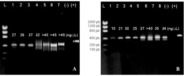
Palumbi (1996) suggested the use of both DMSO and BSA together in reactions, therefore these substances should be used up to 1% of concentration (v/v), otherwise PCR reaction could be hindered due to the enzymatic activity inhibition of Taq DNA polymerase (Innis and Gelfand, 1990; Palumbi, 1996). Nevertheless, some authors (Henegariu et al., 1997; Baum et al., 1998) have employed DMSO at 10% (v/v) in the amplification reactions. This way, several concentrations of DMSO and BSA were tested for the amplifications of the internal spacers ITS1 and ITS2, using both adjuvants either in the same reactions or even just one. As a result, the best amplifications were obtained adding 5% of DMSO and 10% of BSA for the amplification of the spacer ITS1 and 10% of DMSO for the spacer ITS2 (v/v), as demonstrated in Fig. 1.
It became evident that the DMSO and BSA addition inhibited the yield of reactions (Fig. 1). On the other hand, PCR reactions with adjuvants (Fig.1, A1-A3 and B1-B4) only resulted amplicons from nrDNA (Fig. 2).
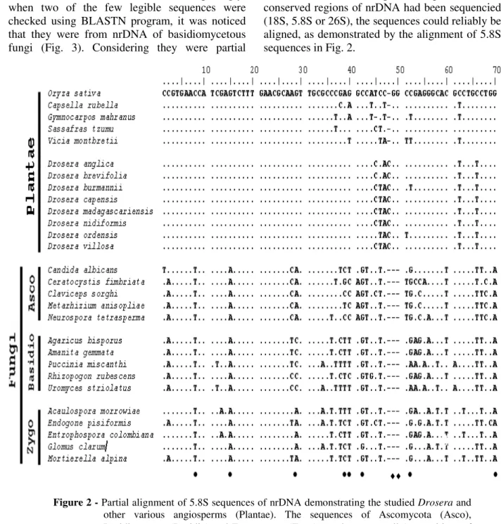
As a result of sequencing of the samples that were not added DMSO and BSA, most of them resulted in non-reliable sequences, caused by the mix of different molecules. Nevertheless, surprisingly, when two of the few legible sequences were checked using BLASTN program, it was noticed that they were from nrDNA of basidiomycetous fungi (Fig. 3). Considering they were partial
sequences of the internal transcribed spacers (ITS1 and ITS2), it was not possible to obtain a reliable alignment with the homologous sequences of Drosera. If at least a partial sequence of the conserved regions of nrDNA had been sequencied (18S, 5.8S or 26S), the sequences could reliably be aligned, as demonstrated by the alignment of 5.8S sequences in Fig. 2.
Figure 2 - Partial alignment of 5.8S sequences of nrDNA demonstrating the studied Drosera and other various angiosperms (Plantae). The sequences of Ascomycota (Asco), Basidiomycota (Basidio) and Zygomycota (Zygo) are shown as well (• - positions of bases that distinguish angiosperm sequences of Fungi; ♦ - indels only found in sequences of Fungi).
The high divergent sequences of internal transcribed spacers obtained from fungi could not be aligned even with sequences of other groups of fungi other than Basidiomycota (Ascomycota, Zygomycota).
obtained (around 300 bp), which could disable robust phylogenetic estimates, it was possible, at least approximately, to provide an approach to
identify their taxonomical position and relation to other groups (Fig. 4).
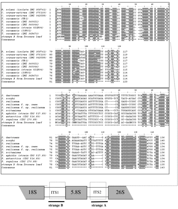
Figure 3 - Partial alignment of the fungi sequences obtained from Drosera leaves with other homologous sequences of Basidiomycota. Strange A corresponds to the spacer ITS2 and strange B to the spacer ITS1 of the nrDNA. Strange A was sequenciated from leaf of Drosera capillaris and strange B from D. anglica. The emphasized nucleotides denote the positions with identity equal or above 80%. The consensus sequence indicates the invariable positions. (C.- Ceratobasidium, P.- Pseudozyma, R.- Rhizoctonia, S.- Sporisorium, T.- Thanatephorus, U.- Ustilago.)
strange A strange B
Phylogenetic analyses demonstrated and confirmed the placement of both sequences in Basidiomycota. With the two “strange sequences” was not possible to produce reliable alignments using data sets of Zygomycota and Ascomycota sequences, as a consequence of too much polymorphisms. On the other hand, when both sequences were added to Basidiomycota sequences
data sets consistent alignments were produced (Fig. 3). Parsimony phylogenetic analyses confirmed that both “strange sequences” were from fungi, possibly strange A, arisen from leaves
of Drosera capillaris, from Thanatephorus
cucumeris (bootstrap=74%; decay index=6) and
strange B from Pseudozyma sp. (bootstrap=100%; decay index=61; Fig.4).
Figure 4 - Phylogenetic analysis of the partial ITS sequences resulted from PCR of Drosera
DISCUSSION
Juniper et al. (1989) suggested the presence of unknown mycelium-forming fungi, apparently in a symbiotic condition, in the absorption zone inside the pitchers of the carnivorous plant Sarracenia (Sarraceniaceae). Pant and Bhatnagar (1977) pointed out the presence of microthyriaceous fungi
in Nepenthes khasiana, as well as other
non-identified fungi in some Nepenthes species, a carnivorous genus of Nepenthaceae. However, the implication of the presence of fungi is not yet fully understood (Juniper et al., 1989). Maybe the fungal presence is related to insectivorous behavior. In the carnivorous plants Nepenthes and
Sarracenia, on the leaves’ surface of Drosera
species (Droseraceae), remains of arthropods in digestion process or even pieces of non-digested insects can be found. Thus, the presence of fungi in a so-rich nutrient area is justified. In addition, according to Zhang et al. (1997), the close associations between the plants and fungi are well known. According to some estimates referred by the same authors, about 80% of vascular plants host fungi, besides co-evolution between the plants and fungi has been suggested (Alexopoulos et al., 1996). Many species called “endophytic” fungi live within plant tissues without causing apparent injury to the host plant, growing as symptomless parasitic fungi (Peixoto Neto et al., 2004; Maheshwari, 2006).
The non-specific amplifications may have serious implications for the phylogenetic approaches. One must recognize the crucial importance of knowing what sequences had been sequencied, as if the amplicons resulted from PCR reactions were from DNA of studied organism. Some authors suggest the use of clones to ITS region (Hillis and Dixon, 1991), thus the cloned molecules can easily be sequencied. However, the use of clones may disguise the high polymorphism. The mix resulted from non-specific amplifications, sometimes consisting of molecules from organisms other than the studied ones (as reported in this study), could bring aberrant molecules as result (from endosimbionts, parasites or even from contamination), which could give unreal phylogenetic approaches. Thereby, perhaps the improvement of PCR protocols, despite being a meticulous and laborious task, could afford better and more reliable results.
The universal primers for ITS region (White et al., 1990) were mainly designed as from comparisons
among sequences of fungi. Thus, their high specificity to these organisms is understandable, which may be even employed in clinical analysis for fungi detection (e.g. Ferrer et al., 2001). In attempt to avoid this problem, many authors have proposed the designing of new primers with specificity to angiosperms. These primers are very similar to the ones designed by White et al. (1990), differing from them only in some bases in order to afford a higher specificity to the plant sequences. The primers ITS.LEU and ITS3B, specially designed for plants, were used in this study, as an attempt to amplify ITS sequences from Drosera, however, with unsatisfactory results. Even these primers, developed for angiosperm sequences, resulted in non-specific amplifications. Nonetheless, the employment of angiosperm specific designed primers could be a very important way to improve the amplifications. The designing of other specific primers, developed from the comparisons of sequences of related taxonomical groups, could be a decisive way to the PCR improvement.
Another alternative way to avoid amplifications of fungi sequences could be the sterilization of the tissue before DNA extraction, as demonstrated by some studies (e.g. Zhang et al., 1997; Guo et al., 2001). Cleaning the surface of the host tissue thoroughly (with ethanol, detergents and reducing agents), could be a simple but important manner to eliminate the phylloplane fungi and other organisms on the host surface (Guo et al., 2001). Nevertheless, endophytic fungi (Alexopoulos et al., 1996) could not be eliminated through this method.
Thus, the protocol optimization could be a very useful manner for PCR improvement, as demonstrated here. In this respect, the increasing of stringency of PCR reactions has proved to be a very important tool to improve the amplifications. Even the use of specific primers, or even the employment of target DNA free of contamination, does not assure specific reactions. Thus, the increasing of stringency is always a favorable way to optimize the amplifications. The use of DMSO and BSA, for this purpose, can be very useful for PCR improvement, as demonstrated in this study.
ACKNOWLEDGMENTS
Baum for his suggestions on PCR protocols; Fundação de Apoio à Pesquisa do Estado de São Paulo (FAPESP) for the financial support of this work and the master degree fellowship of VFOM (Proc. 00/05098-9) and Centro de Estudos de Insetos Sociais - Unesp, Rio Claro for logistical support.
RESUMO
Os espaçadores internos transcritos do DNA nuclear ribossomal (ITS1 e ITS2) de folhas de Drosera (Droseraceae) foram amplificados com o emprego de iniciadores “universais”. A análise demonstrou que a maior parte das amostras continha uma mistura resultante de amplificações não-específicas. Dentre as sequências de DNA obtidas, duas delas foram de fungos basidiomicetos. Sequências homólogas foram obtidas do GenBank e analisadas junto às sequências obtidas de folhas de Drosera. Através das análises filogenéticas de máxima parcimônia foi possível identificar uma seqüência como sendo
de um Ustilaginomycetes e outra de
Heterobasidiomycetes (Basidiomycota).
Possivelmente esses organismos estavam associados às folhas de Drosera e assim não sejam resultantes de contaminação. Com o objetivo de otimizar e buscar uma melhor especificidade das reações de PCR, um protocolo bem sucedido foi demonstrado com o uso de dimetilsulfóxido (DMSO) e soroalbumina bovina (BSA).
REFERENCES
Alexopoulos C.J., Mims C.W. and Blackwell M. (1996), Introductory Mycology 4th.ed., John Wiley and Sons., New York.
Altschul S.F., Madden A.A.S., Zhang J., Zhang Z., Miller W. and Lipman D.J. (1997), Gapped BLAST and PSI-BLAST: a new generation of protein database search programs. Nucleic Acids Res., 25, 3389-3402.
Baldwin BG, Sanderson M.J., Porter M.J., Wojciechowski M.F., Campbell C.S. and Donoghue M.J. (1995), The ITS region of nuclear ribosomal DNA: A valuable source of evidence on angiosperm phylogeny. Ann. Missouri Bot. Gard., 82, 27-277. Baum D.A., Small R.L. and Wendel J.F. (1998),
Biogeography and floral evolution of baobabs (Adansonia, Bombacaceae) as inferred from multiple data sets. Systematic Biology, 47(2), 181-207.
Bobola M.S., Smith D.E. and Klein A.S. (1992), Five major ribosomal repeats represent a large and variable fraction of the total genomic DNA of Picea rubens
and P. mariana. Mol. Biol. Evol., 9, 125-137. Bremer K. (1988), The limits of amino acid sequence
data in angiosperm phylogenetic reconstruction.
Evolution42, 795-803.
Buckler IV E.S. and Holtsford T.P. (1996), Zea ribosomal repeat evolution and substitution patterns.
Mol. Biol. Evol., 13, 612-622.
Buckler IV E.S., Ippolito A. and Holtsford T.P. (1997), The Evolution of Ribosomal DNA: Divergent Paralogues and Phylogenetic Implications. Genetics, 145, 821-832.
Chiang Y.C., Chou C.H., Lee P.R. and Chiang T.Y. (2001), Detection of leaf-associated fungi based on PCR and nucleotide sequence of the ribosomal internal transcribed spacer (ITS) in Miscanthus. Bot. Bull. Acad. Sin., 42, 39-44.
Chomczynski P., Mackey K., Drews R. and Wilfinger W. (1997), DNAzol: A reagent for the rapid isolation of genomic DNA. BioTechniques,22, 550-553. Chomczynski P., Wilfinger W. and Mackey K. (1998),
Isolation of Genomic DNA from Human, Animal, and Plant Samples with DNAzol Reagents.
Biotechnology International, 185-188.
Diels L. (1906), Drosera. In Engler A. (ed.) Das Pflanzenreich, Wilhelm Engelmann, Leipzig, pp. 61-128.
Dubcovsky J. and Dvorák J. (1995), Ribosomal RNA multigene loci: nomads of the Triticeae genomes.
Genetics,140, 1367-1377.
Dvorák J. (1990), Evolution of multigene families: the ribosomal RNA loci of wheat and related species. In Brown AHD and Clegg MT (eds.) Plant Population Genetics, Breeding and Genetics Resources, Sinauer Assoc., Sunderland, MA, pp.83-97.
Felsenstein J. (1985), Confidence limits on phylogenies: an approach using the bootstrap.
Evolution, 39, 783-791.
Ferrer C., Colom F., Frases S., Mulet E., Abad J.L. and Alio J.L. (2001), Detection and identification of fungal pathogens by PCR and by ITS2 and 5.8S ribosomal DNA typing in ocular infections. J. Clin. Microbiol., 39(8), 2873-2879.
Giribet G. and Wheeler W.C. (1999), On gaps.
Molecular Phylogenetics and Evolution, 13 (1), 132-143.
Guo L.D., Hyde K.D. and Liew E.C.Y. (2001), Detection and Taxonomic Placement of Endophytic Fungi within Frond Tissues of Livistona chinensis Based on rDNA Sequences. Molecular Phylogenetics and Evolution,. 20 (1), 1-13.
Henegariu O.N., Heerema A., Dlouhy S.R., Vance G.H. and Vogt P.H. (1997), Multiplex PCR: Critical Parameters and Step-by-Step Protocol.
BioTechniques,23, 504-511.
Hillis D.M. and Dixon M.T. (1991), Ribosomal DNA: molecular evolution and phylogenetic inference. The Quarterly Review of Biology, 66 (4), 411-453. Innis M.A. and Gelfand D.H. (1990), Optimization of
PCRs. In Innis MA, Gelfand DH; Sninsky JJ, White TJ (eds.) PCR Protocols: a guide to methods and applications, Academic Press, San Diego, pp. 3-12. Juniper B.E., Robins R.J. and Joel D.M. (1989), The
Carnivorous Plants, Academic Press, San Diego. Lee S. and Taylor J. (1990), Isolation of DNA from
fungal mycelia and single spores. In: Innis M.A., Gelfand D.H.; Sninsky J.J., White T.J. (eds.) PCR Protocols: a guide to methods and applications, Academic Press, San Diego, pp. 282-287.
Liston A. and Alvarez-Buylla E. (1995), Internal transcribed spacer sequences of conifers: There is a fungus among us. Inoculum, 46, 26. (Abstract). Liston A., Robinson W.A., Oliphant J.M. and
Alvarez-Buylla E.R. (1996), Length variation in the nuclear ribosomal DNA internal transcribed spacer region of non-flowering seeds plants. Syst. Bot., 21, 109-120. Long E.O. and Dawid I.B. (1980), Repeated genes in
eukariotes. Annu. Rev. Biochem., 49, 727-764. Maheshwari R. (2004), What is an endophytic fungus?
Current Science,90 (10), 1309.
Page R.D.M. (1998), TreeView: Tree drawing software for Apple Macintosh and Microsoft Windows, version 1.5.2. Institute of Biomedical and Life Sciences, University of Glasgow, Scotl, UK.
Palumbi S.R. (1996), Nucleic Acids II: The Polymerase Chain Reaction. In Hillis D.M., Moritz C. and Mable B.K. (eds.) Molecular Systematics, 2nd ed., Sinauer Associates, Inc., Sunderland, pp.205-247.
Pant D.D. and Bhatnagar S. (1977), Morphological studies in Nepenthes (Nepenthaceae).
Phytomorphology, 27, 13-34.
Peixoto Neto P.A.S., Azevedo J. L. and Caetano L. C. (2004), Microrganismos endofíticos em plantas: status atual e perspectivas. BLACPMA, 3 (4), 69-72. Rivadavia F., Kondo K., Kato M. and Hasebe M.
(2003), Phylogeny of the sundews, Drosera
(Droseraceae), based on chloroplast rbcL and nuclear 18S ribosomal DNA sequences. American Journal of Botany, 90(1), 123-130.
Sarkar G. and Sommer S.S. (1990), Shedding light on PCR contamination. Nature, 343, 27.
Sekiguchi H. (2001), A single band does not always represent single bacterial strains in denaturing gradient gel electrophoresis analysis. Biotechnology Letters,23 (15), 1205-1208.
Smith D.E. and Klein A.S. (1994), Phylogenetic inferences on the relationship of North American and European Picea species based on nuclear ribosomal 18S sequences and the internal transcribed spacer 1 region. Mol. Phylogenet. Evol.,3, 17-26.
Smith D.E. and Klein A.S. (1996), Erratum. Mol. Phylogenet. Evol, 5, 286-287.
Sorenson M.D. (1996), TreeRot. University of Michigan, Ann Arbor, Michigan, USA.
Suh Y., Thien L.B., Reeve H.E. and Zimmer E.A. (1993), Molecular evolution and phylogenetic implications of internal transcribed spacers sequences of ribosomal DNA in Wintereceae. Am. J. Bot., 80, 1042-1055.
Swofford, D. L, Olsen, G. J., Waddell, P. J. and Hillis, D. M. (1996), Phylogenetic Inference In: Hillis DM, Moritz C and Mable BK (eds.) Molecular Systematics, 2nd ed., Sinauer Associates, Inc., Sunderland, pp.407-425.
Swofford D.L. (1999), PAUP*. Phylogenetic Analysis Using Parsimony (* and Other Methods), version 4.0. Sinauer Associates, Sunderland, Massachusetts. Thompson J.D., Higgins D.G. and Gibson T.J. (1994),
CLUSTAL W: improving the sensitivity of progressive multiple sequence alignment through sequence weighting, position specific gap penalties and weight matrix choice. Nucleic Acids Research,
22, 4673-4680.
White T.J., Bruns T., Lee S. and Taylor J. (1990), Amplification and direct sequencing of fungal ribosomal RNA genes for phylogenetics. In Innis MA, Gelfand DH; Sninsky JJ, White TJ (eds.) PCR Protocols: a guide to methods and applications, Academic Press, San Diego, pp. 315-322.
Wolfe K.H., Li W. and Sharp P.M. (1987), Rates of nucleotide substitution vary greatly among plant mitochondrial, chloroplast, and nuclear DNAs. Proc. Natl. Acad. Sci. USA, 84, 9054-9058.
Zhang W., Wendel J.F. and Clark L.G. (1997), Bamboozled Again! Inadvertment Isolation of Fungal rDNA Sequences from Bamboos (Poaceae: Bambusoideae). Mol. Phylogenet. Evol., 8 (2), 205-217.