Age-related changes of the multidrug
resistance P-glycoprotein function in
normal human peripheral blood T
lymphocytes
1Centro de Terapia Celular, Departamento de Clínica Médica,
Faculdade de Medicina de Ribeirão Preto, Universidade de São Paulo, Ribeirão Preto, SP, Brasil
2Hemocentro de Pernambuco (HEMOPE), Recife, PE, Brasil
C.G. Machado1,2,
R.T. Calado1,
A.B. Garcia1 and
R.P. Falcão1
Abstract
The multidrug resistance P-glycoprotein is a transmembrane efflux pump expressed by lymphocytes and is involved in their cytolytic activity. In the present study, we investigated the age-related changes of P-glycoprotein function in normal peripheral blood lymphocytes. Blood samples from 90 normal volunteers (age range, 0 to 86 years) were analyzed. P-glycoprotein function was assessed by the flow cytometric rhodamine 123 assay. P-glycoprotein function was highest in cord blood and progressively declined with age in peripheral blood T CD4+ and CD8+ cells. In contrast, P-glycoprotein function did not vary with age in CD19+ B or CD16+CD56+ natural killer cells. These data suggest that the decline in P-glycoprotein function in T CD4+ and CD8+ lymphocytes as a function of age may contribute to the decrease in T cell cytolytic activity with aging.
Correspondence
R.P. Falcão
Departamento de Clínica Médica Faculdade de Medicina de Ribeirão Preto, USP Av. Bandeirantes, 3900 14049-900 Ribeirão Preto, SP Brasil
Fax: +55-16-633-1144 E-mail: [email protected]
Research supported by FAPESP (No. 98/14247-6) and CNPq (520.786/96-3). R.T. Calado was the recipient of a FAPESP fellowship (No. 00/13885-0).
Received May 7, 2003 Accepted September 18, 2003
Key words
•P-glycoprotein •Multidrug resistance •Lymphocytes •Rhodamine 123 •Flow cytometry
Introduction
P-glycoprotein (P-gp), encoded by the
MDR1 (ABCB1) gene, is an
energy-depend-ent transmembrane efflux pump for a variety of unrelated hydrophobic amphipathic drugs (1) which actively extrudes toxic compounds from the cell cytoplasm through the plasma membrane. P-glycoprotein is expressed by different normal tissues (2). It is found in epithelial cells of the small and large intes-tine and in the proximal tubules of the kid-ney, in hepatocytes, pancreas, blood-brain and blood-testis barriers, and in hematopoi-etic tissue. In the hematopoihematopoi-etic compart-ment, P-gp is mainly expressed by T, natural killer (NK) and B lymphocytes, as well as
stem cells (3-6).
A strong body of evidence suggests that P-gp also plays a role in cytolytic activity and cytokine secretion by lymphocytes (7-10). The blockade of P-gp function by the MRK-16 monoclonal antibody inhibits T cell-me-diated cytotoxicity. MRK-16 and UCI2 (monoclonal antibodies directed against P-gp) and other drug-pump inhibitors are able to inhibit the transport of interleukin-2 (IL-2), IL-4 and interferon-γ in T lymphocytes.
On the other hand, cellular and humoral immunity progressively declines with age (11-14). T lymphocytes from elderly individuals present decreased in vitro proliferation in
cytokine secretion. Since P-gp mediates lym-phocyte function (7-10), abnormal P-gp activ-ity with aging has been implicated in the de-creased immune function observed in older individuals. Different investigators have ad-dressed this question, but discrepant results have been obtained. Pilarski et al. (15) found that P-gp activity declined with age, while Aggarwal et al. (16) found that P-gp function was higher in the elderly. Until now, this question remains unanswered.
The aim of the present study was to as-sess age-related changes of P-gp function in normal peripheral blood lymphocytes and bone marrow stem cells by means of the flow cytometric rhodamine 123 (Rh123) efflux assay.
Material and Methods
Samples
Ten umbilical cord blood samples were collected from full-term normal newborns immediately after delivery. Venous blood samples were collected from 80 healthy vol-unteers divided into eight age groups: 1 to 11 months (N = 10), 1 to 3 years (N = 10), 4 to 12 years (N = 10), 15 to 18 years (N = 10), 20 to 30 years (N = 10), 35 to 45 years (N = 10), 50 to 65 years (N = 10), and 70 to 86 years (N = 10). All volunteers came from the same geographic region (Northeastern Brazil), and samples were collected after informed con-sent was obtained. This study was approved by the local Ethics Committee.
Peripheral blood mononuclear cells were separated by density gradient centrifugation, washed and resuspended in RPMI 1640 me-dium (Sigma, St. Louis, MO, USA) supple-mented with 5% fetal calf serum to a final concentration of 5 x 10⁶ cells/ml.
Rhodamine 123-efflux assay
P-glycoprotein activity was determined by efflux of Rh123 (Sigma), a fluorescent
dye that is a substrate for P-gp, as previously described (4,17,18). Briefly, 200 µl of puri-fied cell suspension was incubated with Rh123 (final concentration, 200 ng/ml) for 20 min. After washing, cells were incubated in Rh123-free medium in the presence or absence of verapamil (Sigma), a P-gp inhib-itor (final concentration, 10 µM), for 1.5 h at 37ºC. Finally, cells were washed in verapamil-containing RPMI medium and incubated with the respective monoclonal antibodies at 4ºC in the dark for 20 min. Phycoerythrin (PE)-conjugated anti-CD4 (clone SK3, Becton Dickinson, San Jose, CA, USA), peridinin chlorophyll protein-conjugated anti-CD3 (clone SK7, Becton Dickinson), anti-CD8-PE (clone SK1, Becton Dickinson), anti-CD19-PE (clone 4G7, Becton Dickinson), anti-CD16-PE (anti-Leu11c, clone B73.1, Becton Dickinson) and anti-CD56-PE (clone NCAM16.2, Becton Dickinson) antibodies were used. After this period of incubation, cells were washed again and up to 50,000 events were detected and analyzed with a FACScan flow cytometer (Becton Dickinson) equipped with an argon-ion laser with a wavelength setting of 488 nm. The Rh123 efflux index was calculated on the basis of the ratio of mean fluorescence intensity for Rh123 in Rh123 + verapamil/Rh123, according to standard rec-ommendations (19).
The Spearman non-parametric correla-tion test was employed to assess P-gp func-tion changes as a funcfunc-tion of age. In addi-tion, the Kruskal-Wallis non-parametric test followed by Dunn’s multiple comparison test was used to determine differences among age groups in cell subsets. P < 0.05 was considered to be statistically significant.
Results
P-glycoprotein function in peripheral blood T cells
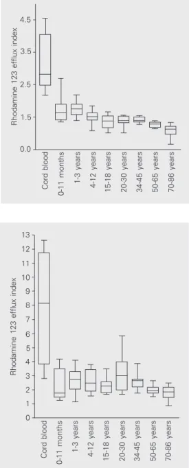
T CD3+CD4+ cells, P-gp function was high-est in cord blood (median, 2.82, first-third quartiles, 2.55-3.44), markedly decreasing with age (20- to 30-year group, 1.39, 1.34-1.51; P < 0.001), and reaching the lowest levels in the 70- to 86-year group (1.13, 1.07-1.18; Spearman r = -0.8; P < 0.0001), as shown in Figure 1. Even when the cord blood group where P-function is highest, was excluded from analysis, the inverse cor-relation was still significant (P < 0.001). Likewise, in T CD3+CD8+ cells, P-gp also decreased with age. The highest levels were observed in the cord blood group (8.14, 4.84-10.04), progressively declining to 3.00 (2.14-3.90; P < 0.05) in the 20- to 30-year group and to 1.83 (1.61-1.95; P < 0.01) in the 70- to 86-year group (Spearman r = -0.42; P < 0.0001), as illustrated in Figure 2. As ob-served for CD4+ T cells, the correlation was still significant (P = 0.006) for CD8+ T cells when the two younger age groups were ex-cluded from analysis.
P-glycoprotein function in peripheral blood B and NK lymphocytes
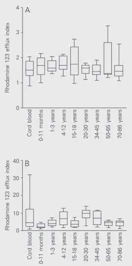
In B CD19+ cells, P-gp activity was low in cord blood (1.51, 1.36-1.68), and did not change significantly with age, the highest median value being detected in the 15- to 18-year group (1.75, 1.62-1.98; Figure 3A). However, these levels were not statistically different from the other age groups (Kruskal-Wallis test, P = 0.49). Similarly, P-gp func-tion did not change significantly with age in NK CD3-CD16+CD56+ lymphocytes (cord blood, 4.48, 2.50-8.51, and 70- to 86-year group, 4.83, 3.72-5.46; Spearman r = 0.16; P = 0.14), as illustrated in Figure 3B.
Discussion
The MDR1 gene-encoded P-gp, involved
in the export of substances from the cell, is expressed by different normal tissues, such as the hematopoietic tissue (2). Peripheral
Figure 1. Variation of P-glyco-protein function in T CD4+ lym-phocytes with age. P-glycopro-tein function is reported as the ratio of rhodamine 123 mean fluorescence in the presence and in the absence of verapamil (rhodamine 123 efflux index). P-glycoprotein functional expres-sion was highest in cord blood, progressively decreasing with age and reaching the lowest lev-els in the 70- to 86-year group.
Cord blood
0-11 months
1-3 years
4-12 years
15-18 years 20-30 years 34-45 years 50-65 years 70-86 years
Rhodamine 123 efflux index
13
10
7
5
2 12
11
8 9
6
4
3
1
0
Figure 2. Variation of P-glyco-protein function in T CD8+ lym-phocytes with age. P-glycopro-tein function is reported as the ratio of rhodamine 123 mean fluorescence in the presence and in the absence of verapamil (rhodamine 123 efflux index). P-glycoprotein functional expres-sion was highest in cord blood, progressively decreasing with age, reaching the lowest values in the 70- to 86-year group.
Rhodamine 123 efflux index
4.5
3.5
2.5
1.5
0.0
Cord blood
0-11 months
1-3 years
4-12 years
15-18 years 20-30 years 34-45 years 50-65 years 70-86 years
blood T, B and NK lymphocytes as well as hematopoietic stem cells are the major he-matopoietic cells expressing P-gp, but the glycoprotein plays different physiological roles in these cells (2-10). P-glycoprotein participates in the cell secretion of cytokines and in the cytotoxic function of killer cells (7-10).
sion in T cells was highest in older individu-als. However, they studied 10 young and 10 elderly subjects only, and their findings may have been the result of a skewed population. In a larger number of healthy volunteers (90 individuals) divided into a broader set of age ranges (nine age groups from newborn to 86 years), we definitively demonstrated that, in T cells, P-gp functional expression is highest in cord blood and is progressively reduced with age. The reduction in P-gp activity with age observed in this study paralleled the reduction in T cell function with aging. These findings could explain, at least in part, the impairment of T cell function observed in the elderly in comparison to young adults (11-14).
Moreover, we found that P-gp function in B CD19+ cells follows a different pattern as a function of age. It was low throughout all age groups, with a slight increase in the 20- to 30-year group. Our results agree with the single previous observation that P-gp function in B CD19+ cells was lower in cord blood and during infancy in comparison to adults (15). In the cited study, the authors speculated that the very high levels of P-gp expression in cord blood T CD4+ and T CD8+ cells might reflect a transient influ-ence of transplacental maternal factors. How-ever, the low P-gp activity observed in cord blood B CD19+ cells does not support this hypothesis.
Likewise, we found that P-gp activity does not seem to be determined by age in NK lymphocytes, since P-gp functional expres-sion remained high in all age groups. To the best of our knowledge, this was the first time that a correlation between age and P-gp func-tion was done in NK lymphocytes. In this cell subset, P-gp seems to mediate cytotoxic-ity, since the blockade of P-gp function by different P-gp inhibitors (verapamil, cyclo-sporine A and PSC833) suppresses NK cell cytotoxic activity in a dose-dependent man-ner (21). It is interesting to correlate these observations with the finding that NK cell
Figure 3. Variation of P-glyco-protein function in B CD19+ (A) and NK CD3-CD16+CD56+ (B) lymphocytes with age. P-glyco-protein function is reported as the ratio of rhodamine 123 mean fluorescence in the pres-ence and in the abspres-ence of verapamil (rhodamine 123 efflux index). P-glycoprotein activity was low in all age groups and was not influenced by age.
Rhodamine 123 efflux index
4
3
2
1
0
Cord blood
0-11 months
1-3 years
4-12 years
15-18 years 20-30 years 34-45 years 50-65 years 70-86 years
Rhodamine 123 efflux index
40
30
20
10
0
Cord blood
0-11 months
1-3 years
4-12 years
15-18 years 20-30 years 34-45 years 50-65 years 70-86 years
A
B
expres-activity does not significantly change with age (22). In contrast, P-gp expression is in-fluenced by genetic factors in NK lympho-cytes. Hitzl et al. (23) found that the MDR1
gene C3435T polymorphism is associated with decreased P-gp function in this cell subset. However, the impact of MDR1 gene
polymorphisms on P-gp expression in he-matopoietic cells is heterogeneous. In a re-cent study, we failed to demonstrate an
asso-ciation between MDR1 gene mutations and
P-gp functional expression in bone marrow stem cells (24). Interestingly, in this cell subset, P-gp expression is directly correlated with age (25).
In conclusion, different hematopoietic cell subsets showed different P-gp activity as a function of age, and these changes might be responsible for the specific characteristics of the immune system in the elderly.
References
1. Higgins CF, Callaghan R, Linton KJ, Rosemberg MF & Ford RC (1997). Structure of the multidrug resistance P-glycoprotein. Semi-nars in Cancer Biology, 8: 135-142.
2. Schinkel AH (1997). The physiological function of drug-transporting P-glycoproteins. Seminars in Cancer Biology, 8: 161-170.
3. Drach D, Zhao S, Drach J, Mahadevia R, Gattringer C, Huber H & Andreeff M (1992). Subpopulations of normal peripheral blood and bone marrow cells express a functional multidrug resistant pheno-type. Blood, 80: 2729-2734.
4. Chaudhary PM, Mechetner EB & Roninson IB (1992). Expression and activity of the multidrug resistance P-glycoprotein in human peripheral blood lymphocytes. Blood, 80: 2735-2739.
5. Ludescher C, Pall G, Irschick EU & Gastl G (1998). Differential activity of P-glycoprotein in normal blood lymphocyte subsets. Brit-ish Journal of Haematology, 101: 722-727.
6. Chaudhary PM & Roninson IB (1991). Expression and activity of P-glycoprotein, a multidrug efflux pump, in human hematopoietic stem cells. Cell, 66: 85-94.
7. Gupta S, Kim C, Tsuruo T & Gollapudi S (1992). Preferential expres-sion and activity of multidrug resistance gene 1 product (P-glycopro-tein), a functionally active efflux pump, in human CD8+ T cells: a role in cytotoxic effector function. Journal of Clinical Immunology, 12: 451-458.
8. Gupta S & Gollapudi S (1993). P-glycoprotein (MDR1 gene product) in cells of the immune system: its possible physiologic role and alteration in aging and human immunodeficiency virus-1 (HIV-1) infection. Journal of Clinical Immunology, 13: 289-301.
9. Drach J, Gsur A, Hamilton G et al. (1996). Involvement of P-glyco-protein in transmembrane transport of interleukin-2 (IL-2), IL-4, and interferon-γ in normal human T lymphocytes. Blood, 88: 1747-1754. 10. Raghu G, Park SW, Roninson IB & Mechetner EB (1996). Monoclon-al antibodies against P-glycoprotein, an MDR1 gene product, inhibit interleukin-2 release from PHA-activated lymphocytes. Experimen-tal Hematology, 24: 1258-1264.
11. Miller RA (1996). The aging immune system: primer and prospec-tus. Science, 273: 70-74.
12. Nagel JE, Chopra RK, Chrest FJ, McCoy MT, Schneider EL, Holbrook NJ & Adler WH (1988). Decreased proliferation, interleukin 2 synthe-sis, and interleukin 2 receptor expression are accompanied by de-creased mRNA expression in phytohemagglutinin-stimulated cells from elderly donors. Journal of Clinical Investigation, 81: 96-109. 13. Fernandes G & Gupta S (1981). Natural killing and
antibody-depend-ent cytotoxicity by lymphocyte subpopulations in young and aging humans. Journal of Clinical Immunology, 1: 141-148.
14. Gupta S (1989). Membrane signal transduction in T cells in aging humans. Annals of the New York Academy of Sciences, 568: 277-282.
15. Pilarski LM, Paine D, McElhaney JE, Cass CE & Belch AR (1995). Multidrug transporter P-glycoprotein 170 as a differentiation antigen on normal human lymphocytes and thymocytes. American Journal of Hematology, 49: 328-335.
16. Aggarwal S, Tsuruo T & Gupta S (1997). Altered expression and function of P-glycoprotein (170 kDa), encoded by the MDR1 gene, in T cell subsets from aging humans. Journal of Clinical Immunol-ogy, 17: 448-454.
17. Calado RT, Garcia AB & Falcão RP (1998). Decreased activity of the multidrug resistance P-glycoprotein in acquired aplastic anaemia: possible pathophysiologic implications. British Journal of Haemato-logy, 102: 1157-1161.
18. Calado RT, Garcia AB, Gallo DAP & Falcão RP (2002). Reduced function of the multidrug resistance P-glycoprotein in CD34+ cells of patients with aplastic anaemia. British Journal of Haematology, 118: 320-326.
19. Marie JP, Huet S, Faussat AM et al. (1997). Multicentric evaluation of the MDR phenotype in leukemia. Leukemia, 11: 1086-1094. 20. Falcão RP & De-Santis GC (1991). Age-associated changes of
memory (CD45RO+) and naive (CD45R+) T cells. Brazilian Journal of Medical and Biological Research, 24: 275-279.
21. Klimecki WT, Taylor CW & Dalton WS (1995). Inhibition of cell-mediated cytolysis and P-glycoprotein function in natural killer cells by verapamil isomers and cyclosporine A analogs. Journal of Clini-cal Immunology, 15: 152-158.
22. Whitman DB (1999). The immunology of aging. Cambridge Scien-tific Abstracts. http://www.csa.com/hottopics/immune-aging/ overview.html (access date January 28, 2002).
23. Hitzl M, Drescher S, van der Kuip H, Schäffeler E, Fischer J, Schwab M, Eichelbaum M & Fromm MF (2001). The C3435T mutation in the human MDR1 gene is associated with altered efflux of the P-glycoprotein substrate rhodamine 123 from CD56+ natural killer cells. Pharmacogenetics, 11: 293-298.
24. Calado RT, Falcão RP, Garcia AB, Gabellini SM, Zago MA & Franco RF (2002). Influence of functional MDR1 gene polymorphisms on P-glycoprotein activity in CD34+ hematopoietic stem cells. Haemato-logica, 87: 564-568.
25. Calado RT, Machado CG, Carneiro J-J, Garcia AB & Falcão RP (2003). Age-related changes of P-glycoprotein-mediated rhodamine 123 efflux in normal human bone marrow hematopoietic stem cells.