Atividade antimicrobiana do óleo-resina de copaíba (
Copaifera langsdorffii)
em
bactérias de significância clínica em úlceras cutâneas
MASSON, D.S.1; SALVADOR, S.L.2; POLIZELLO, A.C.M.2; FRADE, M.A.C.1*
1Universidade de São Paulo, Faculdade de Medicina de Ribeirão Preto, Departamento de Clínica Médica, Divisão de Dermatologia. CEP 14048-900. Ribeirão Preto, Brasil. *E-mail: prof.marcoandrey@gmail.com. 2Universidade de São Paulo, Faculdade de Ciências Farmacêuticas de Ribeirão Preto, Departamento de Análises Clínicas, Toxicológicas e Bromatológicas. CEP14040-903. Ribeirão Preto, Brasil.
Recebido para publicação em 03/05/2011 Aceito para publicação em 12/07/2013
RESUMO: Este trabalho avaliou a atividade antimicrobiana in vitro do óleo-resina da Copaifera langsdorffii, o qual vem sendo utilizado há muitos anos na medicina tradicional popular,
principalmente devido às suas propriedades antiinflamatórias, antibacterianas, cicatrizante
entre outras. O óleo-resina foi testado em bactérias Gram-positivas (Staphylococcus aureus, Streptococcus pyogenes, Enterococcus faecalis) e Gram-negativas (Pseudomonas aeruginosa e Escherichia coli) relacionadas com infecções de úlceras cutâneas. A atividade antimicrobiana foi determinada pelos testes da Concentração Inibitória Mínima (CIM) e Concentração Bactericida Mínima (CBM). O óleo-resina apresentou atividade antimicrobiana in vitro apenas para as bactérias Gram-positivas, com valores de CIM de 200 μg/mL, 400 μg/mL e 1100 μg/mL para S. aureus, S. pyogenes e E. faecalis, respectivamente. Os valores de CBM foram os mesmos que os valores de MIC para S. aureus e S. pyogenes. O valor de CBM para o microrganismo E. faecalis foi de 1200 μg/mL. Considerando que a presença de infecção significativamente impede o processo normal de cicatrização de úlceras cutâneas, acreditamos que o óleo-resina de copaíba, utilizado como componente de formulações tópicas, poderia ser um adjunto importante no tratamento de úlceras cutâneas infectadas, principalmente nos casos de infecção por microrganismos Gram-positivos.
Palavras-chave: Copaifera langsdorffii, óleo-resina de copaíba, atividade antimicrobiana, cicatrização.
ABSTRACT: Antimicrobial activity of copaíba (Copaifera langsdorffii) oleoresin on bacteria
of clinical significance in cutaneous wounds. The present study was designed to evaluate the in vitro antimicrobial activity of Copaifera langsdorffii oleoresin, which has been used in
folk medicine as an anti-inflammatory, antibacterial, healing among others. The oleoresin was
tested against Gram-positive (Staphylococcus aureus, Streptococcus pyogenes, Enterococcus faecalis) and Gram-negative (Pseudomonas aeruginosa and Escherichia coli) bacteria related to infections in cutaneous wounds. Antimicrobial activity was determined by the minimum inhibitory concentration (MIC) and minimum bactericidal concentration (MBC) assays. Copaiba oleoresin showed antimicrobial activity only against the Gram-positive bacteria with MIC of 200
μg/mL, 400 μg/mL and 1100 μg/mL for S. aureus, S. pyogenes and E. faecalis, respectively. MBC values were the same as MIC for S. aureus and S. pyogenes and for E. faecalis it was 1200 μg/mL. Considering that infection significantly impairs the wound healing process, we believe that the use of copaiba oleoresin as a component of a topical formulation could be a valuable adjunct in the treatment of infected wounds, mainly in the case of wounds infected by Gram-positive microorganisms.
Keywords: Copaifera langsdorffii, copaiba oleoresin, antimicrobial activity, wound healing.
11_057 INTRODUCTION
From a microbiological perspective, the primary function of normal, intact skin is to control microbial populations that live on the skin surface
of skin integrity (i.e., a wound) provides a moist, warm, and nutritious environment that is conducive to microbial colonization and proliferation (Bowler et al., 2001). Bacteria, their metabolites and microbial
interactions to wound environment may significantly
impair the process of normal wound healing, and contribute to the antibiotic resistance (Bowler et al., 2001; Wall et al., 2002). Staphylococci, streptococci, enterococci, and facultative Gram-negative bacilli are the most frequent bacterial groups recovered from chronic venous leg ulcers (Davies et al., 2007; Stephens et al., 2003). In the development of infection, a wound fails to heal, and it has deleterious effects on patients by causing increased pain and discomfort that can result in life-threatening illness or even death. Additionally, treatment costs rise, and general wound management practices become more resource demanding (Bowler et al., 2001; Cooper et al., 2002).
The indiscriminate use of antimicrobial drugs has led to multiple bacterial resistance and side effects (Fu et al., 2007). As the development of antibiotics continues and controversy regarding the use of topical antiseptics persists, the need for
identification and development of new agents that
are safe and broadly effective becomes increasingly critical (Bowler et al., 2001). Thus, efforts have been made to develop new compounds outside conventional antibiotic therapy and more natural antimicrobial substances such as plants are desired (Rodrigues et al., 2005).
Brazil is well known for the variety of its tropical plants and, many of them are used as natural medicines by native population. Plants of the Copaifera genus (Leguminosae, Caesalpinioideae), popularly known as copaiba, are large trees that grow in northern Brazil. The exuded material from these trees, traditionally obtained by tapping the trunk of the tree is called copaiba oil, copaiba balsam or oleoresin, a transparent liquid whose color varies from yellow to light brown (Carvalho et al., 2005; Castro-e-Silva et al., 2004; Paiva et al., 2002; Santos et al., 2008a).
Phytochemical studies carried out on the oleoresin from Copaifera langsdorffii revealed the presence of a mixture of diterpenes and sesquiterpenes. The biological activities of Copaifera spp. have been attributed to these groups of compounds. The main sesquiterpene
is β-Caryophyllene and the main diterpenes are
kaurenoic acid and copalic acid (it is considered a characteristic diterpene of the genus Copaifera). Some plant extracts containing diterpenes were
shown to exhibit antimicrobial, anti-inflammatory and
wound healing effects (Paiva et al., 2002; Santos et al., 2008).
The oleoresin of several species of Copaifera
is used in the Amazonian region mainly as a topical
anti-inflammatory and healing agent (Carvalho et
al., 2005). It has been also used in folk medicine as an antineoplastic, and urinary antiseptic, as well as to treat bronchitis, skin diseases, ulcers and syphilis (Brandão et al., 2008). It is also important for its gastroprotective, analgesic, antinociceptive, insect repellant and antimicrobial properties. In cosmetic industry copaiba oleoresin is a component in capillary lotions, shampoos, soaps, creams and bathing foams (Gomes et al. 2008; Paiva et al., 2002; Pieri et al., 2009; Santos et al., 2008b; Veiga Júnior et al. 2001).
Thus, the purpose of this study was to investigate the in vitro antimicrobial activity of Copaifera langsdorffii oleoresin on standard strains: Staphylococcus aureus, Streptococcus pyogenes, Enterococcus faecalis, Pseudomonas aeruginosa and Escherichia coli, which are important microorganisms related with cutaneous wounds infections.
MATERIAL AND METHOD
Plant Material
Copaiba oleoresin was collected from the trunk of the Copaifera langsdorffii tree in Tarauacá, Acre, Brazil (latitude 9o41’0” South, altitude 72o5’0” West), during 1999 and 2000, during the dry season (most likely between May and October). It was obtained from the Central de Cooperativas de Rio Branco by Prof Dr Osvaldo de Freitas (School of Pharmaceutical Sciences of Ribeirão Preto - University of São Paulo), who kindly supplied the oleoresin for this study.
Stock solutions preparation
Specific amounts (0.04; 0.05; 0.1 and 0.15
g) of copaiba oleoresin were solubilized in dimethyl sulfoxide (DMSO), and then appropriate volumes of Brain Heart Infusion (BHI) medium were added
in order to obtain final concentrations of 4 mg/mL,
5 mg/mL, 10 mg/mL and 15 mg/mL, respectively (Valdevite, 2007).
A volume of 300 μL of S. pyogenes and E. faecalis were firstly inoculated into 10 mL thioglycollate medium without dextrose or indicator broth, and then these cultures were incubated at
37ºC for 24 hours. Secondly, from these first cultures, 100 μL were subcultured into 5.0 ml thioglycollate
medium and incubated at 37ºC for 24 hours. Thirdly,
from these previous cultures, 100 μL was cultured
into 10 mL thyoglicollate medium, incubated at 37ºC for 24 hours.
S. aureus, E.coli and P. aeruginosa were firstly seeded into a fresh MH agar slant and incubated at 25ºC for 24 hours. Secondly, each microorganism was cultured into 5.0 mL MH broth and incubated at 37ºC for 24 hours. Thirdly, 100
μL was cultured into 10 mL MH broth, incubated at
37ºC for 24 hours.
All bacteria were cultured for three days because they were obtained from cultures stored in different conditions. Performing this procedure allowed all microorganisms to be reactivated and in the same growth conditions.
Bacterial growth curves
Overnight cultures, described above, were
added (2.5 % v/v) to a final volume of 200 mL of:
MH broth (MHb), BHI and Tryptic Soy Broth (TSB) (S. aureus); BHI and TSB (S. pyogenes); BHI supplemented with glucose and BHI (E. faecalis); MH broth, BHI and TSB (E.coli); MHb, BHI, TSB, Peptone water and Nutrient broth (P. aeruginosa). Before adding the cultures, the absorbance (at 660 nm) of the liquid media (blank), as well as the initial
inocula, was measured. All flasks were incubated at
37oC, under agitation of 100 rpm (Shaker Marconi – Mod. MA420, Brazil). The absorbance of each culture medium was measured at 1 hour intervals throughout the incubation period (Spectronic® 20 Genesys, USA). The bacterial growth and the exponential growth phases were determined from data of absorbance (at 660 nm) versus incubation time (Gabrielson et al., 2002; Leitão et al., 2004).
Inoculum preparation
All bacteria were cultivated for three days, as previously described, and then the most suitable medium was chosen to prepare the inoculum of each microorganism. At half of log phase from exponential growth curves, 1.0 mL was collected in microcentrifuge tubes and centrifuged (Centrifuge Eppendorf 5810-R) at 11500 x g for 2 minutes. The supernatants were discarded and the inocula were prepared by diluting cell mass in 0.9% NaCl solution, adjusted to McFarland scale 0.5 by turbidity reading (Densimat - BioMerieux). Suspensions were further
diluted to provide a final inoculum of 105 CFU mL-1 to be used in the antimicrobial activity assays as
the standard inoculum (Duarte et al., 2007; NCCLS, 2003).
Minimum Inhibitory Concentration
MICs were carried out in 96-well microplates. The concentrations of copaiba oleoresin ranged
from 1.56 to 3000 μg/mL, depending on the
microorganism. BHI broth containing 2% DMSO was
the first solution to be added into wells, this media
was chosen because it allows a suitable growth of all tested microorganisms. The stock solutions of copaiba oleoresin were transferred and diluted into the wells. The standard inoculum was added to the corresponding wells and the microplates were incubated at 37ºC for 15 hours, under agitation (40 rpm). Control wells with cultures without copaiba oleoresin, i.e., wells with culture medium (BHI containing 2% DMSO) and inoculum: negative control, and wells with the culture medium only, without the inoculum: sterility control, were also included (Valdevite, 2007). Tests were performed in triplicate. To overcome the turbidity problem
because of the emulsified oil, and to allow visual
identification of metabolic activity, antimicrobial
activity was detected by adding 50 μL of 0.125% (w/v)
nitrotetrazolium blue chloride solution (Loughlin et
al. 2008). The final concentration of nitrotetrazolium
in each well was 0.025% (v/v). All microplates were again incubated at 37oC for 2 hours under agitation (40 rpm). Then, they were centrifuged at 2000 x g for 10 minutes and images of all microplates were
taken by using a digital scanner. MIC was defined
as the lowest concentration of copaiba oleoresin that inhibited visible growth, as indicated by the nitrotetrazolium staining (Tortora et al., 2005).
Minimum Bactericidal Concentration
MBCs were carried out by inoculating 250
μL of each well from microplates where there was
no visible growth. Solutions from the three closest wells to MIC values were used. BHI agar plate was used for S. pyogenes and E. faecalis, and MH agar plate was used for S. aureus. All Petri plates were incubated at 37ºC for 48 hours. MBC was defined as the lowest concentration of copaiba oleoresin at which incubated microorganisms were completely killed (Fu et al., 2007).
RESULT AND DISCUSSION
Growth profiles of all tested microorganisms
was Nutrient broth.
Assessment of bacterial growth is important in the investigation of antimicrobial effects of a compound (Gabrielson et al., 2002). Before assessing MICs and/or MBCs it is valuable to select a suitable medium and determine the exponential growth phase. During this phase, the cell division is
extremely active (binary fission) and cell metabolism
is increased, justifying the use of this phase as preferred for research and industrial purposes (Tortora et al. 2005).
Copaifera langsdorffii oleoresin exhibited a broad inhibition spectrum only toward Gram-positive bacteria (Table 1). The solvent DMSO showed no effect on the microorganisms growth (data not shown). MIC values of copaiba oleoresin on each microorganism were visualized by coloration development (tetrazolium salt). Wells that showed a blue-colored precipitate indicated microbial growth, and those wells without coloration indicated killed bacteria (Andrews, 2001; Gabrielson et al., 2002). MBC values were the same as MICs to S. aureus and S. pyogenes, but different to E. faecalis. MBC assay was not carried out for P. aeruginosa and E.
coli, because these microorganisms were resistant to copaiba oleoresin in the concentrations studied.
High resolution gas chromatography-mass spectrometry (HRGC-MS) analyses on this same oleoresin found a mixture of sesquiterpenes (75%) and diterpenes (25%). The main compounds
among sesquiterpenes were β-caryophyllene (51%), followed by α-humulene (8.52%); among diterpenes,
the main compounds were 11-acetoxy-copalic acid (5.23%), 11-hydroxy-copalic acid (4.8%), copalic acid (4.69%) and agatic acid (3.32%) (Ramos, 2006). Since sesquiterpenes have been reported to have potent antimicrobial activity and play a critical role in plant defense mechanisms (Teng et al. 2010), they could be related to the bacteriostatic and bactericidal activities observed against the Gram-positive bacteria evaluated in this study.
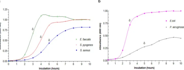
Our results are in agreement with a previous study that investigated the antimicrobial activity of oleoresins from Copaifera martii, Copaifera officinalis and Copaifera reticulata against Gram-positive and Gram-negative microorganisms. This study showed that these species of copaiba did not present antimicrobial activity against Gram-negative FIGURE 1. Bacterial growth curves to (a) S. aureus, S. pyogenes, E. faecalis; (b) E. coli and P. aeruginosa . The arrows represent half of log phase.
a b
MIC and MBC > 3000 μg/mL
TABLE 1. MIC and MBC values of copaiba oleoresin against S. aureus, S. pyogenes, E. faecalis, E. coli and P. aeruginosa.
Bacteria MIC (μg/mL) MBC (μg/mL)
Range MIC Range MBC
Staphylococcus aureus (ATCC 6538) 3.12 - 1500 200 200 - 400 200
Streptococcus pyogenes (ATCC 19615) 1.56 - 1000 400 400 - 1000 400
Enterococcus faecalis (ATCC 10541) 1.56 - 2000 1100 1100 - 1300 1200
Escherichia coli (ATCC 10538) 1000 - 3000 * * *
bacteria. Due to its property of causing morphological and ultra-structural changes, such as cell wall disruption and release of cytoplasmatic content and decreased cellular volume, copaiba oleoresin might affect the bacteria cell wall (Santos et al., 2008a).
Pacheco et al. (2005) also conducted a study on the antimicrobial activity of oleoresins from different trees against Gram-positive and Gram-negative bacteria. They found inactivity of all the samples against Gram-negative bacteria up to 1000 µg/ml.
Gram-positive and Gram-negative bacteria differ on their walls, where Gram-negative bacteria walls represent a stronger barrier than Gram-positive. Copaiba oleoresin seems to modify bacteria cell wall; therefore, a reasonable explanation for no activity against Gram-negative bacteria could be due to their complex walls morphophysiology. Gram-negative walls have a bilayer (containing proteins, phospholipids, and lipopolysaccharides), which is usually considered to be their outermost layer situated above a thin peptidoglycan layer. Together, plasma membrane and the cell wall (outer membrane, peptidoglycan layer, and periplasm) constitute the Gram-negative envelope (Beveridge, 1999).
There is not a consensus on the acceptable bacterial inhibition level for plant materials when compared with standards. A previous study (Duarte et al., 2007) described a classification of plant materials based on MIC results (strong inhibitors:
MIC up to 500 μg/mL; moderate inhibitors: MIC between 600 and 1500 μg/mL; weak inhibitors: MIC above 1600 μg/mL). This classification was
based on antibiotic resistant bacteria, i.e., clinical isolates. Another study (Aligiannis et al., 2001)
suggested a classification based on ATCC strains (strong activity: MIC between 280 and 1270 μg/mL; weak activity: MIC between 1810 and 8850 μg/mL).
Thus, Copaifera langsdorffii oleoresin showed better inhibition levels against S. aureus (MIC 200 μg/mL) and S. pyogenes (MIC 400 μg/mL) than against E. faecalis (MIC 1100 μg/mL), but still effective.
Our results showed that Copaifera langsdorffii oleoresin has antimicrobial activity against Gram-positive bacteria (S. aureus, S. pyogenes and E. faecalis) ATCC strains, and it didn’t have any activity on Gram-negatives (P. aeruginosa and E. coli). In view of the significant healing impairment due to bacterial infection, the use of copaiba oleoresin as a component of a topical formulation could be a valuable adjunct in the treatment of infected wounds, mainly in case of those infected by antibiotic resistant Gram-positive strains.
ACKNOWLEDGMENT
Financial support by CAPES (Brazilian Federal Agency for the Support and Evaluation of
Graduate Education) is gratefully acknowledged. We also thank Professor Dr. Osvaldo de Freitas for kindly supplying the Copaifera langsdorffii oleoresin and Professor Dr. Augusto César Cropanese Spadaro for providing infrastructure during this work.
REFERENCE
ALIGIANNIS, N. et al. Composition and antimicrobial activity of the essential oils of two Origanum species.
Journal of Agricultural Food Chemistry, v.49, n.9, p.4168-4170, 2001.
ANDREWS, J. Determination of Minimum Inhibitory C o n c e n t r a t i o n s . Journal of Antimicrobial
Chemotherapy, v.48, suppl.1, p. 5-16, 2001.
BEVERIDGE, T.J. Structures of Gram-negative cell walls and their derived membrane vesicles. Journal of
Bacteriology, v.181, n.16, p.4275-4733, 1999.
BOWLER, P.G.; DUERDEN, B.I.; ARMSTRONG, D.G. Wound microbiology and associated approaches to wound management. Clinical Microbiological Review, v.14, n.2, p.244-269, 2001.
BRANDÃO, G.L. et al. Brazilian medicinal plants described
by 19th century European naturalists and in the Official
Pharmacopoeia. Journal of Ethnopharmacology, v.120, p.141-148, 2008.
CARVALHO, J.C.T. et al. Topical anti-inflammatory and analgesic activities of Copaifera duckei Dwyer.
Phytotherapy Research, v.19, p.946-950, 2005. CASTRO-E-SILVA JR, O. et al. Antiproliferative activity of
Copaifera duckei oleoresin on liver regeneration in rats.
Phytotherapy Research, v.18, p.92-94, 2004.
COOPER, R.A.; MOLAN, P.C.; HARDING, K.G. The sensitivity to honey of Gram-positive cocci of clinical
significance isolated from wounds. Journal of Applied Microbiology, v.93, p.857–863, 2002.
DAVIES, C.E. et al. A prospective study of the microbiology of chronic venous leg ulcers to reevaluate the clinical predictive value of tissue biopsies and swabs. Wound
Repair and Regeneration, v.15, p.17–22, 2007. DUARTE, M.C.T. et al. Activity of essential oils from
Brazilian medicinal plants on Escherichia coli. Journal
of Ethnopharmacology, v.111, p.197-201, 2007. FU, Y. et al. Antimicrobial activity of clove and rosemary
essential oils alone and in combination. Phytotherapy Research, v.21, p.989-994, 2007.
GABRIELSON, J. et al. Evaluation of redox indicators and the use of digital scanners and spectrophotometer
for quantification of microbial growth in microplates. Journal of Microbiological Methods, v.50, p.63-73, 2002.
GOMES, N.M. et al. Antinociceptive activity of Amazonian copaiba oils. Journal of Ethnopharmacology, v.109, p.486-492, 2008.
LEITÃO, D.P. et al. Comparative evaluation of in vitro
effects of Brazilian Green propolis and Baccharis
dracunculifolia extracts on cariogenic factors of
Streptococcus mutans. Biological and Pharmaceutical
Bulletin, v.27, n.11, p.1831-1839, 2004.
isolates and human fibroblast cells. Letters in Applied Microbiology, v.46, p.428–433, 2008.
NCCLS - National Committee for Clinical Laboratory Standards. Methods for dilution antimicrobial susceptibility tests for bacterial that grow aerobically, Approved Standard. 6ed. NCCLS document M07-A6. National Committee for Clinical Laboratory Standards Wayne, PA, 2003.
PACHECO, T.A.R.; BARATA, L.E.S.; DUARTE, M.C.T. Antimicrobial activity of copaiba (Copaifera spp)
balsams. Revista Brasileira de Plantas Medicinais, v.8, n.esp, p.123-124, 2006.
PAIVA, L.A.F. et al. Investigation on the wound healing activity of oleoresin from Copaifera langsdorffii in rats.
Phytotherapy Research, v.16, p.737-739, 2002. PIERI, F.A.; MUSSI, M.C.; MOREIRA, M.A.S. Óleo de
copaíba (Copaifera sp.): histórico, extração, aplicações industriais e propriedades medicinais. Revista Brasileira de Plantas Medicinais, v.11, n.4, p.465-472, 2009.
RAMOS, M.F.S. Desenvolvimento de microcápsulas
contendo a fracão volátil de copaíba por spray-drying: estudo de estabilidade e avaliação farmacológica. 2006. 132p. Tese (Doutorado - Área de Concentração em Fármacos e Medicamentos) – Faculdade de Ciências Farmacêuticas de Ribeirão Preto, Universidade de São Paulo, Ribeirão Preto. RODRIGUES, K.L. et al. Antimicrobial and healing activity
of kefir and kefiran extract. International Journal of Antimicrobial Agents, v.25, n.5, p.404-408, 2005. SANTOS, A.O. et al. Antimicrobial activity of Brazilian
copaiba oils obtained from different species of the Copaifera genus. Memórias do Instituto Oswaldo
Cruz, v.103, n.3, p.277-281, 2008a.
SANTOS, A.O. et al. Effect of Brazilian copaiba oils on Leishmania amazonensis. Journal of
Ethnopharmacology, v.120, p.204-208, 2008b. STEPHENS, P. et al. Anaerobic cocci populating the deep
tissues of chronic wounds impair cellular wound healing responses in vitro. British Journal of Dermatology,
v.148, p.456–466, 2003.
TENG, Y. et al. In vitro antimicrobial activity of the leaf essential oil of Spiraea alpina Pall. World Journal of
Microbiology and Biotechnology, v.26, p.9-14, 2010. TORTORA, G.F.; FUNKE, B.R.; CASE, C.L. Microbial
growth. In: TORTORA, G.F.; FUNKE, B.R.; CASE, C.L. Microbiology: An introduction, 6th ed. California. Benjamin/ Cummnigs, 2005, p. 154-180.
VALDEVITE, L.M. Estudo do efeito in vitro de extrato das folhas e do óleo-resina de copaíba sobre os fatores de virulência de Streptococcus mutans, relacionados à cárie dental. 2007. 128 f. Dissertação (Mestrado - Área de Concentração em Medicamentos e Cosméticos) – Faculdade de Ciências Farmacêuticas de Ribeirão Preto, Universidade de São Paulo, Ribeirão Preto. VEIGA JÚNIOR, V.F. et al. Phytochemical and
antioedematogenic studies of commercial copaiba oils available in Brazil. Phytotherapy Research, v.15, p.476-480, 2001.
WALL, I.B. et al. Potential role of anaerobic cocci in impaired human wound healing. Wound Repair and