New Insights into Regulation of Proteome and
Polysaccharide in Cell Wall of
Elsholtzia splendens
in
Response to Copper Stress
Tingting Liu, Chaofeng Shen, Yi Wang, Canke Huang, Jiyan Shi*
Institute of Environmental Science and Technology, College of Environmental and Resource Sciences, Zhejiang University Hangzhou, P.R. China
Abstract
Background and Aims:Copper (Cu) is an essential micronutrient for plants. However, excess amounts of Cu are toxic and result in a wide range of harmful effects on the physiological and biochemical processes of plants. Cell wall has a crucial role in plant defense response to toxic metals. To date, the process of cell wall response to Cu and the detoxification mechanism have not been well documented at the proteomic level.
Methods:An recently developed 6-plex Tandem Mass Tag was used for relative and absolute quantitation methods to achieve a comprehensive understanding of Cu tolerance/detoxification molecular mechanisms in the cell wall. LC–MS/MS approach was performed to analyze the Cu-responsive cell wall proteins and polysaccharides.
Key Results:The majority of the 22 up-regulated proteins were involved in the antioxidant defense pathway, cell wall polysaccharide remodeling, and cell metabolism process. Changes in polysaccharide amount, composition, and distribution could offer more binding sites for Cu ions. The 33 down-regulated proteins were involved in the signal pathway, energy, and protein synthesis.
Conclusions: Based on the abundant changes in proteins and polysaccharides, and their putative functions, a possible protein interaction network can provide new insights into Cu stress response in root cell wall. Cu can facilitate further functional research on target proteins associated with metal response in the cell wall.
Citation:Liu T, Shen C, Wang Y, Huang C, Shi J (2014) New Insights into Regulation of Proteome and Polysaccharide in Cell Wall ofElsholtzia splendensin Response to Copper Stress. PLoS ONE 9(10): e109573. doi:10.1371/journal.pone.0109573
Editor:Raffaella Balestrini, Institute for Sustainable Plant Protection, C.N.R., Italy ReceivedJune 5, 2014;AcceptedSeptember 1, 2014;PublishedOctober 23, 2014
Copyright:ß2014 Liu et al. This is an open-access article distributed under the terms of the Creative Commons Attribution License, which permits unrestricted use, distribution, and reproduction in any medium, provided the original author and source are credited.
Data Availability:The authors confirm that all data underlying the findings are fully available without restriction. All relevant data are within the paper and its Supporting Information files.
Funding:The work was supported by the National Natural Science Foundation of China (11179025, 21177109) and Program for New Century Excellent Talents in University (NCET-11-0455). The funders had no role in study design, data collection and analysis, decision to publish, or preparation of the manuscript. Competing Interests:The authors have declared that no competing interests exist.
* Email: shijiyan@zju.edu.cn
Introduction
Copper (Cu) is an essential micronutrient for plants because it is a structural and catalytic component of several proteins or enzymes involved in various physiological metabolisms [1]. However, excess Cu in plants is harmful; it induces phytotoxicity [2] and disturbs the physiological metabolisms, including carbo-hydrate metabolism, protein metabolism, mineral nutrition, cell respiration, and photosynthesis [3]. Given its redox properties, Cu2+
can catalyze the generation of reactive oxygen species (ROS) and Fenton reactions that damage cellular components or interfere with cellular transport processes [4]. Plants cannot avoid the stress conditions from Cu-contaminated areas. Thus, several resistance mechanisms for Cu tolerance have been developed [5], including exclusion, compartmentalization, chelation, and binding to organic ligands, which enable maintenance of Cu homeostasis for plant survival even in a highly contaminated environment [6]. Plant cell wall is the primary site for signal perception and defense response, which is significant in responding to environ-mental stresses [7]. The cell walls of plants that grow on heavy
metal-contaminated areas provide physical barriers against toxic heavy metals and actively participate in plant defense response. The cell wall serves as a repository for high contents of metals; it senses stress signals and transmits them to the cell interior, thereby affecting the cell fate decision [8]. The main chemical components of a plant cell wall include polysaccharides and proteins. Polysaccharides represent up to 90% of the plant cell wall and constitute three different kinds of polymers, namely, cellulose, hemicelluloses, and pectins. Cell wall proteins (CWPs) of plants comprise less than 10% of the cell wall dry weight but have major roles in cell wall structure, metabolism, cell enlargement, signal transduction, defense responses, and many other physiological events [9].
Certain plants can accumulate high content of metal ions in their cell wall through various compounds. Cell wall polysaccha-rides possess crucial roles in heavy metal binding and accumula-tion. Binding of metal ions depends on the number of functional groups in the cell wall polysaccharides. Previous studies confirmed that the essential capacity of the cell wall for binding metal ions
depends mainly on the abundance of polysaccharides in the carboxyl groups [10]. Current information about the regulation of cell wall polysaccharides under metal stress remains limited.
Abiotic stress induces expression of stress-responsive proteins, which lead to cellular adaptation in plant growth under stress conditions. Therefore, studies have been carried out to identify various CWPs involved in stress response, particularly those of regulatory or targeting function proteins [11–17]. Most reports have focused on leaf cell wall proteomes under abiotic stress, such as salt in tobacco [11]; wounding inMedicago[12]; dehydration in chickpea, rice, and maize [9,14,15]; zinc toxicity inHordeum[18]; nickel toxicity in barley [19]; manganese toxicity in cowpea [20,21]; boron deficiency inLupinus albus[22]; and flooding in soybean [23]. Few studies have focused on the root cell wall proteomes under abiotic stress [24–26]. All of these studies broadened our understanding of the complicated regulation of apoplast proteins. An in-depth proteome study of the cell wall can provide extensive information on the intrinsic mechanism of stress response attributed to a possible relationship between protein abundance and plant stress tolerance [27].
Elsholtzia splendensis a plant that can survive in a highly Cu-polluted area. This species can accumulate more than 1000 mg/ kg of Cu in the body [28], and the major proportion (70%) of Cu ions is stored in the root cell wall [29]. Nevertheless, little is known about the defense and detoxification mechanisms in the root cell wall ofE. splendens. A recently developed 6-plex Tandem Mass Tag (TMT) was used for relative and absolute quantitation methods to gain a comprehensive understanding of Cu tolerance/ detoxification molecular mechanisms. LC–MS/MS approach was also conducted to analyze Cu-responsive CWPs and polysaccha-rides in this study. This proteomic study of the apoplast is the first in response to Cu. The results will significantly expand knowledge on the apoplast complexicity in metal-tolerant plants and develop a more comprehensive understanding of the involved cellular reactions in cell defense mechanisms.
Results
Isolation and purification of CWPs
Root CWPs from E. splendens were isolated with sucrose gradients and washed according to the method of Feiz et al. [30]. The activity of G’DH, a typical marker enzyme used to detect contamination of cytoplasmic proteins, was measured to validate the purification method [31]. The G6PDH activity in CaCl2
-extracted CWPs, NaCl--extracted CWPs, and cytosolic protein fractions were 8.36, 5.78, and 107.55 mU/mg protein, respec-tively (Figure 1). The G6PDH activity in CaCl2- or
NaCl-extracted CWPs was 10 times lower than that in cytosolic proteins (p,0.01). These results indicate that the contamination of intracellular proteins was minimal.
The identified proteins were classified into different gene ontology (GO) terms according to their cellular compartment(s), function(s), and biological processes (Table S1). Some of these identified proteins showed a compartment label (apoplast, cell wall, extracellular region, and plant-type cell wall) that is coherent with secreted proteins. The same proteins can also be attributed to various compartments in the GO classification. Three different computer programs (TargetP, Predotar, and WoLF PSORT) were used to predict the protein subcellular localization (Table S1). A protein was secreted when at least two out of the three algorithms predicted extracellular localization. Based on the results, 15 proteins were predicted to be secreted proteins in the fractions (Table S1).
Cu-induced changes in root CWP contents
Plants were grown on a medium supplemented with different CuSO4concentrations to determine changes in CWP abundance
under Cu stress (Figure 2A). Cu toxicity led to a strong increase in CWP contents at low Cu concentrations. The CWP contents increased 1.39-fold at 25mM Cu and 1.96-fold at 50mM Cu. The
highest content of the CWPs was observed at 50mM Cu
concentrations. The plant root began to decompose at 100mM
Cu (Figure 2C). The increase of Cu contents in the cell wall could proportionally increase with the Cu supply in the medium (slope = 0.3, R2
= 0.98) at Cu concentrations below 75mM. The Cu content in the root cell wall continued to increase significantly (slope = 0.45,R2
= 0.99) above 75mM Cu. The highest value of Cu content in the root cell wall reached 1.41 mg/g with the root cell wall dry weight at 100mM Cu. These findings indicate an
increase in Cu binding to cell walls with Cu supply in the medium (Figure 2A). Polypeptide separation by SDS-PAGE (Figure 2B) revealed about 30 polypeptide bands. Band intensities increased with the increase in Cu concentration below 50mM (Figure S1). The staining intensity was the lowest among all the cell wall polypeptides at 100mM Cu concentration.
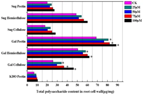
Cu-induced remodeling in root cell wall polysaccharide Evidence generally suggests that cell wall polysaccharides are crucial sites for Cu retention in plants. Therefore, changes in the contents of root cell wall polysaccharide of E. splendens were measured. Treatments with 25, 50, 75, and 100mM Cu significantly increased the expression of CWPs and polysaccha-rides compared with controls (Figure 3). Cu supply enhanced the total sugar contents in various cell wall fractions. Sugar contents were higher in hemicellulose than in pectin and cellulose. Sugar contents in pectin increased 1.22-fold with 25mM Cu. The major components of cell wall fractions are galacturonic acids. The content of galacturonic acids was considerably higher than that in
Figure 1. Evaluation of purity by the G6PDH activity in the cell wall proteins isolated from the roots ofElsholtzia splendens.The activity of G6PDH was assayed in CaCl2-extracted cell wall proteins,
NaCl-extracted cell wall proteins and total soluble proteins. One unit of G6PDH activity is defined as 1mmol of NADPH turnover per min/mg protein. Results are presented as mean6SE of G6PDH activity from three biological replicates. The asterisks indicate significant differences in the G6PDH activity of CaCl2-extracted cell wall proteins,
NaCl-extracted cell wall proteins compared with that of total soluble cytosolic proteins (**p,0.01).
doi:10.1371/journal.pone.0109573.g001
New Insights into Regulation in Plant Cell Wall
the control during Cu treatment, particularly at higher Cu supply. The galacturonic acid values rapidly increased in pectin (1.28-fold), hemicellulose (1.22-(1.28-fold), and cellulose (1.79-fold) at 100mM Cu. A conserved disaccharide residue known as 2-keto-3-deoxyoctonic acid (KDO) is attached to C-3 of the backbone of rhamnogalacturonan II in pectin. The KDO concentration in pectin increased during Cu exposure. Thus, the highest concen-tration of KDO (1.49-fold) was found in the 100mM Cu-treated group.
The results indicated that excess Cu affected the intracellular distribution of polysaccharides in the root cell wall. Given this reason, we focused on the relationship between wall polysaccha-ride composition and CWPs with increasing Cu intensity. The dendrogram proved the connection between the contents of polysaccharides and proteins as matched with different Cu concentrations (Figure 4). This finding suggests that Cu induced changes in the contents of root cell wall polysaccharides and proteins, which altered the intracellular distribution patterns in assigning effects on cellular activities.
Cu-responsive CWP identification using TMT LC-MS/MS This study aimed to gain a better understanding of the metabolic processes and molecular mechanisms involved in
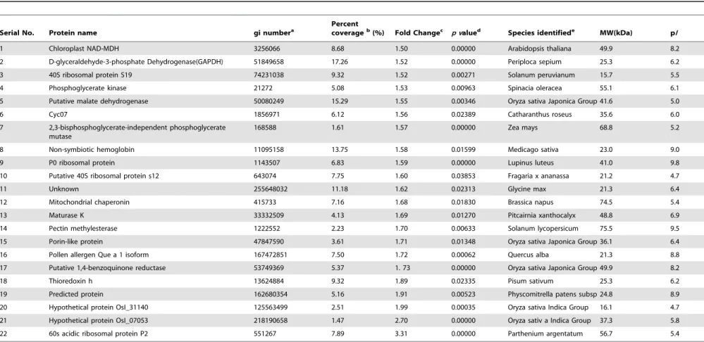
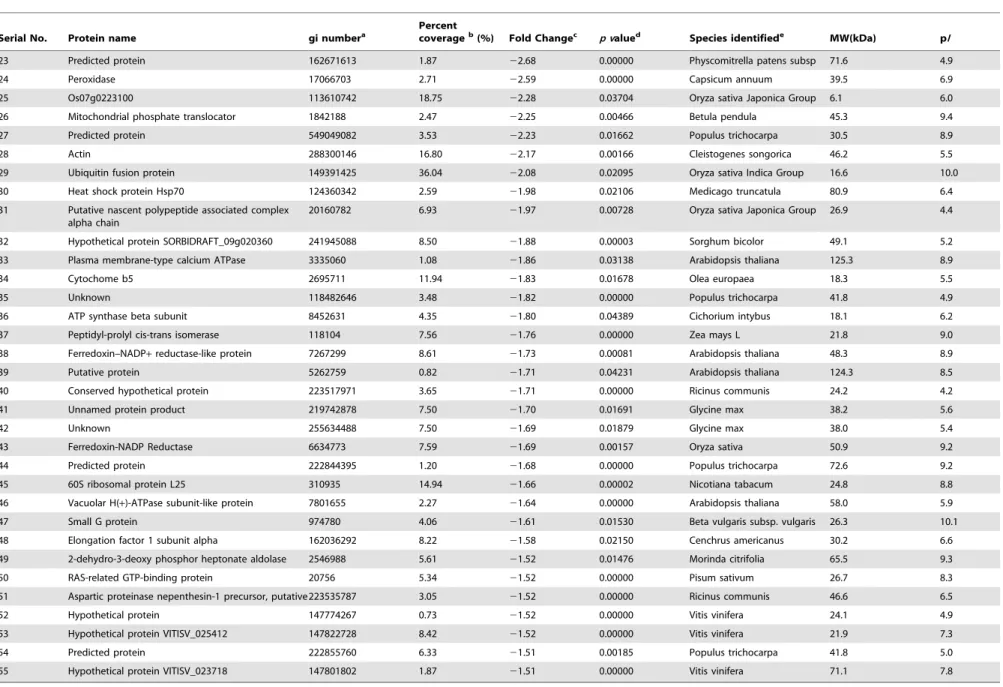
Cu-responsive CWPs. A TMT-based shotgun quantitation approach was used to obtain an overall view of the proteome changes associated with Cu stress in the root cell wall of E. splendens. Unique proteins were successfully identified based on homology searching with a common protein confidence cutoff of 95% and relative quantitative information in control and Cu-treated samples, as indicated by the peak areas of the different TMT tags (Table 1, Table S2). A threshold of 1.5-fold change and ap, 0.05 were set as stringent criteria for significant differences between control and treatment. Fifty five proteins were classified as differentially expressed upon Cu stress using the criteria; 22 and 33 of these proteins showed increasing trends and decreasing abundance, respectively (Table 1, Table 2).
Functional classification of identified CWPs
Database search using Uni-Prot/NCBI accession numbers was conducted to obtain detailed information of differentially ex-pressed proteins. These proteins were categorized into biological process and molecular function using an in-house Perl script according to the extracted GO terms from InterPro (www.ebi.ac. uk/interpro/) or Pfam (pfam.sanger.ac.uk/) (Figure 5). Functional classifications of the 55 differentially expressed proteins (Table 1, Table 2) were classified based on the GO terms that correspond to
Figure 2. Effect of different copper concentration on the cell wall protein and copper ions inElsholtzia splendens’s root cell.A: The content of copper and protein every root cell wall dry weight. B: SDS-PAGE of root cell wall protein under different copper stress. C: Root growth patterns of control and copper-stressed plants during different copper concentration. Data presented are mean6SE (n = 20),*Significant mean differences from control atp= 0.05 in multiple comparison by LSD test.
doi:10.1371/journal.pone.0109573.g002
New Insights into Regulation in Plant Cell Wall
the biological process. The top three categories were those involved in metabolic processes (23.24%), cellular processes (16.20%), and response to stimuli (14.79%) (Figure 5A). These three categories were classified according to the GO molecular annotation into binding (41.38%), catalytic activity (31.03%), and transport activity (11.49%) (Figure 5B). The observed diversity in biological function and processes of the differentially expressed CWPs demonstrated that the response of the root cell wall ofE. splendensto Cu stress was a complex process. Many physiological and biochemical changes were altered to counteract the adverse conditions.
Specific enzymes constituted by some small differentially expressed CWPs participated in various metabolic processes to counteract Cu stress. Classification pathways according to the
KEGG database searches categorized the cell wall enzymes into 12 classes (Table 3, Table S2). The more important category pathways were involved in oxidative phosphorylation, phenylala-nine biosynthesis and metabolism, ascorbate and aldarate metab-olism, and citrate cycle. All these pathways suggested the relatively altered abundance of the enzymes in various metabolic pathways that probably re-optimized the metabolic processes to combat against Cu stress.
Discussion
Cell wall polysaccharide remodeling in root under Cu stress
Cell walls provide a physical barrier to plants grown in Cu-contaminated soil, and cell wall polysaccharides have a crucial role in metal binding and accumulation [7]. The binding ability depends on the number of functional groups in polysaccharides, particularly for abundant carboxyl groups [7]. In the present study, the contents of the various cell wall polysaccharides significantly increased with the increase in Cu concentration. The expression of cell wall polysaccharides, which contain more carboxyl groups, provided more binding sites for Cu ions. Excess Cu also affected the intracellular distribution of polysaccharide in the root cell walls (Figure 3). Similar situations were also described in rice roots under Cd stress [45] andArabidopsisroots under Al stress [46]. The starch and sucrose metabolism-related proteins (Table 3) – Chloroplast NAD-MDH (No. 1, Table 1) – were also markedly up-regulated in response to Cu stress based on our observations. These findings suggest that Cu or Cu-induced oxidative stress may activate some crucial enzymes in the glyconeogenesis pathway, which can alter the contents of cell wall polysaccharides.
Increasing physiological, biochemical, and molecular evidence also showed that the remodeling of the binding properties of root apoplasts was attributed to Cu resistance. Enhancement at the level of low-methylesterified pectins, where the polysaccharides bound more trivalent metal ions, is one of the most remarkable alterations. Pectin methylesterases (PMEs), known as pectinesterases, catalyzed
Figure 3. Effect of different copper concentrations on the contents of root cell wall polysacchride.Sug: sugar, Gal: galacturonic acids, KDO: 2-Keto-3-deoxyoctonic acid. Data presented are mean6SE (n = 3). *Significant mean difference from control atp= 0.05 in multiple comparison by LSD test.
doi:10.1371/journal.pone.0109573.g003
Figure 4. Hierarchical cluster result of cell wall polysaccharide abundance ratio using the average linkage distance between clusters is shown.The color weighting represents normalized levels of each variable from the high (red) to the low (green).
doi:10.1371/journal.pone.0109573.g004
New Insights into Regulation in Plant Cell Wall
Table 1.Root cell wall proteins with significant increased in expression level under 50mM Cu treatment identified by LC-ESI-MS/MS-based proteomics using SIEVE (p,0.05 and fold chang.1.5).
Serial No. Protein name gi numbera
Percent
coverageb(%) Fold Changec p valued Species identifiede MW(kDa) pI
1 Chloroplast NAD-MDH 3256066 8.68 1.50 0.00000 Arabidopsis thaliana 49.9 8.2
2 D-glyceraldehyde-3-phosphate Dehydrogenase(GAPDH) 51849658 17.26 1.52 0.00000 Periploca sepium 25.3 6.2
3 40S ribosomal protein S19 74231038 9.32 1.52 0.00271 Solanum peruvianum 15.7 5.5
4 Phosphoglycerate kinase 21272 5.08 1.53 0.00963 Spinacia oleracea 55.1 6.1
5 Putative malate dehydrogenase 50080249 15.29 1.55 0.00346 Oryza sativa Japonica Group 41.6 5.0
6 Cyc07 1856971 6.12 1.56 0.02389 Catharanthus roseus 35.6 6.0
7 2,3-bisphosphoglycerate-independent phosphoglycerate mutase
168588 1.61 1.57 0.00000 Zea mays 68.8 5.2
8 Non-symbiotic hemoglobin 11095158 13.75 1.58 0.01599 Medicago sativa 23.0 9.0
9 P0 ribosomal protein 1143507 6.83 1.59 0.00000 Lupinus luteus 41.0 9.8
10 Putative 40S ribosomal protein s12 643074 7.75 1.60 0.03853 Fragaria x ananassa 21.2 4.7
11 Unknown 255648032 11.18 1.62 0.02313 Glycine max 21.3 6.4
12 Mitochondrial chaperonin 415733 7.16 1.68 0.01830 Brassica napus 74.5 5.4
13 Maturase K 33332509 4.13 1.69 0.01270 Pitcairnia xanthocalyx 48.8 6.9
14 Pectin methylesterase 1222552 2.23 1.70 0.00633 Solanum lycopersicum 75.5 9.5
15 Porin-like protein 47847590 3.61 1.71 0.01348 Oryza sativa Japonica Group 36.1 6.4
16 Pollen allergen Que a 1 isoform 167472851 7.50 1.72 0.00062 Quercus alba 21.3 8.8
17 Putative 1,4-benzoquinone reductase 53749369 5.37 1. 73 0.00000 Oryza sativa Japonica Group 49.9 8.2
18 Thioredoxin h 13624884 9.32 1.89 0.02335 Pisum sativum 25.3 6.2
19 Predicted protein 162680354 5.16 1.91 0.00523 Physcomitrella patens subsp 24.8 8.9
20 Hypothetical protein OsI_31140 125563499 2.51 1.99 0.00035 Oryza sativa Indica Group 16.1 4.7
21 Hypothetical protein OsI_07053 218190658 1.47 2.70 0.00000 Oryza sativ a Indica Group 37.3 5.8
22 60s acidic ribosomal protein P2 551267 7.89 3.31 0.00000 Parthenium argentatum 56.7 5.4
agi no., gene identification number as in GenBankTM. bCoverage., sequence coverage.
cFold change. d
p value, indicates the significance of up- or down- regulation of spots according to the F-test through analysis of variance (p,0.05).
eSpecies identified by Mascot search using NCBI.
doi:10.1371/journal.pone.0109573.t001
New
Insights
into
Regulation
in
Plant
Cell
Wall
PLOS
ONE
|
www.ploson
e.org
5
October
2014
|
Volume
9
|
Issue
10
|
Table 2.Root cell wall proteins with significant decreased in expression level under 50mM Cu treatment identified by LC-ESI-MS/MS-based proteomics using SIEVE (p,0.05 and fold chang.1.5).
Serial No. Protein name gi numbera
Percent
coverageb(%) Fold Changec p valued Species identifiede MW(kDa) pI
23 Predicted protein 162671613 1.87 22.68 0.00000 Physcomitrella patens subsp 71.6 4.9
24 Peroxidase 17066703 2.71 22.59 0.00000 Capsicum annuum 39.5 6.9
25 Os07g0223100 113610742 18.75 22.28 0.03704 Oryza sativa Japonica Group 6.1 6.0
26 Mitochondrial phosphate translocator 1842188 2.47 22.25 0.00466 Betula pendula 45.3 9.4
27 Predicted protein 549049082 3.53 22.23 0.01662 Populus trichocarpa 30.5 8.9
28 Actin 288300146 16.80 22.17 0.00166 Cleistogenes songorica 46.2 5.5
29 Ubiquitin fusion protein 149391425 36.04 22.08 0.02095 Oryza sativa Indica Group 16.6 10.0
30 Heat shock protein Hsp70 124360342 2.59 21.98 0.02106 Medicago truncatula 80.9 6.4
31 Putative nascent polypeptide associated complex alpha chain
20160782 6.93 21.97 0.00728 Oryza sativa Japonica Group 26.9 4.4
32 Hypothetical protein SORBIDRAFT_09g020360 241945088 8.50 21.88 0.00003 Sorghum bicolor 49.1 5.2
33 Plasma membrane-type calcium ATPase 3335060 1.08 21.86 0.03138 Arabidopsis thaliana 125.3 8.9
34 Cytochome b5 2695711 11.94 21.83 0.01678 Olea europaea 18.3 5.5
35 Unknown 118482646 3.48 21.82 0.00000 Populus trichocarpa 41.8 4.9
36 ATP synthase beta subunit 8452631 4.35 21.80 0.04389 Cichorium intybus 18.1 6.2
37 Peptidyl-prolyl cis-trans isomerase 118104 7.56 21.76 0.00000 Zea mays L 21.8 9.0
38 Ferredoxin–NADP+reductase-like protein 7267299 8.61 21.73 0.00081 Arabidopsis thaliana 48.3 8.9
39 Putative protein 5262759 0.82 21.71 0.04231 Arabidopsis thaliana 124.3 8.5
40 Conserved hypothetical protein 223517971 3.65 21.71 0.00000 Ricinus communis 24.2 4.2
41 Unnamed protein product 219742878 7.50 21.70 0.01691 Glycine max 38.2 5.6
42 Unknown 255634488 7.50 21.69 0.01879 Glycine max 38.0 5.4
43 Ferredoxin-NADP Reductase 6634773 7.59 21.69 0.00157 Oryza sativa 50.9 9.2
44 Predicted protein 222844395 1.20 21.68 0.00000 Populus trichocarpa 72.6 9.2
45 60S ribosomal protein L25 310935 14.94 21.66 0.00002 Nicotiana tabacum 24.8 8.8
46 Vacuolar H(+)-ATPase subunit-like protein 7801655 2.27 21.64 0.00000 Arabidopsis thaliana 58.0 5.9
47 Small G protein 974780 4.06 21.61 0.01530 Beta vulgaris subsp. vulgaris 26.3 10.1
48 Elongation factor 1 subunit alpha 162036292 8.22 21.58 0.02150 Cenchrus americanus 30.2 6.6
49 2-dehydro-3-deoxy phosphor heptonate aldolase 2546988 5.61 21.52 0.01476 Morinda citrifolia 65.5 9.3
50 RAS-related GTP-binding protein 20756 5.34 21.52 0.00000 Pisum sativum 26.7 8.3
51 Aspartic proteinase nepenthesin-1 precursor, putative223535787 3.05 21.52 0.00000 Ricinus communis 46.6 6.5
52 Hypothetical protein 147774267 0.73 21.52 0.00000 Vitis vinifera 24.1 4.9
53 Hypothetical protein VITISV_025412 147822728 8.42 21.52 0.00000 Vitis vinifera 21.9 7.3
54 Predicted protein 222855760 6.33 21.51 0.00185 Populus trichocarpa 41.8 5.0
55 Hypothetical protein VITISV_023718 147801802 1.87 21.51 0.00000 Vitis vinifera 71.1 7.8
agi no., gene identification number as in GenBankTM. bCoverage., sequence coverage.
cFold change.
dp value, indicates the significance of up- or down- regulation of spots according to the F-test through analysis of variance (p,0.05). eSpecies identified by Mascot search using NCBI.
doi:10.1371/journal.pone.0109573.t002
New
Insights
into
Regulation
in
Plant
Cell
Wall
PLOS
ONE
|
www.ploson
e.org
6
October
2014
|
Volume
9
|
Issue
10
|
the demethylesterification of the homogalacturonan pectin domain in the cell wall. Demethylesterification of the pectin increases the abundance of free carboxylic acid groups on the galacturonic acid residue [47]. A flax PME has been recently implicated in wall remodeling following Cd treatment [48]. Interestingly, this study identified PMEs (No. 14, Table 1) as Cu-induced differentially expressed proteins; the protein had changes in abundance of 1.7-fold (p#0.5). Catalyzed ROS by excess Cu2+can also damage or cause degradation of essential complex molecules in cell wall polysaccharide. Generating hydroxyl radicals from H2O2possesses
a direct role in cell wall loosening through polysaccharide cleavage [49,50]. Hydroxyl radicals (˙OH) may cause non-enzymic scission of polysaccharides in vivo (e.g., in plant cell walls) [50]. Basing on these findings, we concluded that the chemical composition and distribution of plant cell wall polysaccharides are a factor in the
outcome of plant–metal interaction, which elucidated a possible role in a novel Cu-resistance mechanism.
Cu-induced oxidative stress and antioxidant defense in cell wall
Cu is a redox active metal that catalyzes ROS production, such as superoxide (O2˙), hydrogen peroxide (H2O2), and hydroxyl
radicals (OH?) through Habere Weiss and Fenton reactions [51]. ROS can act as signaling molecules for stress response. However, ROS can cause damage to many cellular components above a certain threshold. Most Cu-tolerance mechanisms are primarily involved in protecting the cellular structure. An important method is the control of the level of ROS or the limitation of damage caused by ROS. In the current study, significant changes in the abundance of some antioxidant and defense-related proteins suggested that ROS can be involved in a Cu-induced oxidative
Figure 5. Functional cataloguing of 55 differentially expressed cell wall proteins in Elsholtzia splendens’s root based on GO annotation.The pie charts show the distribution of 55 differentially expressed cell wall proteins on of the Cu –responsive proteins into their functional classes in percentage. A: Biological Process Ontology, B: Molecular Function Ontology.
doi:10.1371/journal.pone.0109573.g005
New Insights into Regulation in Plant Cell Wall
stress response. Analysis ofE. splendensroot CWPs confirmed the associated gene expression for enzymes involved in ROS scavenging and oxidative phosphorylation (Table 3), such as peroxidase (No. 24, Table 2), peptidyl-prolyl cis-trans isomerase (No. 37, Table 2), and phosphoglycerate kinase (No. 4, Table 1). Among these proteins, peptidyl-prolyl cis-trans isomerase, as a protein chaperone, possess complementary and sometimes over-lapping roles in protecting the proteins [15].
Excess Cu generates oxidative stress, thereby hindering some important metabolic process, such as up-regulation of antioxidant and stress-related regulatory proteins, that help maintain cellular homeostasis [52]. Maturase K (No. 13, Table 1) changed in abundance of 1.69-fold. Maturase K has an important role in plant growth, cell division, and expansion as well as in protecting metabolic processes against H2O2and other toxic derivatives of
oxygen. Hydroquinone formation was accompanied by the oxidation of two moles of NADPH and the presence of an inducible 1,4-benzoquinone reductase. Thus, putative 1,4-benzo-quinone reductase (No. 17, Table 1) plays an essential role in antioxidant defense pathway [53]. Studies also reported about wall-bound malate dehydrogenase (No. 5, Table 1) [54,55], which can regenerate NAD(P)H that is needed by cell wall peroxidases for free radical generation associated with lignin polymerization. Thioredoxin (No. 18, Table 1) is also involved in redox regulation by reducing disulfides on the target protein for detoxifying lipid
hydroperoxides or repairing oxidized proteins and relaying the signal to mitogen-activated protein kinase pathway of stress signaling [56]. Phosphoglycerate kinase (No. 4, Table 1) can also interact with cytosolic catalase and has a role in relieving oxidative stress. Interestingly, down-regulation of key metabolic enzymes revealed that oxidant protection conferred by these proteins was also regulated during Cu treatment. Ferredoxin-NADP reductase (No. 43, Table 2) and ferredoxin-NADP reductase-like protein (No. 38, Table 2), known to sequester highly reactive Fe3+
and prevent formation of toxic˙ OH species, were also identified [57]. Vacuolar H(+)-ATPase subunit-like protein (No. 46, Table 2) was involved in ascorbate and aldarate metabolism. L-ascorbic acid is characterized by plant tissues, and ascorbate is one of the most important antioxidant molecules [58]. Furthermore, 2-dehydro-3-deoxy-phosphorheptonate aldolase synthase (No. 49, Table 2) may be involved in aromatic pathways in the secondary metabolites, which are known to act as defense responses to abiotic stress [59].
Cu stress-activated signaling pathways
The communication between the cytoskeleton and the CWPs is one of the most characterized features of cellular mechanisms that enable cells to respond effectively to various extracellular signals. Several candidate components involved in signal transduction were identified in this study, including Hsp70, small G protein, Table 3.Category of differentially expressed cell wall proteins refers to the entry on the classification of pathways from KEGG database.
No. Pathway Enzyme Enzyme ID Change Folds Protein Name
1 Phenylpropanoid biosynthesis??? phenylalanine metabolism
Lactoperoxidase ec:1.11.1.7 22.59 Peroxidase
21.69 Ferredoxin-NADP Reductase
21.66 Predicted protein
2 Phenylalanine, tyrosine and tryptophan biosynthesis
Synthase ec:2.5.1.54 21.52 Hypothetical protein VITISV_025412
2.70 Hypothetical protein OsI_07053 3 Nitrogen metabolism (6S)-tetrahydrofolate
aminomethyltransferase dehydratas
ec:2.1.2.10 1.72 Pollen allergen Que a 1 isoform
ec:4.2.1.1 21.83 Cytochome b5
4 One carbon pool by folate (6S)-tetrahydrofolate aminomethyltransferase
ec:2.1.2.10 1.72 Pollen allergen Que a 1 isoform
5 Starch and sucrose metabolism Synthase ec:2.4.1.34 1.50 Chloroplast NAD-MDH
6 Oxidative phosphorylation ATPase ec:3.6.3.6 21.76 Peptidyl-prolyl cis-trans isomerase
Dehydrogenase ec:1.3.5.1 1.53 Phosphoglycerate kinase
7 Ascorbate and aldarate metabolism Oxidase ec:1.1.3.8 21.64 Vacuolar H(+)-ATPase subunit-like protein 8 Glyoxylate and dicarboxylate
metabolism
(Si)-synthase ec:2.3.3.1 21.86 Plasma membrane-type calcium ATPase
9 Sphingolipid metabolism Phosphodiesterase ec:3.1.4.12 21.52 2-dehydro-3-deoxyphosphoheptonate
aldolase
10 Citrate cycle (TCA cycle) (Si)-synthase ec:2.3.3.1 21.86 Plasma membrane-type calcium ATPase
dehydratas ec:1.3.5.1 1.53 Phosphoglycerate kinase
11 Glycine, serine and threonine metabolism
(6S)-tetrahydrofolate aminomethyltransferase
ec:2.1.2.10 1.72 Pollen allergen Que a 1 isoform
12 Methane metbolisma Lactoperoxidase ec:1.11.1.7 22.59 Peroxidase
21.69 Ferredoxin-NADP Reductase
21.66 Predicted protein
doi:10.1371/journal.pone.0109573.t003
New Insights into Regulation in Plant Cell Wall
and cytochrome. The biosynthesis and accumulation of HSP proteins (No. 30, Table 2) can generally contribute to the protection and repair of cells under stress [60]. These proteins may be involved in cell wall biogenesis [61]. Small G proteins (No. 47, Table 2) transduced signals from receptors to control a wide range of cellular functions, particularly for regulating Ca2+ channel expression at the cell surface [62]. These proteins are clustered into distinct families but all act as molecular switches, which are active in their GTP-bound form but inactive when GDP-bound [63]. RAS-related GTP-binding protein (No. 50, Table 2) also has a primarily role in modulating cellular functions that involve actin cytoskeleton (No. 28, Table 2), such as establishing cell polarity and morphology [64]. Cytochrome has been identified in the stem cell wall ofMedicago sativa[65] and germinating embryos of Oryza sativa [66]. This first proteomic study showed that cytochromes (No. 6, Table 1; No. 34, Table 2) were Cu-responsive proteins in the root cell wall ofE. splendens. More preliminary evidence suggested that porins can form aqueous transmembrane channels for transporting solutes and macromolecules across the extracellular surface [67] [68]. These pores are regulated by ATP and GTP with a gating mechanism that modulates the pore size and ion selectivity [54]. In this case, Cu resistance mechanism may be induced by active apoplastic permeability because of up-regulated porin-like proteins (No. 15, Table 1).
Cu stress-activated energy pathways and protein synthesis
ATP is an essential metabolite in cell walls associated with energy conversion [69]. The plasma membrane calcium ATPase (PMCA) (No. 33, Table 2) or Ca2+
pump transports Ca2+ ions out of the cells using the stored energy in ATP. Control of Ca2+ concentration is significant in the cytosol [70]. PMCA down-regulation at the protein levels was observed during the experiment, which may represent an adaptive mechanism to facilitate removal of Ca2+
in the maintenance of calcium homeostasis in abiotic stress. The vacuolar H(+)-ATPase (No. 46, Table 2) functions as a primary proton pump that generates electrochemical gradients of protons across the trans-membrane region [71], which provides the primary driving force for transporting numerous ions and metabolites against their electrochemical gradients [72]. Glyceraldehy3-phosphate de-hydrogenase (GAPDH) (No. 2, Table 1), as a classical cellular enzyme involved in glycolysis, is also differentially expressed in response to Cu exposure, where GAPDH is specifically targeted at the cell wall [73]. Mitochondrial phosphate translocator (No. 26, Table 2), involved in ATP/ADP transportation, is also identified in the cell wall. Amino acids and amino acid-derived molecules can be chelated with metal ions by high affinity ligands [74]. Pollen allergen Que a 1 isoform (No. 16, Table 1), which is involved in glycine, serine, and threonine metabolism (Table 3), was up-regulated in the root cell wall of E. splendens, whereas aspartic proteinase nepenthesin-1 precursor (No. 51, Table 2) was down-regulated by Cu stress. Different expression levels of amino acid-related proteins in the cell wall elucidated their possible role in Cu tolerance.
Other Cu stress-responsive proteins and their potential functions
The 2,3-bisphosphoglycerate independent phosphoglycerate mutase (No. 7, Table 1) was up-regulated in the study, which was also differentially expressed in rice anthers under cold stress [75]. Previous evidence indicated that the expression of
non-symbiotic hemoglobins (nsHbs) from cotton is induced with Verticillium wilt fungus [76]. The proteins also had a role in the defense responses against pathogen invasions inArabidopsis[76]. Our studies showed that the nsHbs (No. 8, Table 1) were up-regulated in the cell wall of E. splendensroot under Cu stress, although their physiological function is yet to be determined. A significant amount of the unidentified proteins were classified as hypothetical or predictable. These proteins were annotated in databases as unknown, hypothetical, or putative proteins because of the theoretical translation of open reading frame sequences [77]. The roles of these proteins remain to be investigated.
Overview of regulated CWP functions
This study provides insights into the functional role of the cell wall ofE. splendens under Cu stress, where several CWPs were identified with possible roles in Cu tolerance/detoxification. Cu stress conditions can alter the composition (polysaccharide and protein) of the cell wall both qualitatively and quantitatively. Approximately 40% of the differentially expressed CWPs showed higher abundance in response to Cu stress involved in antioxidant defense, cell wall polysaccharide remodeling, and metabolism process. Up to 60% of the CWPs were in low abundance in response to Cu stress that is involved in signal, energy, and protein synthesis. Polysaccharide analysis confirmed the cell wall remod-eling under Cu stress. The amount, composition, and distribution of the cell wall polysaccharides are consequential for plant adaptation to enhance Cu ion levels. Proteome analysis of cell wall confirmed that most proteins were associated with an antioxidant defense response. Hsp 70, small G protein, and RAS-related GTP-binding protein also have essential roles in signal transduction across the cell wall and through the cytoskeleton. Literature provides fundamental information about the role of polysaccharide composition of plant cell wall in metal tolerance and complementary evidence on continuous crosstalk between CWPs and the cytoskeleton. Therefore, knowledge has been expanded on plant stress-related signaling pathways in the cell wall. Cu regulation of these proteins may also not solely respond on abundance changes. Post-translational modification and dynamics are interesting subjects for future investigation. The present study is the first cell wall proteome and polysaccharide investigation of plants in response to Cu and important for the understanding of the plant cell wall response to environmental heavy metal stresses. We propose a possible protein interaction network (Figure 6) that provides new insights into Cu stress response in the root cell wall and facilitates further functional research of target proteins that are associated with Cu response based on the abundant changes in these proteins, as well as polysaccharides and their putative functions.
Materials and Methods
Ethics statement
No specific permissions were required for collecting E. splendensseeds from deposited Cu-mining soil in Zhuji County, Zhejiang Province, China.E. splendensis neither endangered nor protected. Authors maintained the population at sustainable levels. All plant work was conducted according to relevant national and international guidelines.
Plant material and Cu treatment
E. splendens seeds were collected from plants that grew on deposited Cu-mining soil in Zhuji County, Zhejiang Province, China. The seeds were washed with deionized water and soaked in distilled water to germinate in a controlled dark condition (25uC)
New Insights into Regulation in Plant Cell Wall
for 48 h. The solution was changed to one-quarter-strength complete nutrient solution after germination. At the fourth leaf stage, uniform seedlings were transferred into vessels filled with full-strength nutrient solution that contained macronutrients (in mM): 1.0 Ca(NO3)2, 0.5 MgSO4, and 0.5 K2HPO4; and
micronutrients (in mmol/L): 27.0 Fe(III)–EDTA, 23.0 H3BO3,
0.8 CuSO4, 0.5 Na2MoO4, 0.5 ZnSO4, and 4.5 MnSO4. The pH
was adjusted to 5.8 with 0.1 M HCl and NaOH. Nutrient solutions were renewed every 3 d and aerated continuously through 0.2mm filters. Plants were grown in a hydroponic
nutrient solution chamber with a 16 h, 25uC day and an 8 h, 20uC night regimen at 60% to 70% relative humidity. Light conditions in the growth chamber were fixed at 5mmol to 10mmol
photons m22
s21
. Plants were exposed to various concentrations of Cu (25, 50, 75, and 100mM) after 28 d of growth, which was
added as sulfate for 48 h. Each treatment (15 plants) was conducted in triplicate, and the control plants (CK) were grown in pure nutrient solution for comparison. Plant roots were rinsed with distilled water at harvest and then immersed in 5 mmol/L of Ca(NO3)2for 20 min to remove the putative adsorbed Cu
2+[32].
Roots were separated, pooled, and rinsed with deionized water; plant parts were washed, immediately frozen in liquid nitrogen, and stored at280uC for analysis [33].
Cell wall preparation
Cell wall purification and CWP extraction were prepared as described by Feiz et al. [34] with slight modifications. All procedures were conducted at 4uC unless mentioned otherwise. Fifteen independent root preparations were pooled to yield one biological replicate. Root tissues (4 g) were homogenized in pre-cold extraction buffer [5 mM acetate, 0.4 M sucrose, 1 mM phenylmethylsulfonyl fluoride (PMSF), pH 4.6, 125 mL] with a chilled mortar and pestle. After adding 0.4 g of polyvinylpoly-pyrrolidone (PVPP), the mixture was incubated at 4uC for 30 min with stirring. Cell walls were separated from soluble cytoplasmic fluid by centrifuging the homogenate at 1,0006gfor 15 min. The pellet was washed by suspension in 125 mL of 5 mM acetate buffer at pH 4.6 with 0.6 M sucrose. The mixture was centrifuged at 1,0006g for 15 min. The pellet was further purified by resuspension in 125 mL of 5 mM acetate buffer at pH 4.6 with
Figure 6. Pathways involved in cell defense, signaling, and cell wall remodeling under copper stress in the root cell wall ofElsholtzia splendens. Proteins identified in this study are displayed on the corresponding metabolic pathways and the number indicates the protein identification number. Gal, galacturonic acids; Methyl-gal, methylated galacturonic acids; KDO, 2-keto-3-deoxyoctonic acid; Rha, rhamnose; PME, Pectin methylesterase; Xyl, xylanase; Glu, glucose; MD, malate dehydrogenase; Trx, thioredoxin; MAPKK, mitogen-activated protein kinase kinase; PPI, peptidyl-prolyl cis-trans isomerase; PPK, phosphoglycerate kinase; MatK, maturase K; 1,4-BR, putative 1,4-benzoquinone reductase; oxidation; FNR, ferredoxin-NADP reductase; HQ, hydroquinone; DHQ, Durohydroquinone; DAHP, 3-deoxy-D-arabino-heptulosonic acid-7-phosphate; E4P, perythrose-4-phosphate; PEP, phosphoenolpyruvate; RGP, RAS-related GTP-binding protein; PLP, porin-like proteins; Cyc, cytochrome; PMCA, plasma membrane calcium ATPase; V-ATPases, vacuolar H(+)-ATPases; MPT, mitochondrial phosphate translocator; GADPH glyceraldehyde-3-phosphate dehydrogenase; RP, ribosomal protein; UF, ubiquitin fusion protein; EF, elongation factor 1 subunit alpha; PQ, Pollen allergen Que a 1 isoform; AP, aspartic proteinase nepenthesin-1 precursor; nsHbs. non-symbiotic hemoglobin; OsARF, Os070223100; iPGM, 2,3-bisphosphoglycerate-independent phosphoglycerate mutase.
doi:10.1371/journal.pone.0109573.g006
New Insights into Regulation in Plant Cell Wall
1 M sucrose and centrifuged at 1,0006gfor 15 min. The residue
was washed thoroughly with 750 mL of 5 mM acetate buffer at pH 4.6 while filtered on a layer of Miracloth (Merck, Darmstadt, Germany). The supernatant was discarded, and the final pellet was freeze-dried overnight. The freeze-dried cell wall materials were then stored at280uC for further use. Most intracellular proteins were removed from the cell walls given the advantage of sucrose gradients and extensive washing with low ionic strength acidic buffer. PVPP was treated with acid to increase polymerization and to remove metal ions and contaminants.
Cell wall composition extraction and analysis
Isolation of cell wall material. Cell wall materials were extracted according to Zhong and Lauchli (1993) [35]. The pectin fraction was extracted twice with 0.5% ammonium oxalate buffer that contained 0.1% NaBH4(pH 4) in boiling water bath for 1 h
each and pooled the supernatants. Pellets were subsequently subjected to triple extractions with 4% KOH that contained 0.1% NaBH4 at room temperature for 24 h, followed by similar
extraction with 24% KOH that contained 0.1% NaBH4. The
supernatants from the 4% and 24% KOH extractions were collected and thus yielded the hemicellulose fractions. The remaining pellet from the 24% KOH extraction was then lyophilized, weighed, and considered to be the cellulose fraction [36].
Protein extraction and digestion. A portion (2 g) of the roots was homogenized with 1 mL of PBS (pH 7.6) that contained 65 mM K2HPO4, 2.6 mM KH2PO4, 400 mM NaCl, and 3 mM
NaN3in a mortar and pestle. The homogenate was centrifuged
twice at 15,0006gfor 10 min, and the supernatant was collected as the total soluble protein. CWPs were extracted from the root cell wall fraction in two successive steps. The first step used CaCl2
solution (5 mM sodium acetate buffer, pH 4.6, 0.2 M CaCl2, and
1 mM PMSF; CaCl2can efficiently extract CWPs that exert weak
electrostatic interactions with other cell wall components [37]). The second step involved two extractions with NaCl solution (5 mM sodium acetate buffer, pH 4.6, 1 M NaCl, and 1 mM PMSF; NaCl was also used to extract the strong ionically bound proteins) [34]. For each extraction, the sample was incubated with vortexing at 4uC, and the supernatant was collected after centrifugation at 4,0006gfor 15 min. The supernatants from all the extraction steps were pooled and concentrated to 4 mL by centrifugation at 1,5006gat 10uC using a 3 kDa molecular weight
cut-off Amicon Spin Tube (Millipore, MA). The protein sample was buffer-exchanged with ultrapure water.
CWP extracts were dissolved in 100mL of lysis buffer (7 M
urea, 2 M thiourea, 5 mM EDTA, 10 mM DTT, and 1 mM PMSF). The supernatant was transferred to a new tube, reduced with 10 mM DTT for 1 h at 56uC, and alkylated with 55 mM iodoacetamide for 45 min at room temperature in darkness. The protein was precipitated with four volumes of pre-chilled acetone for 30 min at220uC. The pellet was dissolved in 0.5 M TEAB after centrifugation and sonicated for 5 min. The centrifugation step was repeated, and the supernatant was collected. Approxi-mately 100mg of proteins from each sample was digested with trypsin (Promega) overnight at 37uC in a 1:20 trypsin-to-protein mass ratio.
Analysis of Cu, protein, and cell wall polysaccharide content. Cell wall fraction was dried at 70uC to a constant weight and then digested at 145uC for 24 h in an acid mixture of HNO3:HClO4 (3:1, v:v). Cu concentrations were measured by
Elmer flame atomic absorption spectrometry (AAS-3600). Protein contents were determined by the Bradford method [38] with bovine serum albumin as the standard. CWPs were dissolved
with 400mL of SDS sample lysis buffer by boiling for 5 min and then loaded onto a 12% acrylamide mini-gel (5 cm to 8 cm) for 1D SDS-PAGE.
The contents of total sugars, galacturonic acids, and KDO were determined colorimetrically by sulfate-phenol [39], hydroxydiphe-nyl [40], and thiobarbituric acid [41], respectively.
Labeling TMT reagents
Peptide was desalted by Strata XC18 SPE column (Phenom-enex) and vacuum-dried after trypsin digestion. Peptide was reconstituted in 0.5 M TEAB and processed according to the protocol for 6-plex TMT reagent kits. In brief, two units of TMT reagent (defined as the amount of reagent required to label 100mg of protein) was thawed and reconstituted in 41mL of acetonitrile.
Peptide samples from 50mM Cu were treated for 48 h, and CK samples were labeled with TMT tags 126, 128, and 130 as well as TMT tags 127, 129, and 131. Both samples were incubated at room temperature for 2 h. The peptide mixtures were then pooled, desalted, and dried by vacuum centrifugation.
LC–ESI–MS/MS analysis by Q Exactive
The labeled peptide was resuspended in buffer A (2% ACN, 0.1% FA) and centrifuged at 20,0006gfor 2 min. The superna-tant was transferred into a sample tube and loaded onto an Acclaim PepMap 100 C18 trap column (75mm62 cm; Dionex) by
EASY nLC1000 nano UPLC (Thermo). The peptide was then eluted onto an Acclaim PepMap RSLC C18 analytical column (50mm615 cm; Dionex). An 85 min gradient program was run at
300 nL/min, which started from 3% to 35% B (80% ACN, 0.1% FA), followed by 5 min linear gradient to 90% B, and maintained at 90% B for 5 min.
The peptides were subjected to NSI source, followed by tandem mass spectrometry (MS/MS) in Q Exactive (Thermo) coupled online to the UPLC. Intact peptides were detected in the Orbitrap at a resolution of 70,000. Peptides were selected for MS/MS using 27% NCE with 12% stepped NCE; ion fragments were detected in the Orbitrap at a resolution of 17,500. A data-dependent procedure that alternated between one MS scan followed by 20 MS/MS scans was applied to the top 20 precursor ions above a threshold ion count of 3E4 in the MS survey scan with 5.0 s dynamic exclusion. The applied electrospray voltage was 1.8 kV. Automatic gain control was used to prevent overfilling of the ion trap; 1E5 ions were accumulated for generation of MS/MS spectra. The m/z scan range was 350 Da to 2000 Da for MS scans.
Proteomic database search
The instrument data file (.raw) was merged and transformed to an.mgf file by Proteome Discoverer (ver. 1.3.0.339; Thermo). Peptide and protein identifications were performed using the Mascot search engine (ver. 2.3.02; Matrix Science). Derived protein sequences from plants in the NCBI were collected, and a database containing 1,596,443 sequences was created. In the Mascot search engine version 2.3.02 software, all parameters were set as follows. Database searching was restricted to tryptic peptides. Carbamidomethyl (C), TMT 6-plex (N-term), and TMT 6-plex (K) were selected as fixed, and deamidated (NQ), GlnRpyro-Glu (N-term Q), and oxidation (M) were selected as
variable modifications, where two missed cleavages were allowed with precursor error tolerance at 10 ppm and fragment deviation at 0.02 Da. The Mascot search results were quantified using Mascot 2.3.02 with the following criteria: protein ratio type = median, minimum unique peptides = 1, peptide threshold type = at least homolog. Peptides were not quantified if peptide score was
New Insights into Regulation in Plant Cell Wall
too low or the deviation was too large. The final ratios of protein were then normalized by taking the median of all the proteins quantified. The complete list of identified peptides was encoded in an Excel (Microsoft) database for grouping of results into proteins and calculation of ratios and coefficient of variation.
To obtain the differentially expressed proteins, we selected the proteins according to the following principles. (a) The expression change of protein folds is more than 1.5. (b) The protein quantitative T-test p value is less than 0.05. A total of 55 differentially expressed proteins were obtained in our bioinfor-matics analysis data.
Bioinformatics studies and statistical analysis
Data were the average of at least three independent exper-imental replicates. One-way ANOVA (Duncan’s test) and LSD test with p,0.05 as the significance level were performed to analyze the differences between the control and treatments. All data were presented as the mean value6standard error and were analyzed using SPSS statistical software package (version 16.0).
The clustering of content abundance profiles was performed using Cluster 3.0. Euclidean metric was used for computing the distance between points and cluster centroids, which is a typical choice for K-means clustering analysis. The output was visualized using TreeView after hierarchical clustering.
GO information was used to categorize the biological processes of identified proteins from TMT data sets. GO annotations were extracted from the Uni-Prot database and were matched with corresponding gene locus identifiers embedded in the NCBI RefSeq database. Proteins were then classified based on their biological process using the Web Gene Ontology Annotation Plot [42] (http://wego.genomics.org.cn/cgi-bin/wego/index.pl).
Func-tionally analyzed Cu stress-induced CWPs in biosynthesis path-ways were identified through the Kyoto Encyclopedia of Genes and Genomes (KEGG) [43]. These pathways were classified into hierarchical categories according to the KEGG website (http:// www.genome.jp/kegg/). Subcellular locations were predicted using TargetP (http://www.cbs.dtu.dk/services/TargetP/), Pre-dotar (http://urgi.versailles.inra.fr/prePre-dotar/prePre-dotar.html), and WoLF PSORT (http://wolfpsort.org/) (http://www.genscript. com/psort/wolf_psort.html)software applications as previously described [44].
Supporting Information
Figure S1 SDS–PAGE of root cell wall proteins under different copper stress.
(DOC)
Table S1 Protein subcellular localization and go anno-tation of 55 significantly differentially expressed pro-teins.
(XLS)
Table S2 Details of 55 significantly differentially ex-pressed proteins identified by TMT using ANOVA including theirp–value.
(XLS)
Author Contributions
Conceived and designed the experiments: TTL JYS. Performed the experiments: TTL CFS. Analyzed the data: TTL CKH JYS. Contributed reagents/materials/analysis tools: YW. Wrote the paper: TTL.
References
1. Maksymiec W (1998) Effect of copper on cellular processes in higher plants. Photosynthetica 34: 321-342.
2. Teisseire H, Guy V (2000) Copper-induced changes in antioxidant enzymes activities in fronds of duckweed (Lemna minor)Plant science 153: 65-72. 3. Vinit-Dunand F, Epron D, Alaoui-Sosse´ B, Badot PM (2002) Effects of copper
on growth and on photosynthesis of mature and expanding leaves in cucumber plants. Plant science 163: 53-58.
4. Bona E, Marsano F, Cavaletto M, Berta G (2007) Proteomic characterization of copper stress response inCannabis sativaroots. Proteomics 7: 1121-1130. 5. Hall J (2002) Cellular mechanisms for heavy metal detoxification and tolerance.
Journal of Experimental Botany 53: 1-11.
6. Cobbett C, Goldsbrough P (2002) Phytochelatins and metallothioneins: roles in heavy metal detoxification and homeostasis. Annual review of plant biology 53: 159-182.
7. Krzesłowska M (2011) The cell wall in plant cell response to trace metals: polysaccharide remodeling and its role in defense strategy. Acta Physiologiae Plantarum 33: 35-51.
8. Jarvis MC (2009) Plant cell walls: supramolecular assembly, signalling and stress. Structural Chemistry 20: 245-253.
9. Zhu J, Chen S, Alvarez S, Asirvatham VS, Schachtman DP, et al. (2006) Cell wall proteome in the maize primary root elongation zone. I. Extraction and identification of water-soluble and lightly ionically bound proteins. Plant physiology 140: 311-325.
10. Vorwerk S, Somerville S, Somerville C (2004) The role of plant cell wall polysaccharide composition in disease resistance. Trends in plant science 9: 203-209.
11. Dani V, Simon WJ, Duranti M, Croy RR (2005) Changes in the tobacco leaf apoplast proteome in response to salt stress. Proteomics 5: 737-745. 12. Soares NC, Francisco R, Vielba JM, Ricardo CP, Jackson PA (2009) Associating
wound-related changes in the apoplast proteome of Medicago with early steps in the ROS signal-transduction pathway. Journal of Proteome Research 8: 2298-2309.
13. Jamet E, Canut H, Boudart G, Pont-Lezica RF (2006) Cell wall proteins: a new insight through proteomics. Trends in plant science 11: 33-39.
14. Bhushan D, Pandey A, Choudhary MK, Datta A, Chakraborty S, et al. (2007) Comparative proteomics analysis of differentially expressed proteins in chickpea extracellular matrix during dehydration stress. Molecular & Cellular Proteomics 6: 1868-1884.
15. Pandey A, Rajamani U, Verma J, Subba P, Chakraborty N, et al. (2010) Identification of extracellular matrix proteins of rice (Oryza sativaL.) involved in
dehydration-responsive network: a proteomic approach. Journal of Proteome Research 9: 3443-3464.
16. Floerl S, Druebert C, Majcherczyk A, Karlovsky P, Ku¨es U, et al. (2008) Defence reactions in the apoplastic proteome of oilseed rape (Brassica napus var. napus) attenuate Verticillium longisporum growth but not disease symptoms. Bmc Plant Biology 8: 129.
17. Goulet C, Goulet C, Goulet MC, Michaud D (2010) 2-DE proteome maps for the leaf apoplast of Nicotiana benthamiana. Proteomics 10: 2536-2544. 18. Brune A, Urbach W, Dietz K-J (1994) Zinc stress induces changes in apoplasmic
protein content and polypeptide composition of barley primary leaves. Journal of Experimental Botany 45: 1189-1196.
19. Blinda A, Koch B, Ramanjulu S, Dietz K (1997) De novo synthesis and accumulation of apoplastic proteins in leaves of heavy metal-exposed barley seedlings. Plant, Cell & Environment 20: 969-981.
20. Fecht-Christoffers MM, Braun H-P, Lemaitre-Guillier C, VanDorsselaer A, Horst WJ (2003) Effect of manganese toxicity on the proteome of the leaf apoplast in cowpea. Plant physiology 133: 1935-1946.
21. Fecht-Christoffers MM, Fu¨hrs H, Braun H-P, Horst WJ (2006) The role of hydrogen peroxide-producing and hydrogen peroxide-consuming peroxidases in the leaf apoplast of cowpea in manganese tolerance. Plant physiology 140: 1451-1463.
22. Alves M, Francisco R, Martins I, Ricardo C (2006) Analysis ofLupinus albus
leaf apoplastic proteins in response to boron deficiency. Plant and Soil 279: 1-11. 23. Komatsu S, Kobayashi Y, Nishizawa K, Nanjo Y, Furukawa K (2010) Comparative proteomics analysis of differentially expressed proteins in soybean cell wall during flooding stress. Amino Acids 39: 1435-1449.
24. Kataoka T, Furukawa J, Nakanishi T (2003) The decrease of extracted apoplast protein in soybean root tip by aluminium treatment. Biologia plantarum 46: 445-449.
25. Kong F-J, Oyanagi A, Komatsu S (2010) Cell wall proteome of wheat roots under flooding stress using gel-based and LC MS/MS-based proteomics approaches. Biochimica et Biophysica Acta (BBA)-Proteins and Proteomics 1804: 124-136.
26. Zhou L, Bokhari SA, Dong C-J, Liu J-Y (2011) Comparative proteomics analysis of the root apoplasts of rice seedlings in response to hydrogen peroxide. Plos One 6: e16723.
27. Hossain Z, Nouri M-Z, Komatsu S (2011) Plant cell organelle proteomics in response to abiotic stress. Journal of Proteome Research 11: 37-48.
New Insights into Regulation in Plant Cell Wall
28. Yang MJ, Yang XE, Ro¨mheld V (2002) Growth and nutrient composition of
Elsholtzia splendens Nakaiunder copper toxicity. Journal of plant nutrition 25: 1359-1375.
29. Lou L, Shen Z, Li X (2004) The copper tolerance mechanisms ofElsholtzia haichowensis, a plant from copper-enriched soils. Environmental and Experi-mental Botany 51: 111-120.
30. Feiz L, Irshad M, Pont-Lezica RF, Canut H, Jamet E (2006) Evaluation of cell wall preparations for proteomics: a new procedure for purifying cell walls from Arabidopsis hypocotyls. Plant Methods 2: 10.
31. Kwon H-K, Yokoyama R, Nishitani K (2005) A proteomic approach to apoplastic proteins involved in cell wall regeneration in protoplasts of Arabidopsis suspension-cultured cells. Plant and Cell Physiology 46: 843-857. 32. Harrison S, Lepp N, Phipps D (1979) Uptake of Copper by excised roots II.
Copper desorption from the free space. Zeitschrift fu¨r Pflanzenphysiologie 94: 27-34.
33. Li F, Shi J, Shen C, Chen G, Hu S, et al. (2009) Proteomic characterization of copper stress response inElsholtzia splendensroots and leaves. Plant Molecular Biology 71: 251-263.
34. Feiz L, Irshad M, Pont-Lezica RF, Canut H, Jamet E (2006) Evaluation of cell wall preparations for proteomics: a new procedure for purifying cell walls from Arabidopsis hypocotyls. Plant Methods 2.
35. Zhong H, Lauchli A (1993) Changes of cell wall composition and polymer size in primary roots of cotton seedlings under high salinity. Journal of Experimental Botany 44: 773-778.
36. Yu M, Shen R, Liu J, Chen R, Xu M, et al. (2009) The role of root border cells in aluminum resistance of pea (Pisum sativum) grown in mist culture. Journal of Plant Nutrition and Soil Science 172: 528-534.
37. Jamet E, Boudart G, Borderies G, Charmont S, Lafitte C, et al. (2008) Isolation of plant cell wall proteins. 2D PAGE: Sample Preparation and Fractionation: Springer. pp. 187-201.
38. Bradford MM (1976) A rapid and sensitive method for the quantitation of microgram quantities of protein utilizing the principle of protein-dye binding. Analytical biochemistry 72: 248-254.
39. Dubois M, Gilles KA, Hamilton JK, Rebers P, Smith F (1956) Colorimetric method for determination of sugars and related substances. Analytical chemistry 28: 350-356.
40. Dronnet V, Renard C, Axelos M, Thibault J-F (1996) Heavy metals binding by pectins: selectivity, quantification and characterisation. Progress in Biotechnol-ogy 14: 535-540.
41. Mohnen D (2008) Pectin structure and biosynthesis. Current opinion in plant biology 11: 266-277.
42. Ye J, Fang L, Zheng H, Zhang Y, Chen J, et al. (2006) WEGO: a web tool for plotting GO annotations. Nucleic acids research 34: W293-W297.
43. Kanehisa M, Goto S (2000) KEGG: kyoto encyclopedia of genes and genomes. Nucleic acids research 28: 27-30.
44. Day A, Fe´nart S, Neutelings G, Hawkins S, Rolando C, et al. (2013) Identification of cell wall proteins in the flax (Linum usitatissimum) stem. Proteomics.
45. Xiong J, An L, Lu H, Zhu C (2009) Exogenous nitric oxide enhances cadmium tolerance of rice by increasing pectin and hemicellulose contents in root cell wall. Planta 230: 755-765.
46. Yang JL, Zhu XF, Peng YX, Zheng C, Li GX, et al. (2011) Cell wall hemicellulose contributes significantly to aluminum adsorption and root growth in Arabidopsis. Plant physiology 155: 1885-1892.
47. Pelloux J, Rusterucci C, Mellerowicz EJ (2007) New insights into pectin methylesterase structure and function. Trends in plant science 12: 267-277. 48. Paynel F, Schaumann A, Arkoun M, Douchiche O, Morvan C (2009) Temporal
regulation of cell-wall pectin methylesterase and peroxidase isoforms in cadmium-treated flax hypocotyl. Annals of Botany 104: 1363-1372. 49. Liszkay A, Kenk B, Schopfer P (2003) Evidence for the involvement of cell wall
peroxidase in the generation of hydroxyl radicals mediating extension growth. Planta 217: 658-667.
50. Fry S, Dumville J, Miller J (2001) Fingerprinting of polysaccharides attacked by hydroxyl radicals in vitro and in the cell walls of ripening pear fruit. Biochem J 357: 729-737.
51. Thounaojam TC, Panda P, Mazumdar P, Kumar D, Sharma G, et al. (2012) Excess copper induced oxidative stress and response of antioxidants in rice. Plant Physiology and Biochemistry 53: 33-39.
52. Ahsan N, Lee D-G, Lee S-H, Kang KY, Lee JJ, et al. (2007) Excess copper induced physiological and proteomic changes in germinating rice seeds. Chemosphere 67: 1182-1193.
53. Spain JC, Wyss O, Gibson DT (1979) Enzymatic oxidation of p-nitrophenol. Biochemical and Biophysical Research Communications 88: 634-641. 54. Gross GG (1977) Cell wall-bound malate dehydrogenase from horseradish.
Phytochemistry 16: 319-321.
55. Li Z-C, McClure JW, Hagerman AE (1989) Soluble and bound apoplastic activity for peroxidase,b-D-glucosidase, malate dehydrogenase, and nonspecific arylesterase, in barley (Hordeum vulgareL.) and oat (Avena sativaL.) primary leaves. Plant physiology 90: 185-190.
56. Apel K, Hirt H (2004) Reactive oxygen species: metabolism, oxidative stress, and signal transduction. Annu Rev Plant Biol 55: 373-399.
57. Jensen KA, Houtman CJ, Ryan ZC, Hammel KE (2001) Pathways for extracellular Fenton chemistry in the brown rot basidiomycete Gloeophyllum trabeum. Applied and Environmental Microbiology 67: 2705-2711. 58. Howard L, Talcott S, Brenes C, Villalon B (2000) Changes in phytochemical
and antioxidant activity of selected pepper cultivars (Capsicum species) as influenced by maturity. Journal of Agricultural and Food Chemistry 48: 1713-1720.
59. Keith B, Dong X, Ausubel FM, Fink GR (1991) Differential induction of 3-deoxy-D-arabino-heptulosonate 7-phosphate synthase genes in Arabidopsis thaliana by wounding and pathogenic attack. Proceedings of the National Academy of Sciences 88: 8821-8825.
60. Timperio AM, Egidi MG, Zolla L (2008) Proteomics applied on plant abiotic stresses: role of heat shock proteins (HSP). Journal of proteomics 71: 391-411. 61. Wang SB, Hu Q, Sommerfeld M, Chen F (2004) Cell wall proteomics of the
green algaHaematococcus pluvialis(Chlorophyceae). Proteomics 4: 692-708. 62. Be´guin P, Nagashima K, Gonoi T, Shibasaki T, Takahashi K, et al. (2001)
Regulation of Ca2+channel expression at the cell surface by the small G-protein kir/Gem. Nature 411: 701-706.
63. Rittinger K, Walker PA, Eccleston JF, Nurmahomed K, Owen D, et al. (1997) Crystal structure of a small G protein in complex with the GTPase-activating protein rhoGAP. Nature 388: 693-697.
64. Hall A (1994) Small GTP-binding proteins and the regulation of the actin cytoskeleton. Annual review of cell biology 10: 31-54.
65. Watson BS, Lei Z, Dixon RA, Sumner LW (2004) Proteomics ofMedicago sativacell walls. Phytochemistry 65: 1709-1720.
66. Zhang H, Lian C, Shen Z (2009) Proteomic identification of small, copper-responsive proteins in germinating embryos ofOryza sativa. Annals of Botany 103: 923-930.
67. Odou M, Singer E, Romond M, Dubreuil L (1998) Isolation and character-ization of a porin-like protein of 45 kilodaltons from Bacteroides fragilis. Fems Microbiology Letters 166: 347-354.
68. Massari P, Ho Y, Wetzler LM (2000) Neisseria meningitidis porin PorB interacts with mitochondria and protects cells from apoptosis. Proceedings of the National Academy of Sciences 97: 9070-9075.
69. Chivasa S, Ndimba BK, Simon WJ, Lindsey K, Slabas AR (2005) Extracellular ATP functions as an endogenous external metabolite regulating plant cell viability. The Plant Cell Online 17: 3019-3034.
70. Carafoli E, Lim D (2009) Plasma Membrane Calcium ATPase. Handbook of Neurochemistry and Molecular Neurobiology: Springer. pp. 581-596. 71. Nelson H, Nelson N (1990) Disruption of genes encoding subunits of yeast
vacuolar H (+)-ATPase causes conditional lethality. Proceedings of the National Academy of Sciences 87: 3503-3507.
72. Sze H, Ward JM, Lai S (1992) Vacuolar H+-translocating ATPases from plants: structure, function, and isoforms. Journal of bioenergetics and biomembranes 24: 371-381.
73. Petersen J, Brinkmann H, Cerff R (2003) Origin, evolution, and metabolic role of a novel glycolytic GAPDH enzyme recruited by land plant plastids. Journal of molecular evolution 57: 16-26.
74. Sharma SS, Dietz K-J (2006) The significance of amino acids and amino acid-derived molecules in plant responses and adaptation to heavy metal stress. Journal of Experimental Botany 57: 711-726.
75. Imin N, Kerim T, Rolfe BG, Weinman JJ (2004) Effect of early cold stress on the maturation of rice anthers. Proteomics 4: 1873-1882.
76. Qu Z-L, Zhong N-Q, Wang H-Y, Chen A-P, Jian G-L, et al. (2006) Ectopic expression of the cotton non-symbiotic hemoglobin gene GhHbd1 triggers defense responses and increases disease tolerance in Arabidopsis. Plant and Cell Physiology 47: 1058-1068.
77. Zhou X, Li J, Cheng W, Liu H, Li M, et al. (2010) Gene structure analysis of rice ADP-ribosylation factors (OsARFs) and their mRNA expression in developing rice plants. Plant Molecular Biology Reporter 28: 692-703.
New Insights into Regulation in Plant Cell Wall