COMPUTED TOMOGRAPHY IN THE ASSESSMENT OF ANGIOINVASIVE
PULMONARY ASPERGILLOSIS IN PATIENTS WITH ACUTE LEUKEMIA*
Renata Carneiro Leão1
, Edson Marchiori2
, Rosana Rodrigues3
, Arthur Soares Souza Jr.4
, Emerson L. Gasparetto5, Dante L. Escuissato6
OBJECTIVE: The aim of this study was to evaluate the main findings of computed tomography in patients presenting acute leukemia complicated by angioinvasive aspergillosis. MATERIALS AND METHODS: Com-puted tomography images of 19 patients were retrospectively studied for the presence of consolidations, nodules and masses, with or without presentation of halo sign, cavitation and air crescent sign. RESULTS: Consolidation was the most frequent finding, occurring in 12 of the 19 cases, most of them presenting the halo sign; cavitation was found in 5 of 12 cases, one of them with air crescent sign. Nodules and masses occurred respectively in six and four cases, most of them with halo sign. Cavitation was found in only one case of mass. Other findings observed were: crazy-paving pattern (two cases), patchy areas of ground-glass attenuation opacity (three cases) and pleural involvement (seven cases) under the form of effusion or thick-ening. CONCLUSION: Areas of consolidation, mass or nodule, even a solitary one, presenting halo sign on CT images evaluated in an appropriate clinical context are highly suggestive of angioinvasive aspergillosis. Keywords: Angioinvasive pulmonary aspergillosis; Acute leukemia; Computed tomography.
Tomografia computadorizada na avaliação da aspergilose pulmonar angioinvasiva em pacientes com leuce-mia aguda.
OBJETIVO: O objetivo deste trabalho foi avaliar os principais achados na tomografia computadorizada de pa-cientes portadores de leucemia aguda complicada com aspergilose pulmonar angioinvasiva. MATERIAIS E MÉ-TODOS: Foram estudadas, retrospectivamente, tomografias computadorizadas de 19 pacientes, avaliando-se a presença de consolidações, nódulos e massas, com ou sem sinal do halo, escavação e sinal do crescente aéreo. RESULTADOS: Áreas de consolidação foram o achado mais comum, ocorrendo em 12 dos 19 casos. A maioria delas apresentou o sinal do halo, enquanto escavação foi encontrada em 5 dos 12 casos com conso-lidações, sendo um deles com sinal do crescente aéreo. Nódulos e massas ocorreram em, respectivamente, seis e quatro casos, a maioria com sinal do halo. Escavação foi encontrada em apenas um caso de massa. Outros achados observados foram pavimentação em mosaico (dois casos), áreas de vidro fosco esparsas (três casos) e envolvimento pleural (sete casos), sob a forma de derrame ou espessamento. CONCLUSÃO: Áreas de consolidação, massas ou nódulo, mesmo solitário, com sinal do halo, quando vistos na tomografia computa-dorizada em um contexto clínico apropriado, são altamente sugestivos de aspergilose angioinvasiva. Unitermos: Aspergilose pulmonar angioinvasiva; Leucemia aguda; Tomografia computadorizada. Abstract
Resumo
* Study developed at Hospital Universitário Clementino Fraga Filho Radiodiagnosis Service – Universidade Federal do Rio de Janeiro, Rio de Janeiro, RJ, Brazil.
1. MD, Radiologist at Clínicas Luiz Felippe Mattoso, IRM and Centro de Mastologia do Rio de Janeiro.
2. Titular Professor of Radiology at Universidade Federal Fluminense, Adjunct Coordinator for the Course of Post-Gradu-ation in Radiology at Universidade Federal do Rio de Janeiro.
3. MD, Radiologist at Hospital Universitário Clementino Fraga Filho Radiodiagnosis Service – Universidade Federal do Rio de Janeiro, and at Clínica Lab’s.
4. Adjunct Professor of Radiology at Faculdade de Medicina de São José do Rio Preto, SP.
5. Adjunct Professor of Radiology at Universidade Federal do Rio de Janeiro.
6. Assistant Professor of Radiology at Universidade Federal do Paraná, Curitiba, PR.
pergillosis and sensitivity pneumonitis, which are the most frequent forms of hy-persensitivity reaction against the
Aspergil-lus, aspergilloma, semi-invasive
aspergillo-sis and invasive aspergilloaspergillo-sis. The disease is characterized by a spectrum of clinical and radiological findings directly related to the immunologic condition of the host or to the presence of structural pulmonary disease(1–3).
The invasive form of pulmonary as-pergillosis occurs primarily in deeply immunocompromised individuals, espe-cially in patients presenting malignant
he-matological disease, most commonly acute leukemia. Invasive pulmonary aspergillo-sis may be bronchoinvasive or angioinva-sive, clinically very similar, but presenting different radiological and histological as-pects. The angioinvasive form is the most frequent one(4–6).
Computed tomography (CT), especially high resolution computed tomography (HRCT), has been systematically utilized to support the early diagnosis of angio-invasive aspergillosis, allowing an effec-tive antifungal treatment and consequential improvement of the disease prognosis(7–9).
The most significant radiological find-ing is the halo sign, characterized by a halo of ground-glass attenuation surrounding the nodule, mass or consolidation, highly indicative of angioinvasive aspergillosis,
INTRODUCTION
Pulmonary aspergillosis is a mycotic infection caused by species of Aspergillus, usually A. fumigatus, whose main presen-tations are allergic bronchopulmonary
as-Mailing address: Prof. Dr. Edson Marchiori. Rua Thomaz Cameron, 438, Valparaíso. Petrópolis, RJ, Brazil 25685-120. E-mail: [email protected]
detectable only early in the course of the disease, i.e. during the neutropenic period. Cavitation frequently develops later in the course of immunological recovery(4,10–13). The present study objective was to evaluate the main findings on chest CT images of 19 patients with acute leukemia associated with angioinvasive pulmonary aspergillosis.
MATERIALS AND METHODS
The retrospective analysis covered 19 chest CT of patients presenting acute leu-kemia with angioinvasive pulmonary as-pergillosis diagnosis confirmed by anato-mopathological studies, clinical radiologi-cal correlation or an appropriate therapeu-tic response. These cases were randomly gathered from files of several institutions in Rio de Janeiro, São Paulo and Salvador – Brazil.
In this study, an adequate standardiza-tion of protocols was unfeasible because
CT imaging was performed in different institutions. The greatest part of the CT examinations applied the high-resolution technique, consisting of very narrow X-ray beam collimation (1-2 mm thick slices) at 10 mm intervals, from the pulmonary apex to the diaphragmatic cupula, and a so called “high spatial reconstruction algorithm, in a supine patient. Images were recorded on radiological films with windows ranging between 1,200 and 1,600 UH and center between –450 and –650 UH for evaluating pulmonary parenchyma, and windows ranging between 250 and 500 UH, and cen-ter between 30 and 50 UH for evaluating mediastinum, depending on the equipment utilized in each institution.
The images were independently ana-lyzed by two radiologists and decisions were made by consensus.
The criteria for definition of tomo-graphic patterns are in compliance with the Fleischner Society Definitions(14). The
ter-minology applied was that of the
consen-sus elaborated by Colégio Brasileiro de Radiologia (Brazilian College of Radiol-ogy)(15).
RESULTS
The study covered 19 cases of angioin-vasive pulmonary aspergillosis in patients with acute leukemia, 12 female and seven male, age range between two and 72 years, mean age 36 years.
Consolidation occurred in 12 of the 19 cases (63.2%) (Figure 1), three of them (25%) associated with nodules. In eight cases (66%), consolidations were multiple and in four (33.3%), solitary. As for local-ization, consolidations were peripheral in ten of 12 cases (83.3%) and central in five (41.6%) and, in three of these 12 cases, both (peripheral and central) were associ-ated (25%). Air bronchogram was observed in six patients (50%). Halo sign was found in nine of the 12 cases of consolidation (75%), while cavitation occurred in six of
these cases (50%), two with air crescent sign (16.7%).
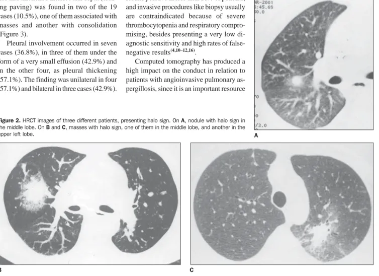
Nodules were found in six of the 19 cases (31.6%) (Figure 2A), multiple in five (83.3%) and solitary in only one case (16.7%). In four (66.7%) of the six cases, nodules presented well-defined borders. Small nodules occurred in all of the six cases (100%), and in one of them small and large nodules were associated. Halo sign was observed in five of the six cases (83.3%), while in none of them cavitation was found.
Masses with irregular contour were found in four of the 19 cases (21.1%) (Fig-ures 2B and 2C), three of them being soli-tary. Halo sign surrounding these masses was observed in three (75%) of the four cases, while cavitation occurred in only one of these cases (25%).
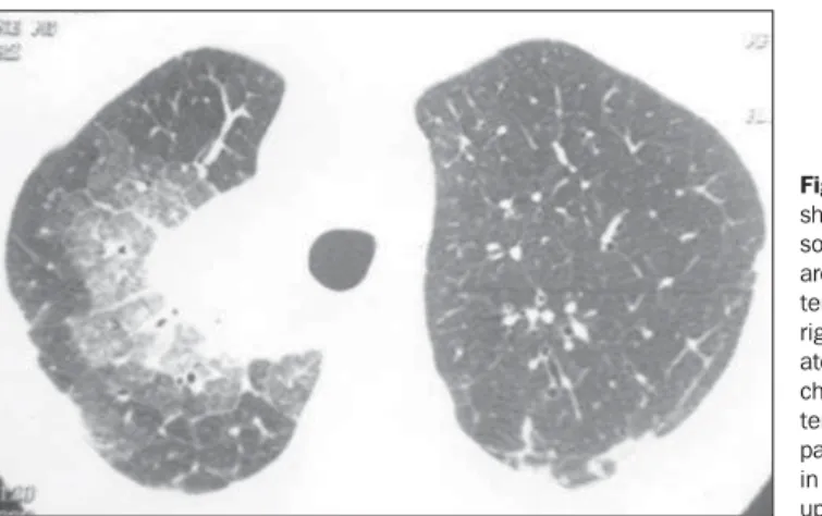
Areas of ground-glass attenuation non-associated with nodules and consolidations were found in three of the 19 cases (15.8%). Superimposition of ground-glass opaci-ties and thickened interlobular septa (craz-ing pav(craz-ing) was found in two of the 19 cases (10.5%), one of them associated with masses and another with consolidation (Figure 3).
Pleural involvement occurred in seven cases (36.8%), in three of them under the form of a very small effusion (42.9%) and in the other four, as pleural thickening (57.1%). The finding was unilateral in four (57.1%) and bilateral in three cases (42.9%).
DISCUSSION
All of the 19 patients with angioinva-sive pulmonary aspergillosis were immu-nocompromised as a result of chemo-therapy for treatment of acute leukemia which is in compliance with the literature since it describes extended granulocytope-nia in patients undergoing treatment with chemotherapy as the most usual context where the angioinvasive pulmonary as-pergillosis develops(4,7,10,16,17). There are studies demonstrating that about 50% of neutropenic patients undergoing treatment with chemotherapy, or submitted to bone marrow transplantation, present severe fun-gal infection at some phase of their evolu-tion(16,18).
Clinical and radiographic presentations of angioinvasive pulmonary aspergillosis are similar to those of other infectious pneumonias. Additionally, by the time when the antifungal therapy could change the patient’s survival, the sputum culture results positive in less than 10% of patients, and invasive procedures like biopsy usually are contraindicated because of severe thrombocytopenia and respiratory compro-mising, besides presenting a very low di-agnostic sensitivity and high rates of false-negative results(4,10–12,16).
Computed tomography has produced a high impact on the conduct in relation to patients with angioinvasive pulmonary as-pergillosis, since it is an important resource
for the early diagnosis of this disease, es-pecially when invasive procedures offer high risks. Compared to CT, HRCT pro-vides a more clear definition of lesions, particularly ground-glass opacities present in the early phases of the disease(1,4,8,10–12,16–
18)
.
Recent studies have reported the rel-evance of HRCT for the early diagnosis of angioinvasive pulmonary aspergillosis, through the detection of the typical halo sign. This sign is highly suggestive for the diagnosis of angioinvasive pulmonary as-pergillosis in an early phase when radio-graphic findings are non-specific and the sputum culture presents negative results(4,9–
11,16). Although non-pathognomonic and
possibly associated with a range of infec-tious and non-infecinfec-tious processes, the halo sign is highly suggestive of angioinvasive pulmonary aspergillosis when detected in neutropenic patients, and some authors consider it sufficiently characteristic to
jus-Figure 2. HRCT images of three different patients, presenting halo sign. On A, nodule with halo sign in the middle lobe. On B and C, masses with halo sign, one of them in the middle lobe, and another in the upper left lobe.
C B
tify the commencement of the antifungal therapy(4,8,10,18,19).
Another finding considered as sugges-tive of angioinvasive pulmonary as-pergillosis is the air crescent sign when associated with an appropriate clinical pre-sentation. However, contrarily to the halo sign, the air crescent sign appears later in the course of the disease and is seen in the phase of the infection resolution, coincid-ing with the improvement of neutropenia. This fact makes the clinical value of this finding limited in the early diagnosis of the disease, but is reported as an indication of good prognosis(4,8,10,11,16,18,19).
Won et al.(19) have found consolidation
in 80% of leukemic patients with angioin-vasive pulmonary aspergillosis, such con-solidations being associated with nodules in 20% of the cases, a data that is similar to those found in this study, where consoli-dations occurred in 12 of 19 cases (63%), and associated with nodules in 25% of cases. Consolidations were predominantly peripheral in 83.3% in this casuistic, while
(28.5%), both cavitary, one of them with air crescent sign. Kuhlman et al.(4) have found cavitary consolidations in five of the seven cases (71.4%).
In our study, nodules occurred in 31.6% of cases, 83.4% being multiple and 16.7% solitary, which is different from the study of Kuhlman et al.(9), whose percentages were respectively 55% and 45%.Well de-fined nodules were found in 66.7% of our cases, differently from studies like that of Franco et al.(16), where all the nodules
pre-sented regular contours.
A ground-glass halo was found in 83.4% of cases presenting nodules, which is in agreement with the studies of Kuhlman et al.(11) and Franco et al.(16),
where the halo sign in nodules was found respectively in 88.8% and 80% of cases. On the other hand, Mori et al.(18) and Won et al.(19) have found this sign respectively
in only 19% and 40% of cases. No cavita-tion was found in any case, differently from the findings of Kuhlman et al.(11), where cavitation was observed in five of seven
(25%), while Kuhlman et al.(4) observed
percentages of up to 71%.
Pleural involvement was observed in seven of our patients (35%), similarly to the majority of the referred authors(7,20,21). In
five cases, the pleural involvement oc-curred in the form of an effusion, while in the other two cases only pleural thickening was observed, probably with no relation-ship with the fungal lesion.
The mosaic pattern of attenuation (crazy paving) (thickened interlobular septa asso-ciated with ground-glass opacity) occurred in two cases (10.5%), which is in agree-ment with the findings of Franco et al.(16), who have found this aspect in only one of the eight cases studied (12.5%). Data in the literature about this finding in patients with angioinvasive pulmonary aspergillosis are scarce, difficulting its analysis.
The clinical diagnosis of angioinvasive pulmonary aspergillosis is difficult and, generally, the prognosis is poor, with very high mortality rates. The role of computed tomography, especially HRCT, has become significant in the whole course of the dis-ease, from its early detection, indicating the commencement of the antifungal therapy, to the monitoring of possible recurrences during the chemotherapy treatment(10,12,16).
So, in patients with neutropenia under the risk of angioinvasive pulmonary aspergillo-sis, the systematic use of HRCT is justified since this is the best way to establish an early and probably specific diagnosis, once the halo sign is detected. Because of the short period during the course of the dis-ease in which the halo sign is detectable by HRCT, the scan should be performed as soon as the clinical suspicion is raised, al-lowing an early diagnosis with a
REFERENCES
1. Klein DL, Gamsu G. Thoracic manifestations of aspergillosis. AJR Am J Roentgenol 1980;134: 543–552.
2. Pozes AS, Maranhão B, Marchiori E, Zanardi PN, Martins EML, Vabo KA. Aspergilose pulmonar semi-invasiva: relato de caso. Rev Imagem 2002; 24:197–201.
3. Yousem SA. The histological spectrum of chronic necrotizing forms of pulmonary aspergillosis. Hum Pathol 1997;28:650–656.
4. Kuhlman JE, Fishman EK, Burch PA, Karp JE, Zerhouni EA, Siegelman SS. Invasive pulmonary aspergillosis in acute leukemia. The contribution of CT to early diagnosis and aggressive manage-ment. Chest 1987;92:95–99.
5. Nuño CG, Alfonso PP, Vazquez JCR, Gomez MMR, Prats IP, Gonzáles JG. Aspergilosis pulmo-nar: un nuevo enfoque en la reemergencia. Acta Médica 2000;9:67–72.
6. Webb WR, Müller NL, Naidich DP. High-resolu-tion CT of the lung. 3rd ed. New York: Raven, 2001.
7. Aquino SL, Kee ST, Warnock ML, Gamsu G. Pul-monary aspergillosis: imaging findings with pathologic correlation. AJR Am J Roentgenol 1994;163:811–815.
8. Caillot D, Couaillier JF, Bernard A, et al. Increas-ing volume and changIncreas-ing characteristics of
inva-sive pulmonary aspergillosis on sequential tho-racic computed tomography scans in patients with neutropenia. J Clin Oncol 2001;19:253–259. 9. Logan PM, Primack SL, Miller RR, Müller NL.
Invasive aspergillosis of the airways: radio-graphic, CT, and pathologic findings. Radiology 1994;193:383–388.
10. Kuhlman JE, Fishman EK, Burch PA, Karp JE, Zerhouni EA, Siegelman SS. CT of invasive pul-monary aspergillosis. AJR Am J Roentgenol 1998;150:1015–1020.
11. Kuhlman JE, Fishman EK, Siegelman SS. Inva-sive pulmonary aspergillosis in acute leukemia: characteristic findings on CT, the CT halo sign, and the role of CT in early diagnosis. Radiology 1985;157:611–614.
12. Marchiori E, Valiante PM, Souza Jr AS. Nódulos com sinal do halo na aspergilose pulmonar angio-invasiva: correlação da tomografia computadori-zada de alta resolução com a anatomopatologia. Radiol Bras 2002;35:195–198.
13. Persegani MK, Marchiori E, Rodrigues R, et al. O “sinal do halo” na tomografia computadorizada de alta resolução do tórax. Rev Imagem 2001;23: 225–231.
14. Austin JHM, Müller NL, Friedman PJ, et al. Glos-sary of terms for CT of the lungs: recommendations of the Nomenclature Committee of the Fleischner Society. Radiology 1996;200:327–331. 15. Souza Jr AS, Araújo Neto C, Jasinovodolinsky D,
et al. Terminologia para a descrição de
tomogra-fia computadorizada do tórax. Radiol Bras 2002; 35:125–128.
16. Franco IO, Marchiori E, Souza Jr AS, Melo AVF, Crespo SJV, Irion K. Aspergilose pulmonar an-gioinvasiva: aspectos na tomografia computado-rizada. Rev Imagem 2002;24:77–82. 17. Kenney HH, Agrons GA, Shin JS; Armed Forces
Institute of Pathology. Best cases of the AFIP. In-vasive pulmonary aspergillosis: radiologic and pathologic findings. RadioGraphics 2002;22: 1507–1510.
18. Mori M, Galvin JR, Barloon TJ, Gingrich RD, Stanford W. Fungal pulmonary infections after bone marrow transplantation: evaluation with radiography and CT. Radiology 1991;178:721– 726.
19. Won HJ, Lee KS, Cheon JE, et al. Invasive pul-monary aspergillosis: prediction at thin-section CT in patients with neutropenia – a prospective study. Radiology 1998;208:777–782. 20. Santos MLO, Marchiori E, Vianna AD, Souza AS,
Moraes HP. Opacidades em vidro fosco nas doen-ças pulmonares difusas: correlação da tomogra-fia computadorizada de alta resolução com a ana-tomopatologia. Radiol Bras 2003;36:329–338. 21. Franquet T, Müller NL, Giménez A, Guembe P,