Intrathoracic manifestations of collagen vascular
diseases on high-resolution chest computed tomography*
Manifestações intratorácicas das doenças do colágeno na tomografia computadorizada de alta resolução do tórax
C. Isabela S. Silva1, Nestor L. Müller2
Intrathoracic manifestations of collagen vascular diseases are very common. The frequency of intrathoracic manifestations and the patterns of abnormality are variable depending on the type of collagen vascular disease and may simultaneously involve one or more of the following: lung parenchyma, airways, pulmonary vessels, pericardium, and pleura. The most common pulmonary manifestations are diffuse interstitial pneumonia and pulmonary hypertension which together represent the main causes of morbidity and mortality of these patients. Pulmonary, airway and pleural involvement may also be secondary to the disease therapy, or result from bacterial pneumonia or opportunistic infection. In the present review, the authors summarize the main intrathoracic manifestations of collagen vascular diseases and the differential diagnosis on high-resolution chest computed tomography.
Keywords: Lung; Collagen vascular disease; Interstitial pneumonia; Pulmonary hypertension; Computed tomography.
As manifestações intratorácicas das doenças do colágeno são bastante comuns. O padrão e a freqüência de comprometimento dependem do tipo específico de doença do colágeno, que pode envolver um ou vários compartimentos simultaneamente, tais como parênquima, vias aéreas, artérias pulmonares, pleura, e peri-cárdio. As manifestações mais importantes incluem as pneumonias intersticiais difusas e a hipertensão pul-monar, que em conjunto representam as principais causas de mortalidade e morbidade nesses pacientes. O acometimento pulmonar, pleural e de via aérea pode ser também secundário a terapêutica instituída ou ser decorrente de processos infecciosos bacterianos ou por germes oportunistas, por causa da imunossupres-são. Nesta revisão os autores sumarizam as manifestações intratorácicas e o diagnóstico diferencial das principais doenças do colágeno na tomografia computadorizada de alta resolução do tórax.
Unitermos: Pulmão; Doença do colágeno; Pneumonia intersticial; Hipertensão pulmonar; Tomografia com-putadorizada.
Abstract
Resumo
* From the Department of Radiology, University of British Co-lumbia, Vancouver General Hospital, Vancouver, Canada.
1. MD, PhD, Research Associate, University of British Colum-bia, Vancouver General Hospital, Vancouver, Canada.
2. Professor and Chairman, Department of Radiology at Uni-versity of British Columbia, Vancouver General Hospital, Vancouver, Canada.
Mailing address: Dr C. Isabela S. Silva. Department of Radiol-ogy, University of British Columbia, Vancouver General Hospital. 3350-950 West 10th Avenue, Vancouver, BC V5Z 4E3 Canada. E-mail: [email protected]
Received May 5, 2008. Accepted after revision May 27, 2008.
rheumatoid arthritis, progressive systemic sclerosis, systemic lupus erythematosus, dermatopolymyositis, mixed connective tissue disease and Sjögren’s syndrome(1,2,6). Chest radiography represents the imaging method most frequently utilized in the ini-tial evaluation of intrathoracic manifesta-tions of collagen vascular diseases. Chest radiography, however, has low sensitivity and specificity. High-resolution computed tomography (HRCT) is the method of choice for assessment of pulmonary abnor-malities in collagen vascular diseases, of-fering the best correlation with histologic findings, disease severity, prognosis, evalu-ation of disease progression, and differen-tial diagnosis(1). The aim of this manuscript is to review the main intrathoracic manifes-tations of collagen vascular diseases on HRCT.
Silva CIS, Müller NL. Intrathoracic manifestations of collagen vascular diseases on high-resolution chest computed tomogra-phy. Radiol Bras. 2008;41(3):189–197.
specific type of collagen vascular disease and may include one or more pulmonary compartments such as alveolus, intersti-tium, vessels, lymphatic tissue, airways and pleura(1–7). The most frequent pulmonary manifestations are diffuse interstitial pneumonias(1,2,6) and pulmonary hyperten-sion(1,2,8) which as a whole represent the main causes of mortality and morbidity in these patients(1,2). It is important to note that pulmonary abnormalities in patients with collagen vascular disease may be due not only to the underlying disease, but also re-sult from its treatment, and include drug reaction and infection by bacteria or oppor-tunistic organisms, such as Pneumocystis jiroveci, and atypical mycobacteria as a result of immunosuppression(1,2). The col-lagen vascular diseases that most com-monly result in interstitial lung disease are
INTRODUCTION
MAIN INTRATHORACIC
MANIFESTATIONS OF COLLAGEN VASCULAR DISEASES
1. Diffuse interstitial pneumonias
Interstitial pneumonias represent the most common intrathoracic manifestations of collagen vascular disease(1,2). Collagen vascular diseases may be associated with virtually all patterns of diffuse interstitial pneumonia and may progress to pulmonary fibrosis(1–3,6). The histological classifica-tion is similar to that of idiopathic intersti-tial pneumonias including: a) patterns of chronic progression – usual interstitial pneumonia, non-specific interstitial monia and lymphocytic interstitial pneu-monia; b) pattern of subacute progression – organizing pneumonia (also known as bronchiolitis obliterans organizing pneu-monia —BOOP); c) pattern of acute
pro-gression of pulmonary injury – diffuse al-veolar damage(3,9,10).
High-resolution CT manifestations of interstitial pneumonias in patients with col-lagen vascular diseases also are similar to the ones described for patients with idio-pathic disease (Table 1)(1–3,9,11). Collagen vascular diseases most frequently associ-ated with interstitial pneumonia are: rheu-matoid arthritis, progressive systemic scle-rosis (scleroderma) and dermatopolymyo-sitis. In general, non-specific interstitial pneumonia is the most common pattern of interstitial pneumonia in patients with col-lagen vascular diseases (Figure 1)(1,2). In pa-tients with rheumatoid arthritis, however, it seems that the pattern of usual intersti-tial pneumonia (Figure 2) is the most fre-quent one(1,7). On the other hand, lympho-cytic interstitial pneumonia, although rare, is most common in patients with Sjögren’s
syndrome (Figure 3)(1,2,12), while the diffuse alveolar damage pattern is more frequent in patients with dermatopolymyositis and systemic lupus erythematosus. Table 2 shows the frequency of each interstitial pneumonia pattern found in the main col-lagen vascular diseases discussed in the present review.
In general, chronic progressive intersti-tial pneumonias in collagen vascular dis-eases have a better prognosis than those of idiopathic nature, with a better five year survival rate (Figure 4)(13). However, cases of unfavorable progression in patients with collagen vascular disease and interstitial pulmonary involvement with no response to the therapy are not infrequent. Similarly to the idiopathic form of disease, the non-specific interstitial pneumonia pattern in collagen vascular disease has a better prog-nosis than the pattern of usual interstitial
Table 1 HRCT findings in interstitial pneumonias.
Pattern Usual interstitial pneumonia Non-specific interstitial pneumonia Organizing pneumonia (BOOP) Lymphocytic interstitial pneumonia Diffuse alveolar damage Typical findings
Reticular pattern, bronchiectasis and traction bronchiectasis, honeycombing, absent or minimal ground-glass opacities
Ground-glass opacities, mild reticulation, traction bronchiectasis and bronchiolec-tasis, absent honeycombing in the early stages
Consolidation is the typical finding, ground-glass opacity usually is associated with areas of consolidation, perilobular pattern or reversed halo sign may be found
Ground-glass opacities associated with sparse cysts ranging from 1–3 cm in di-ameter
Ground-glass opacity, “crazy paving” pat-tern, bilateral areas of consolidation may be present mainly in dependent regions
Typical distribution
Peripheral, basal and posterior pre-dominance
Lower lobes and peripheral regions. Findings may be diffuse. 20–50% of patients show less severe involvement of the pulmonary parenchyma imme-diately adjacent to the pleural surface (subpleural sparing)
Bilateral symmetrical or asymmetrical, peripheral and/or peribronchovascular in 60–80% of cases, predominance in lower lobes
May be diffuse or predominate in the lower lobes; cysts may have subpleu-ral and periphesubpleu-ral distribution
Bilateral and diffuse, rarely multifocal
Unusual findings
Asymmetrical distribution, major in-volvement of upper lobes
Extensive reticular pattern, consolida-tion (may represent areas of co-exist-ing organizco-exist-ing pneumonia – BOOP), peribronchovascular distribution
Focal consolidation mimicking mass may progress to fibrosis
Isolated pulmonary cysts, ground-glass opacity as an isolated finding, septal thickening, and centrolobular nodules
May progress to pulmonary fibrosis. Patients who survive may have HRCT pattern similar to non-specific intersti-tial pneumonia
Table 2 Frequency of interstitial pneumonias in collagen vascular diseases.
Pattern
Usual interstitial pneumonia Non-specific interstitial pneumonia Organizing pneumonia (BOOP) Lymphocytic interstitial pneumonia Diffuse alveolar damage
Rheumatoid arthritis +++ ++ ++ + + Progressive systemic sclerosis + +++ + – + Dermatopolymyositis + +++ +++ – ++ Sjögren’s syndrome + ++ – ++ + Mixed connective tissue disease + ++ + – – Systemic lupus erythematosus + ++ + + ++
pneumonia(13). The concomitant presence of more than one pattern of interstitial pneumonia, particularly the association between non-specific interstitial pneumo-nia and organizing pneumopneumo-nia (BOOP) is not rare in collagen vascular disease, and is most common in patients with dermato-polymyositis and mixed connective tissue disease (Figure 5). Generally, the pattern of diffuse alveolar damage is associated with poor prognosis, both in patients with idio-pathic disease and patients with collagen vascular disease(10).
Interstitial pneumonias may precede the clinical onset of the collagen vascular dis-ease for a period of three months up to five
years, usually with a pattern of non-specific interstitial pneumonia(14)or acutely with a pattern of diffuse alveolar damage. Patients with collagen vascular disease, especially rheumatoid arthritis, scleroderma and dermatopolymyositis, may progress with acute exacerbation of fibrotic interstitial pneumonia(15). Most frequently, acute pul-monary injury manifests histologically as diffuse alveolar damage superimposed on underlying pulmonary fibrosis (generally usual interstitial pneumonia or non-specific interstitial pneumonia patterns)(15). HRCT findings of acute exacerbation of chronic progressive interstitial pneumonia in pa-tients with collagen vascular disease consist in diffuse areas of ground-glass attenuation,
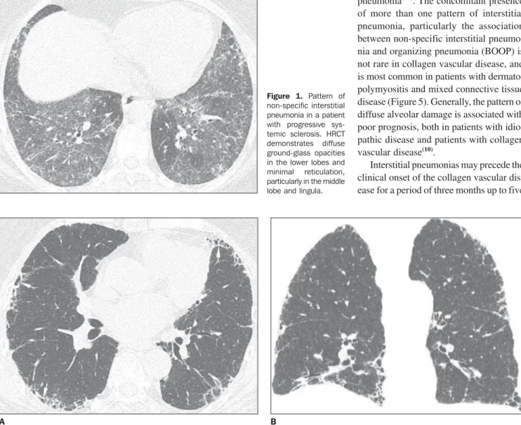
Figure 1. Pattern of non-specific interstitial pneumonia in a patient with progressive sys-temic sclerosis. HRCT demonstrates diffuse ground-glass opacities in the lower lobes and minimal reticulation, particularly in the middle lobe and lingula.
Figure 3. Pattern of lymphocytic interstitial pneumonia in a patient with Sjögren’s syn-drome. HRCT image demonstrates bilateral ground-glass opacities and a few pulmonary cysts.
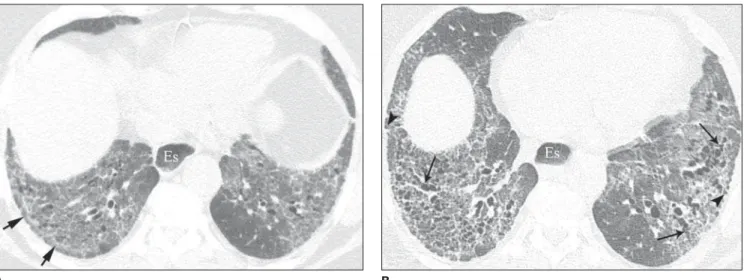
Figure 2. Pattern of usual interstitial pneumonia in a patient with rheumatoid arthritis. Axial (A) and coronal (B) HRCT images demonstrate findings of archi-tectural distortion with peripheral reticulation and honeycombing (arrows) intermingled with relatively normal areas of pulmonary parenchyma. Note the more extensive involvement of the lung bases (B).
with or without associated smooth inter-and intralobular lines resulting in a crazy paving pattern associated with signs of fi-brosis from the pre-existing interstitial pneumonia (distortion of pulmonary archi-tecture, traction bronchiectasis, reticulation and honeycombing) (Figure 6)(15,16). Areas of focal or bilateral dependent consolida-tion may be present. The main differential diagnosis for ground-glass attenuation at
HRCT in patients with collagen vascular disease and fibrotic interstitial pneumonia includes opportunistic infections (particu-larly by Pneumocystis) and drug-induced lung disease (Figure 7)(1–3). Consolidation may be due to organizing pneumonia or an infectious process (by bacteria, mycobac-teria or fungi).
It is important to note that, in smoking patients with collagen vascular disease,
smoking-related interstitial pneumonias such as respiratory bronchiolitis with inter-stitial pulmonary disease and desquama-tive interstitial pneumonia may also be found(2,9).
2. Pulmonary vascular disease
Pulmonary vasculopathy in collagen vascular diseases generally manifests as pulmonary hypertension, diffuse pulmo-nary hemorrhage (capillaritis) or small and medium vessel vasculitis(2,8). Pulmonary venoocclusive disease and pulmonary cap-illary hemangiomatosis, although rare, have been described in some patients with col-lagen vascular disease such as scleroderma and systemic lupus erythematosus(8,17).
Pulmonary hypertension in collagen vascular disease may result from a range of different pathophysiological mechanisms including loss of pulmonary capillary net-work as a result of fibrotic interstitial dis-ease; intrinsic pulmonary arteriopathy with vascular remodelling and pulmonary arte-rial hypertension; left ventricular diastolic dysfunction with pulmonary venous hyper-tension; and chronic thromboembolic dis-ease associated with hypercoagulability and antiphospholipides (patients with sys-temic lupus erythematosus)(8,18). It may occur as an isolated finding (Figure 8) or occurring in association with fibrotic
inter-Figure 4. High-resolution CT follow-up of non-specific interstitial pneumonia in a patient with progressive systemic sclerosis receiving corticosteroid therapy. HRCT image (A) demonstrates diffuse ground-glass opacities, extensive reticulation and subtle signs of fibrosis characterized by irregularities of the pleuropul-monary interface and presence of some irregular and tortuous bronchi and bronchioles in the areas of ground-glass attenuation. Note the less extensive in-volvement of the posterior pulmonary regions immediately adjacent to the pleural surface (arrows; subpleural sparing) - a typical finding of non-specific inter-stitial pneumonia -, as well of esophageal dilatation (Es), a common finding in patients with progressive systemic fibrosis. HRCT image (B) acquired two years later demonstrates, despite treatment, substantial progression of fibrosis, with extensive traction bronchiectasis (long arrows) and bronchiolectasis, and inter-val development of mild subpleural honeycombing (arrowheads).
A B
stitial pneumonia. The incidence of pulmo-nary hypertension in collagen vascular dis-eases is variable, depending on the type of collagen vascular disease, on the concomi-tant presence of interstitial disease and on the diagnostic method utilized(8,18). Pulmo-nary hypertension is most common in scle-roderma (approximately 25% of patients), systemic lupus erythematosus (5–10%) and mixed connective tissue disease(8,18).
Increase in the diameter of the main pul-monary artery trunk (> 2.9 cm) (Figure 8),
main, lobar and segmental pulmonary ar-teries represent typical findings of pulmo-nary hypertension at HRCT(3). Artery wall calcification may be identified in patients with severe and longstanding hypertension, as well as thrombi, especially in patients with systemic lupus erythematosus and antiphospholipid antibodies. A mosaic per-fusion pattern may be present. Rarely, centrilobular nodules and diffuse or patchy ground-glass opacities are identified at HRCT (Figure 9). These abnormalities may
be multifactorial and result from the pul-monary hypertension itself (proliferation of small muscular arteries, chronic passive congestion which may lead to engorgement of the capillary bed, deposition of choles-terol granulomas, focal areas of hemor-rhage) or may be associated with the pres-ence of pulmonary capillary hemangioma-tosis (Figure 9) (exaggerated multifocal capillary proliferation within the alveolar walls). The concomitant presence of inter-lobular septal thickening in patients with
Figure 6. Acute exacerbation of usual interstitial pneumonia in a patient with rheumatoid arthritis. HRCT image (A) demonstrates typical findings of usual interstitial pneumonia characterized by peripheral reticulation, pulmonary architectural distortion and minimal subpleural honeycombing, without the presence of ground-glass opacities. HRCT image (B) acquired two months later, when the patient presented with respiratory failure in the absence of precipitating fac-tors, such as respiratory infection, cardiac decompensation or use of medication, demonstrates extensive areas of ground-glass attenuation and inter- and intralobular septa thickening (“crazy paving” pattern), and tortuous and irregular bronchi. These abnormalities correspond to findings of acute idiopathic pul-monary injury (diffuse alveolar damage) in a patient with pulpul-monary fibrosis and collagen vascular disease.
A B
Figure 7. Drug-induced lung disease secondary to the use of metotrexate in a patient with rheumatoid arthritis and no previous parenchymal abnormalities. HRCT image demonstrates diffuse bilateral ground-glass opacities and mini-mal reticulation.
pulmonary hypertension can be secondary to right cardiac failure or, less frequently, pulmonary venoocclusive disease.
The main HRCT manifestations of dif-fuse pulmonary hemorrhage (capillaritis) in collagen vascular disease consist in diffuse ground-glass opacities (Figure 10), “crazy paving” pattern, patchy consolidation, and centrilobular nodules, or lobular areas of ground-glass opacities(3). The main dif-ferential HRCT diagnosis includes: pulmo-nary infections, especially pneumonia by
Pneumocystis, diffuse alveolar damage (Figure 6) and drug-induced lung disease (Figure 7)(1).
3. Pulmonary nodules or masses
The differential diagnosis of pulmonary nodules or masses in patients with collagen vascular disease is broad and includes
op-portunistic infectious processes (Nocardia,
Cryptococcus,Aspergillus, Histoplasma, atypical mycobacteria), neoplasms (carci-noma, lymphoma), metabolic process (amyloidosis), and immunological process (rheumatoid nodules)(19). The main causes of pulmonary nodules and masses found at HRCT of patients with collagen vascular disease are summarized as follows:
a) Rheumatoid nodules – Rare, but may be found in patients with rheumatoid arthritis, subcutaneous nodules and posi-tive rheumatoid factor(1,20). Sizes range be-tween 0.5 cm and 7 cm in diameter, and may be single or multiple. These nodules rarely develop in the pleura or pericardium. Rheumatoid nodules may cavitate (up to 50%), causing hemoptysis or pneumotho-rax, or become infected(19-21). Spontaneous resolution may occur. Calcification is
rare(19). An unusual association between rheumatoid nodules and pneumoconiosis (particularly in coal-mining workers) in patients with rheumatoid disease is called Caplan’s syndrome(19).
b)Pulmonary lymphoma – Most fre-quently occurring in patients with Sjögren’s syndrome, it also can be found in patients with rheumatoid arthritis(19,22). At HRCT, pulmonary lymphomas may present as nod-ules (generally with > 1.0 cm in diameter), consolidation or, rarely, ground-glass opacities(12,22).
c) Amyloidosis – The parenchymal nodular presentation of amyloidosis may occur in patients with Sjögren´s syndrome and lymphocytic interstitial pneumo-nia(22,23). The nodules may be single or multiple, may have foci of calcification and localization in the pulmonary parenchyma
Figure 10. Diffuse pul-monary hemorrhage in a patient with systemic lupus erythematosus. HRCT image demon-strates diffuse ground-glass opacities pre-dominating in the right lung and nodular opaci-ties.
or within pulmonary cysts. The diffuse pre-sentation of pulmonary amyloidosis is rare, but may be found in patients with rheuma-toid arthritis and Sjögren’s syndrome(12, 19,22,24). At HRCT, diffuse pulmonary
amy-loidosis is characterized by diffuse micro-nodules and interlobular septal thickening. d)Pulmonary carcinoma – Some stud-ies have shown an increased incidence of pulmonary carcinoma in patients with col-lagen disease and concomitant fibrotic in-terstitial vascular disease(1,19); however, this relationship is not well established(25,26).
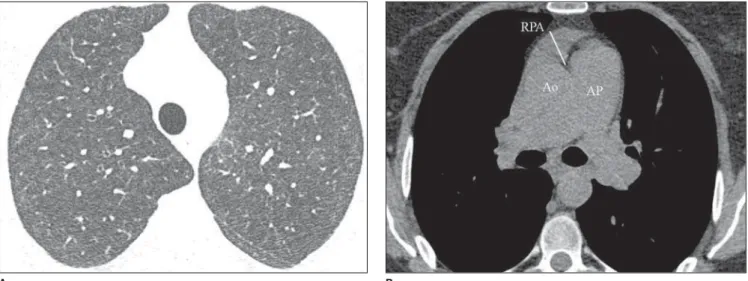
Figure 9. Pulmonary capillary hemangiomatosis in a patient with progressive systemic sclerosis and severe pulmonary hypertension. HRCT image (A) demon-strates diffuse ground-glass opacities and centrilobular nodules mimicking hypersensitivity pneumonitis. Image photographed at soft tissue windows (B) dem-onstrates increase in the caliber of the pulmonary artery (AP) trunk and presence of fluid in the anterior pericardial recess (RPA) with more than 10 mm in thickness a finding shows to correlate with poor prognosis. Ao, ascending aorta.
The incidence of neoplasms (including lung cancer) in patients with scleroderma is estimated to range from 3.6% to 10.7%(27). Recently, a study has observed that the incidence of lung cancer in patients with scleroderma is not related to the con-comitant presence of pulmonary fibrosis, scleroderma subtype, or to the immuno-logical status of the patient; on the other hand, smoking patients with scleroderma have a seven times higher risk for develop-ing lung cancer than non-smokdevelop-ing pa-tients(28). At HRCT, pulmonary carcinoma may manifest as a spiculated nodule or pulmonary mass (Figure 11). The main dif-ferential diagnosis includes rheumatoid nodule (in rheumatoid arthritis), infectious processes (especially fungi), focal amyloi-dosis and pulmonary lymphoma (particu-larly in patients with Sjögren’s syndrome) and localized organizing pneumonia (BOOP) (Figure 12).
4. Diffuse pulmonary consolidation
In patients with systemic lupus erythe-matosus, diffuse consolidation may result from lupus pneumonitis or diffuse pulmo-nary hemorrhage (Figure 10)(1,19). Drug-in-duced lung disease and infection should always be considered in the differential
diagnosis. The presence of predominant consolidation in dependent pulmonary re-gions, particularly in association with centrilobular nodules, may correspond to aspiration pneumonia, a quite common finding in patients with scleroderma and dermatopolymyositis caused by esophageal dysmotility(19,29).
5. Airway
a) Large airways – bronchiectasis
Bronchiectasis occurs most commonly in rheumatoid arthritis (up to 30%), and also may be found in Sjögren’s syndrome and systemic lupus erythematosus(1,3,19). At HRCT, it is more common in the lower lobes, and of cylindrical type (Figure 13)(7). The cause for bronchiectasis in patients with collagen vascular disease is multifac-torial; it may be due to repeated infections (particularly in rheumatoid arthritis), in-volvement of the small airways or airway dryness (Sjögren’s syndrome)(1–3,7).
b) Small airways–- bronchiolitis
– Bronchiolitis obliterans (constrictive bronchiolitis) occurs most commonly in women with rheumatoid arthritis and less frequently in patients with Sjögren’s syn-drome(1–3). Main HRCT findings include
areas of decreased attenuation and vascu-larity, with or without redistribution of the blood flow towards non-involved regions, resulting in mosaic perfusion pattern (Fig-ure 13)(3,7). Expiratory HRCT demonstrates air trapping. Dilated bronchi (bronchiecta-sis) (Figure 13) and thickened bronchial walls also can be found(3,7). It is important to note that bronchiolitis obliterans may also result from penicilamine therapy in patients with rheumatoid arthritis(19). Pul-monary hypertension also may result in mosaic perfusion (attenuation) at HRCT; however, at expiratory HRCT, air trapping is not usually found and, differently from bronchiolitis obliterans, an increase in the pulmonary artery caliber is observed.
– Follicular bronchiolitis represents bronchiolocentric lymphoid hyperplasia most frequently found in rheumatoid arthri-tis, Sjögren’s syndrome and scleroderma(1– 3,30). The HRCT may be normal or show
centrilobular and peribronchovascular nod-ules(31). The main differential diagnosis at HRCT includes infectious process and as-piration (most frequently found in depen-dent pulmonary regions).
– Exudative bronchiolitis (cellular),
generally of infectious nature, character-ized at HRCT as centrilobular nodulesor
Figure 11. Pulmonary carcinoma in a patient with progressive systemic scle-rosis and pulmonary fibscle-rosis. Coronal HRCT image demonstrates a tortuous and dilated esophagus (arrowheads), spiculated nodule (arrow) in the periphery of the right lung base and signs of advanced-stage pulmonary fibrosis, character-ized by honeycombing, reticulation and traction bronchiectasis mainly in the lung bases.
branching nodular opacities (tree-in-bud pattern) with bilateral and asymmetrical distribution(3).
6. Other intrathoracic manifestations of collagen vascular diseases
Collagen vascular diseases may also be associated with other intrathoracic abnor-malities summarized as follows:
a) Lymphonodomegaly
Enlargement o mediastinal lymph nodes is frequently found in patients with col-lagen vascular disease; it is most commonly associated with the presence of interstitial lung disease, and is found in approximately 70% of patients with scleroderma(19,32). Usually, lymph nodes measure up to 1.5 cm in short axis diameter, and only one or two nodal stations are involved. Enlarged hilar and mediastinal lymph nodes may, how-ever, be found in any patient with collagen vascular disease, including in the absence of interstitial disease, from varied causes such as: sarcoidosis, infection, lymphoma, metastasis, inflammatory response to the presence of reflux or aspiration pneumo-nia(19,32).
b) Abnormalities of the pleura and pericardium
Pleural involvement (effusion or thick-ening) is quite frequent particularly in sys-temic lupus erythematosus and rheumatoid arthritis(1,19,33). The presence of concomi-tant pericardial effusion is common. More
than half of patients with systemic lupus erythematosus, as the disease progresses, develop pleural effusion, generally bilateral and with a serous or serosanguinolent ap-pearance(33). Pleural effusion, as well as pericardial effusion, may also be a result from the use of procainamide and hydrala-zine in patients with systemic lupus erythe-matosus(19). In rheumatoid arthritis, pleural thickening is found in 70% of patients and pleural effusion in approximately 5% of patients(19).
In many patients (up to 50%), pericar-dial abnormalities (pericarpericar-dial effusion, pericardial adherence and fibrotic or fibrin-ous pericarditis) are asymptomatic(34). Peri-cardial effusion, frequently in association with pleural effusion, is one of the most common intrathoracic manifestations of systemic lupus erythematosus. Generally, pericardial effusion has a good prognosis. However, it is important to note that the presence of pericardial effusion associated with pulmonary hypertension (defined by means of echocardiography) in patients with collagen vascular disease, particularly in cases where fluid is detected in the an-terior pericardial recess, measuring > 10 mm (Figure 9B), is associated with a poor prognosis(34,35).
c) Diaphragmatic dysfunction
This is an uncommon condition, but may be found in patients with systemic lupus erythematosus and, occasionally, in patients with dermatopolymyositis(1,3),
re-sulting in dyspnea, restrictive pattern of pulmonary function in the absence of pul-monary abnormalities and diaphragmatic elevation with adjacent basal atelectasis.
d) Esophageal dysmotility
This is a common condition in patients with scleroderma and dermatopolymyo-sitis(19). Asymptomatic esophageal dilata-tion may be identified in 40% to 80% of patients with scleroderma (Figure 4)(29). At HRCT, the coronal diameter of the esoph-ageal lumen in patients with scleroderma ranges between 1.2 cm and 4.0 cm (mean 2.3 cm)(29). Esophageal dysmotility may lead to aspiration pneumonia.
CONCLUSION
Intrathoracic manifestations of collagen vascular diseases are common and are clinically important. The main pulmonary manifestations include diffuse interstitial pneumonia and pulmonary hypertension which, as a whole, represent the main causes of morbidity and mortality in these patients. The pattern and frequency of pa-renchymal, vascular, pericardial, pleural and airway involvement, as well as dia-phragmatic and esophageal abnormalities depend on the specific type of collagen vascular disease. Awareness of the several patterns of involvement found at HRCT, as well as their clinical/radiological correla-tion is essential. Intrathoracic manifesta-tions of collagen vascular diseases may be
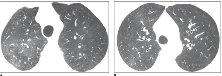
Figure 13. Airway abnormalities in a patient with rheumatoid arthritis. HRCT images (A, B) demonstrate dilated bronchi (arrows on B) and signs of small airway involvement (bronchiolitis obliterans) characterized by areas of decreased attenuation and vascularity with blood flow redistribution to non-involved regions resulting in a pattern of mosaic perfusion (attenuation).
multicompartmental, be due to the under-lying disease or be secondary to treatment and thus include drug reaction and oppor-tunistic infection.
REFERENCES
1. Devaraj A, Wells AU, Hansell DM. Computed tomographic imaging in connective tissue dis-eases. Semin Respir Crit Care Med. 2007;28: 389–97.
2. Woodhead F, Wells AU, Desai SR. Pulmonary complications of connective tissue diseases. Clin Chest Med. 2008;29:149–64, vii.
3. Tanaka N, Newell JD, Brown KK, et al. Collagen vascular disease-related lung disease: high-reso-lution computed tomography findings based on the pathologic classification. J Comput Assist Tomogr. 2004;28:351–60.
4. Tansey D, Wells AU, Colby TV, et al. Variations in histological patterns of interstitial pneumonia between connective tissue disorders and their relationship to prognosis. Histopathology. 2004; 44:585–96.
5. Primack SL, Müller NL. Radiologic manifesta-tions of the systemic autoimmune diseases. Clin Chest Med. 1998;19:573–86, vii.
6. Lamblin C, Bergoin C, Saelens T, et al. Intersti-tial lung diseases in collagen vascular diseases. Eur Respir J Suppl. 2001;32:69S–80. 7. Tanaka N, Kim JS, Newell JD, et al. Rheumatoid
arthritis-related lung diseases: CT findings. Radiology. 2004;232:81–91.
8. Hoeper MM. Pulmonary hypertension in collagen vascular disease. Eur Respir J. 2002;19:571–6.
9. Lynch DA, Travis WD, Muller NL, et al. Idio-pathic interstitial pneumonias: CT features. Radiology. 2005;236:10–21.
10. Won Huh J, Soon Kim D, Keun Lee C, et al. Two distinct clinical types of interstitial lung disease associated with polymyositis-dermatomyositis. Respir Med. 2007;101:1761–9.
11. Desai SR, Veeraraghavan S, Hansell DM, et al. CT features of lung disease in patients with systemic sclerosis: comparison with idiopathic pulmonary fibrosis and nonspecific interstitial pneumonia. Radiology. 2004;232:560–7.
12. Parambil JG, Myers JL, Lindell RM, et al.
Inter-stitial lung disease in primary Sjögren syndrome. Chest. 2006;130:1489–95.
13. Park JH, Kim DS, Park IN, et al. Prognosis of fi-brotic interstitial pneumonia: idiopathic versus collagen vascular disease-related subtypes. Am J Respir Crit Care Med. 2007;175:705–11.
14. Sato T, Fujita J, Yamadori I, et al. Non-specific interstitial pneumonia; as the first clinical presen-tation of various collagen vascular disorders. Rheumatol Int. 2006;26:551–5.
15. Silva CI, Müller NL, Fujimoto K, et al. Acute exacerbation of chronic interstitial pneumonia: high-resolution computed tomography and patho-logic findings. J Thorac Imaging. 2007;22:221–9.
16. Horoupian N, English J, Müller NL. Accelerated deterioration of usual interstitial pneumonia with acute development of honeycomb cysts in rheuma-toid arthritis. J Thorac Imaging. 2004;19:127–30.
17. Kishida Y, Kanai Y, Kuramochi S, et al. Pulmo-nary venoocclusive disease in a patient with sys-temic lupus erythematosus. J Rheumatol. 1993; 20:2161–2.
18. Handa T, Nagai S, Miki S, et al. Incidence of pulmonary hypertension and its clinical relevance in patients with interstitial pneumonias: compari-son between idiopathic and collagen vascular disease associated interstitial pneumonias. Intern Med. 2007;46:831–7.
19. Rockall AG, Rickards D, Shaw PJ. Imaging of the pulmonary manifestations of systemic disease. Postgrad Med J. 2001;77:621–38.
20. Tanoue LT. Pulmonary involvement in collagen vascular disease: a review of the pulmonary mani-festations of the Marfan syndrome, ankylosing spondylitis, Sjögrens syndrome, and relapsing polychondritis. J Thorac Imaging. 1992;7:62–77.
21. Kobayashi T, Satoh K, Ohkawa M, et al. Multiple rheumatoid nodules with rapid thin-walled cav-ity formation producing pneumothorax. J Thorac Imaging. 2005;20:47–9.
22. Jeong YJ, Lee KS, Chung MP, et al. Amyloidosis and lymphoproliferative disease in Sjögren syn-drome: thin-section computed tomography find-ings and histopathologic comparisons. J Comput Assist Tomogr. 2004;28:776–81.
23. Desai SR, Nicholson AG, Stewart S, et al. Benign pulmonary lymphocytic infiltration and amyloi-dosis: computed tomographic and pathologic
fea-tures in three cases. J Thorac Imaging. 1997;12: 215–20.
24. Sumiya M, Ohya N, Shinoura H, et al. Diffuse interstitial pulmonary amyloidosis in rheumatoid arthritis. J Rheumatol. 1996;23:933–6.
25. Pearson JE, Silman AJ. Risk of cancer in patients with scleroderma. Ann Rheum Dis. 2003;62:697– 9.
26. Hill CL, Nguyen AM, Roder D, et al. Risk of can-cer in patients with scleroderma: a population based cohort study. Ann Rheum Dis. 2003;62: 728–31.
27. Wooten M. Systemic sclerosis and malignancy: a review of the literature. South Med J. 2008;101: 59–62.
28. Pontifex EK, Hill CL, Roberts-Thomson P. Risk factors for lung cancer in patients with sclero-derma: a nested case-control study. Ann Rheum Dis. 2007;66:551–3.
29. Bhalla M, Silver RM, Shepard JA, et al. Chest CT in patients with scleroderma: prevalence of asymptomatic esophageal dilatation and medias-tinal lymphadenopathy. AJR Am J Roentgenol. 1993;161:269–72.
30. Hayakawa H, Sato A, Imokawa S, et al. Bronchi-olar disease in rheumatoid arthritis. Am J Respir Crit Care Med. 1996;154:1531–6.
31. Howling SJ, Hansell DM, Wells AU, et al. Folli-cular bronchiolitis: thin-section CT and histologic findings. Radiology. 1999;212:637–42. 32. Wechsler RJ, Steiner RM, Spirn PW, et al. The
relationship of thoracic lymphadenopathy to pulmonary interstitial disease in diffuse and limited systemic sclerosis: CT findings. AJR Am J Roentgenol. 1996;167:101–4.
33. Kim JS, Lee KS, Koh EM, et al. Thoracic involve-ment of systemic lupus erythematosus: clinical, pathologic, and radiologic findings. J Comput Assist Tomogr. 2000;24:9–18.
34. Fischer A, Misumi S, Curran-Everett D, et al. Pericardial abnormalities predict the presence of echocardiographically defined pulmonary arterial hypertension in systemic sclerosis-related inter-stitial lung disease. Chest. 2007;131:988–92. 35. Baque-Juston MC, Wells AU, Hansell DM.