ABSTRACT
Morphometric study of the root anatomy in
123!"#$
%&3'()*4++"5
1- DDS, MSc, PhD, Department of Diagnosis and Surgery, Araraquara Dental School, UNESP – Univ. Estadual Paulista, Araraquara, SP, Brazil. 2- DDS, Private Practice, Araraquara, SP, Brazil.
3- DDS, MSc student, Department of Diagnosis and Surgery, Araraquara Dental School, UNESP - Univ. Estadual Paulista, Araraquara, SP, Brazil. 4- DDS, MSc, PhD, Adjunct Professor, Department of Morphology, Araraquara Dental School, UNESP - Univ. Estadual Paulista, Araraquara, SP, Brazil. 5- DDS, MSc, PhD, Associate Professor, Department of Diagnosis and Surgery, Araraquara Dental School, UNESP - Univ. Estadual Paulista, Araraquara, SP, Brazil.
, - Prof. Joni Augusto Cirelli - Disciplina de Periodontia, Departamento de Diagnóstico e Cirurgia, Faculdade de Odontologia de Araraquara/Universidade Estadual Paulista - UNESP - Rua Humaitá, 1680 - 2º andar - Araraquara - SP - 14801-903 - Brasil - Phone: +55-16-3301-6375 - Fax: +55-16-3301-6369 - e-mail: [email protected]
$-$089:;<<:= >"-++";?;<9<=,"-"8;?;<9<
F
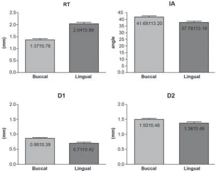
urcation involvement in periodontal disease has been a challenge for the dentist. Objective: The aim of this study was to investigate root dimensions in the furcation were used to obtain the following measurements on the buccal and lingual surfaces of each tooth: root trunk height (RT), horizontal interadicular distance obtained 1 mm (D1) and 2 mm (D2) below the fornix and interadicular angle (IA). Results: Mean± standard deviation of buccal and lingual furcation measurements were, respectively, 1.37±0.78 mm and 2.04±0.89 mm for RT; 0.86±0.39 mm and 0.71±0.42 mm for D1; 1.50±0.48 mm and ! "!#$%%$ &'*+ differences were found between all measured parameters for buccal and lingual sides (p<0.05, paired t test). Conclusions: In conclusion, the lingual furcation of mandibular - / = suggesting more limitation for instrumentation and worse prognosis to lingual furcation involvements in comparison to buccal lesions.Key words: Furcation defects. Periodontics. Tooth root.
INTRODUCTION
> ? + += resulting in marginal alveolar bone resorption and attachment loss7. As the destruction of the periodontium progresses apically, the furcation of multirooted teeth is exposed, leading to irreversible bone loss in the interadicular area15.
@ B + E @ furcation as “the anatomic area of a multirooted tooth where the roots diverge” and furcation invasion refers to the “pathologic resorption of bone within a furcation”2.
Effective instrumentation of furcation defects have always been a challenge for dentists15,29 due to the limited accessibility through the furcation
of furcated teeth. Several studies have focused on anatomical features of molar teeth2,3,5,6,13,17,22,25,31. The main anatomical considerations are: root trunk and furcation entrance dimensions, root surface area, and root separation distances.
According to previous studies, root trunk dimensions play an important role in the periodontal disease process due to its significant relation to both prognosis and treatment of the tooth13. Concerning the furcation entrance dimensions, a > width values equal to or less than 0.75 mm. These values are smaller than the width of common curettes, which means that such instruments do not clean appropriately the dental surface in the furcation entrance area2,4,8,26. Thus, the effectiveness to instrument the furcation entrance area is compromised because such curettes do not ' + is root separation area in furcation region, which corresponds to the portion where the roots are separated by alveolar bone. The measurement of this area tends to increase apically demonstrating how divergent the roots can be31. Interadicular separation higher than or equal to 2 mm has been associated with the improvement of the furcation healing after regenerative therapies18.
The aim of the present study was to investigate the morphology of the furcation area of mandibular = based on the limited information regarding this comparison.
MATERIAL AND METHODS
The experimental sample consisted of 233 K @ Bank of the Laboratory of Anatomy, Department of Morphology, Araraquara Dental School, UNESP, Brazil. For sample selection, teeth should present good conditions, in other words, teeth with caries, fused roots, calculus or restorative treatment that could interfere in the area of interest were excluded. The reasons for tooth extraction and any information about possible periodontal treatment before extraction could not be identified. The present study was approved by the Research Ethics Committee of UNESP (protocol number 13/05).
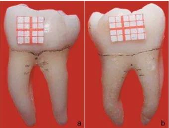
After selection of teeth, reference points were marked on the buccal and lingual surfaces of each tooth with a 0.3 mm graphite pencil, under a WX#YW += Heerbrugg, Switzerland). Initially, a line was drawn on the cementoenamel junction (CEJ), and a point mark indicated the fornix of the furcation entrance. In each root, two other dots were performed, at 1 mm and 2 mm apical to the fornix point. After, -\-\=
standardize tooth position and facilitate points and lines visualization. A small piece of millimeter paper - \ - - adjustment and stereometric measurements conversion from pixels to millimeter after taking the photomicrographs (Figure 1).
P h o t o m i c r o g r a p h s w e r e t a k e n a t 1 5 x magnification using a digital camera DXC-107 A/107 AP (Sony Electronics Inc., Tokyo, Japan). The images were transferred to a microcomputer and image analysis software (Jandel Sigma Scan Pro, Jandel Corporation, San Rafael, CA, USA) was used for stereometric analysis.
In the digital images, the following lines were drawn: line 1) a horizontal line tangent to the highest points of the CEJ of each root; line 2) a line parallel to the previous one, passing through the point of the fornix; lines 3) and 4) horizontal lines binding the points of each root, corresponding to 1 mm and 2 mm apical to the fornix. Lines 1 and 2 were used to obtain the measurement of the root trunk (RT). Lines 3 and 4 determined the
Figure 1- Buccal (a) and lingual (b) surfaces of a mandibular molar with line and reference points
Figure 2- Reference lines 1, 2, 3 and 4 for Stereometric analysis
1
interadicular distance.
After the lines were drawn, the following measurements were taken (Figure 2):
RT (root trunk)= distance between the fornix and the highest point of the CEJ; D1= distance between the mesial and distal roots 1 mm apical to the fornix; D2= distance between the mesial and distal roots 2 mm apical to the fornix; IA (interadicular
angle)= angle of separation formed by the buccal and lingual roots of the furcation.
For the IA measurement, two lines were drawn along the inner wall of the mesial and distal root. The angle formed between the intersection of these two lines was recorded as the IA. Also, the average of three measurements corresponding to 1 mm in the grid paper on each digital image was obtained.
Figure 4- Pearson’s correlation analysis between buccal and lingual surfaces for root trunk height (RT) (a), D1 (b), D2 (c)
Figure 3- Mean and standard deviation (SD) values of root trunk height (RT), D1 and D2 linear measurements (mm) and !! "
This value allowed the conversion of all linear measurements from pixel to millimeters.
All measurements obtained in this study had B >= allowing parametric statistical analysis. Hence, all data were expressed as the mean±standard deviation of buccal and lingual furcations and comparative analysis between both sides was performed. For the evaluation of the variables RT, interadicular distance at 1 mm (D1) and 2 mm (D2) and IA statistical analysis was performed using a paired t test, comparing buccal and lingual
measures. Correlations between buccal and lingual measurements were calculated using the Pearson’s @> -set as p<0.05.
RESULTS
The results are presented by Figures 3 and ! *+ - between all measured parameters for buccal and lingual sides (p<0.05). The buccal RT was + `@Y{| The interadicular distance increased apically for both sides and the buccal side of the furcation presented greater D1 and D2 distances than the lingual side (p<0.001). The IA of the lingual side - + side (p<0.001).
DISCUSSION
Thorough knowledge of root anatomy is mandatory in periodontal therapies as it is intimately associated with the establishment of an accurate diagnosis and the correct choice of the treatment modality to provide optimal @ the present study was that the lingual furcation is anatomically different from the buccal furcation for all measurements evaluated, which probably affects disease establishment and prognosis.
The severity of furcation involvements is directly associated with the relationship between the amount of attachment loss and the RT length16. In the present study, morphometric analysis of > longer lingual root trunk in comparison with buccal /= -}@ accordance with those of previous studies9,10,13,14. Short RT is more likely to develop early furcation involvement and attachment loss in the presence of periodontal disease because it has less surface area for periodontal attachment23. Even though, once the disease is installed, reduced RT lengths tend to lead to satisfactory periodontal treatment outcomes because of their easier access1,13-15,17,19,24,29. Also,
short RT has been associated with longer individual roots and, consequently, greater potential for corrective therapy12,15. However, in the initial stages of the periodontal disease, long RT has a more favorable prognosis compared to the short one, because it protects the furcation from disease involvement. On the other hand, if the furcation is affected, the prognosis is poorer for longer RT, because the access for instrumentation is hampered15,24 and the roots are shorter indicating reduced chance of repair after periodontal therapy. In addition, it has been reported that there is no root trunk longer than 6 mm, which implies that if you have 6 mm of attachment loss in a multirooted tooth, you are probably dealing with a tooth with furcation involvement5.
The furcation entrance measure is extremely important in anticipating the success of periodontal therapy. In this study, the lingual furcation was statistically narrower than the buccal furcation based on the interadicular distances (D1 and D2) and on the interadicular angle differences. This is + width and relationship between the buccal and lingual surfaces. Narrow furcation implies an + entrances for complete root debridement leading to a poor periodontal outcome2,4,8,24. On the other hand, longer root trunk ends up compensating for this characteristic of the lingual side because it makes the furcation access more difficult, preventing early periodontal involvement. This is a positive relationship between root trunk dimension and interadicular distance/angle found in this study, since these features reduce the chance of dental plaque accumulation in the furcation entrance.
Curettes are the manual instruments commonly used during periodontal therapy to produce a smooth and biologically acceptable surface and to permit satisfactory healing11,13. The blades of these instruments play an important role since they must present a width that allows correct and effective root debridement. However, narrow furcation entrance dimensions may complicate the periodontal treatment of furcation involvements4 > YB+ curette) present width of 0.95-1.2 mm and do 2,26. Considering the furcation entrance as well as the blade width of periodontal instruments, various studies have found + + + furcations2,26.
- > >= to access and perform a correctly instrumentation during the periodontal treatment. A recent study evaluating the radiographic characteristics of furcation involvements showed that narrower root furcations may have better outcomes after nonsurgical periodontal therapy because they are less exposed to contaminants and have less root irregularities28.
Regarding regenerative therapy, Pepelassi, et al.18 (1991) have stated that interadicular separation of 2 mm or greater provides more favorable regenerative healing. On the other hand, Pontoriero, et al.20,21 (1988,1989) have found that furcation width – interadicular separation area – greater than 4 mm2 and entrance height of 3 mm or greater failed to heal with complete defect closure. This means that there may be limitation values for interadicular separation – size and height of the furcation defect - that promotes a favorable healing on regenerative therapies.
The positive correlation obtained in this study between buccal and lingual sides suggests that teeth with difficult access to one surface will +>+ side. This finding has implications for clinical therapeutic practice.
Reports by other studies confirm that the +~ + is extremely complex and must be thoroughly understood to improve success rate of periodontal therapy15,22,26 The results of this study call attention to the importance of the furcation dimensions in these teeth to a better clinical practice, involving diagnosis, prevention and treatment of periodontal disease.
CONCLUSIONS
@ + the interadicular width. According to the present = > + narrower interadicular distance and greater root trunk height in the lingual surface than in the buccal surface. These observations have implications for clinical practice in the treatment planning and prognosis determination of furcation involvements in patients with periodontal disease.
ACKNOWLEDGEMENTS
The authors wish to thank Mr. Marcelo Brito Conti, laboratory technician from the Department of Morphology, for technical support.
REFERENCES
1- Al-Shammari KF, Kazor CE, Wang HL. Molar root anatomy and management of furcation defects. J Clin Periodontol. 2001;28(8):730-40.
2- Bower RC. Furcation morphology relative to periodontal treatment. Furcation entrance architecture. J Periodontol. 1979;50(1):23-7.
3- Bower RC. Furcation morphology relative to periodontal treatment. Furcation root surface anatomy. J Periodontol. 1979;50(7):366-74.
! H = X = H }= K H E H permanent molars. J Periodontol. 1991;62(5):308-11.
`= B ` E "#Y!|! #B= '`` + in pathogenesis and treatment of periodontal disease. J Am Dent Assoc. 1980;101(4):627-33.
%KJ+WK ->+ with nonsurgical debridement? Periodontol 2000. 2005;37:72-87. K BW= H *}= = @ HH @ + furcation entrance in Chinese molars. Furcation entrance dimensions. J Clin Periodontol. 1994;21(7):451-6.
K BW=H=@HH= '*'- of molar furcation involvement based on the root trunk and horizontal and vertical bone loss. Int J Periodontics Restorative Dent. 1998;18(3):257-65.
K BW=@HHH> bifurcational ridge correlated with molar furcation involvements. J Periodontol. 1997;68(7):687-93.
11- Jones SJ, Lozdan J, Boyde A. Tooth surfaces treated in situ with periodontal instruments. Scanning electron microscopic studies. Br Dent J. 1972;132(2):57-64.
12- Kapin SH, Eskow RN. Furcation invasions: correlating a + - E &&& Sectioning teeth in the treatment of furcation invasions. Compend Contin Educ Dent. 1984;5(8):612-4, 617, 619.
B=B-K=-=/ H== Kerns LL. Root trunk dimensions of 5 different tooth types. Int J Periodontics Restorative Dent. 1999;19(1):82-91.
!B'=KW=`W' + of the furcation region of mandibular molars. Compend Contin Educ Dent. 1998;19(2):113-6,118-20.
+=J X= *' in the etiology and management of maxillary and mandibular molars with furcation involvement. Int J Periodontics Restorative Dent. 1991;11(5):398-409.
16- Matthews DC, Tabesh M. Detection of localized tooth-related factors that predispose to periodontal infections. Periodontol 2000. 2004;34:136-50.
% E = E B= * '= E ' root furcation: morphometric and morphologic analysis. Int J Periodontics Restorative Dent. 1998;18(5):488-501.
E=}=B-K=}H} \++ tricalcium phosphate composite graft facilitates osseous healing in advanced periodontal furcation defects. J Periodontol. 1991;62(2):106-15.
19- Plagmann HC, Holtorf S, Kocher T. A study on the imaging of complex furcation forms in upper and lower molars. J Clin Periodontol. 2000;27(12):926-31.
20- Pontoriero R, Lindhe J, Nyman S, Karring T, Rosenberg E, *> } B && involved mandibular molars. A clinical study. J Clin Periodontol. 1988;15(4):247-54.
E K}=E =XJE=@ analysis of the maxillary permanent molar teeth and its relation to furcation involvement. Braz Oral Res. 2004;18(3):187-91. E K}=XJE=E =@ = AJ. Root trunk height as a risk factor for periodontal furcation
> >\+ in vitro study. J Int Acad
Periodontol. 2007;9(3):89-95.
24- Roussa E. Anatomic characteristics of the furcation and management of marginal periodontitis. Clin Anat. 1998;11(3):177-86.
*`=J&=BK=B+= '=W CW. Morphometric analysis of the furcation anatomy of mandibular molars. J Periodontol. 2004;75(6):824-9.
#* =E *H=E /@=*=EBW= Santos FA. Molar furcation entrance and its relation to the width of curette blades used in periodontal mechanical therapy. Int J Dent Hyg. 2009;7(4):263-9.
% *> B= W E> involvements in patients referred for periodontal treatment. J Clin Periodontol. 1996;23(12):1093-9.
28- Vale HF, Del Peloso Ribeiro E, Bittencourt S, Nociti FH, Jr, * '= H X ` involvements in mandibular molars as prognostic indicators of healing after nonsurgical periodontal therapy. J Am Dent Assoc. 2009;140(4):434-40.
29- Waerhaug J. The furcation problem. Etiology, pathogenesis, diagnosis, therapy and prognosis. J Clin Periodontol. 1980;7(2):73-95.