P
O
ABSTRACT
RESUMO
AIRPOLISHING EFFECT ON BOVINE ENAMEL AND THE
POSTERIOR REMINERALIZING EFFECT OF SALIVA.
AN
IN VITRO
STUDY
EFEITO DO JATO DE BICARBONATO DE SÓDIO SOBRE O ESMALTE BOVINO E
POSTERIOR EFEITO REMINERALIZADOR DA SALIVA. ESTUDO IN VITRO
Helena Zaramella Vono RIBEIRO1, José Eduardo de Oliveira LIMA2, Bernardo Gonzalez VONO3,
Maria Aparcida de Andrade Moreira MACHADO2, Salete Moura Bonifácio da SILVA2
1- MSc, Graduate Studant, Department of Pediatric Dentistry, Bauru School of Dentistry, University of Sao Paulo. 2- PhD, Assistant Professor, Department of Pediatric Dentistry, Bauru School of Dentistry, University of Sao Paulo. 3- PhD, Titular Professor, Department of Pediatric Dentistry, Bauru School of Dentistry, University of Sao Paulo.
Corresponding address: Helena Zaramella Vono Ribeiro - Praça Salim Haddad Neto, 13-10 Ap. 1402, Vila Universitária Cep.: 17012-503 - Tel.: (14)32270642 - [email protected]
Received: May 8, 2005 - Modification: January 30, 2006 - Accepted: March 24, 2006
urpose: The aim of the present study was to evaluate the alterations of surface microhardness and wear caused by the sodium bicarbonate jet on bovine enamel and the further remineralizing effect of artificial saliva. Methods: Fifteen enamel samples (4,0mm x 4,0mm) were used, which constituted the groups: no treatment (MI); treatment with sodium bicarbonate jet (MII and DI); treatment with sodium bicarbonate jet and immersion in saliva for one hour (MIII and DII), 24 hours (MIV and DIII) and 7 days (MV and DIV). Microhardness tests were carried out using a microdurometer in groups M and wear tests by a rugosimeter in groups D. The data were assessed by the one criterion variance analysis and Tukey test. Results: The mean value of microhardness, in KHN, in groups MI, MII, MIII, MIV and MV were 359,80; 335,46; 369,20; 377,73 and 341,86, respectively, whereas the mean values in µm, of wear for group DI, DII, DIII and DIV were 0,564; 0,519; 0,441 and 0,428, respectively. Conclusions: The sodium bicarbonate jet caused a wear and a reduction in microhardness on the enamel surface; saliva promoted the recovery of initial condition surface microhardness and reduced the wear; the repairing effect of saliva on the surface microhardness alterations occurred within one hour of treatment, having no significant statistical difference from the effect obtained in 24 hours; the best saliva repairing effect on the wear occurred with treatment of 24 hours.
Uniterms: Airpolishing; Tooth abrasion; Microhardness; Tooth remineralization.
bjetivo: A finalidade do trabalho foi avaliar as alterações da microdureza e o desgaste provocado pelo jato de bicarbonato de sódio em esmalte bovino e o posterior efeito remineralizador da saliva artificial. Métodos: Utilizaram-se 15 espécimes de esmalte (4,0mm x 4,0mm) que constituíram os grupos: sem tratamento (MI); tratamento com jato de bicarbonato de sódio (MII e DI); tratamento com jato de bicarbonato de sódio e imersão em saliva artificial por uma hora (MIII e DII), 24 horas (MIV e DIII) e sete dias (MV e DIV). Foram realizados testes de microdureza com um microdurômetro nos grupos M e testes de desgaste com um rugosímetro nos grupos D. Resultados: Os dados foram avaliados pela Análise de Variância a um critério e pelo Teste de Tukey. O valor das médias da microdureza, em KHN, nos grupos MI, MII, MIII, MIV e MV foram 359,80; 335,46; 369,20; 377,73 e 341,86; respectivamente, enquanto que os valores médios, em µm, do desgaste para o grupo DI, DII, DIII e DIV foram 0,564; 0,519; 0,441 e 0,428, respectivamente. Conclusões: o jato de bicarbonato de sódio causou desgaste e diminuição da microdureza superficial; a saliva promoveu o retorno da microdureza superficial à condição inicial e reduziu o desgaste; o efeito reparador da saliva sobre as alterações na microdureza superficial já ocorreu com uma hora de tratamento, não havendo diferença estatisticamente significante do efeito obtido com 24 horas; o melhor efeito reparador da saliva sobre o desgaste ocorreu com 24 horas de tratamento.
INTRODUCTION
On the development of dental caries, bacterial plaque, plays an essential role, and thus, both the chemical and mechanical plaque control methods have been enphasized in modern dentistry.
One of the available professional and prophylactic methods consists in employing a sodium bicarbonate jet, which acts promoting a mechanical remotion of plaque and also a polishing from the dental surfaces (professional prophylaxis).
When a professional prophylaxis is carried out, a wear of the dental surface occurs. Several studies4,12,13,16,19 have
quantified the ocurrence of sound enamel wear, but there is no consence as to the results, although it is known that the wear is higher when the prophylaxis is performed on the previously demineralized enamel3,5,8. Besides, there is a lack
of information about the consequences related to this procedure on the superficial microhardness of enamel.
However, it is known that when there is a mineral loss on a tooth, a remineralization by the action of saliva occurs
1,10,17. Saliva contains in its composition the main components
of dental structure, as calcium and phosphate having a protective action on enamel and dentine 11.
Although it is undoubtfull the benefit for caries prevention resulting from the plaque control, it is known that: the mechanical plaque remotion causes a certain wear of the enamel surface which quantification is not completely clarified yet; there is a lack of information on what happens to the enamel microhardness submitted to the prophylaxis; the enamel alterations originated from prophylaxis can be minimized by the remineralizator power of saliva, but it is still questionable how much of repair occurs and in which time span it occurs.
The present study intended to contribute to a better understanding of those aspects having as a purpose: to evaluate in vitro whether the application of the sodium bicarbonate jet on bovine enamel promotes wear and reduction of its surface microhardness and which is the effect of artificial saliva, in different periods of action, on the repairing of the possible occurred alterations.
MATERIAL AND METHODS
After the remotion of the roots from 30 incisors bovine, the crowns were imbedded in thermoactivated godiva in a cristal acrilic plate, which has been coupled to a precision cutting device (ISOMET Low Speed Saw). With the aid of two duble faced diamond discs – XLI 2205, “high concentration”, and a stainless steel spreader(7cm diameter, 4mm wide, and central orifice 1.3 cm) between the discs, 30 enamel specimens with 4mm X 4mm taken from the plane portion of the crown, making a double secction in cervico-incisal direction and other in the mesiodistal direction.
Then, the enamel blocks were fixed with sticking wax in the center of a cristal acrilic disc (30mm X 8mm), with the purpose of firstly perform the dentine planification.The set
disc/tooth was adapted in a metalographic polishing device ( APL4, Arotec, Cotia, SP). For the planification a carbide silicon sandpaper granulation 320 (Extec Corp.) was used.
Following, the blocks were inverted and now fixed with the enamel placed on the upside face. The set was adapted to the polishing device and the enamel was polish with a carbide silicone sandpaper (Extec corp.) granulation 600, and further with a granulation 1200 sandpaper. In order to finish the polishing, a wet felt with a diamond suspention of 1µm (Buehler), was used.
RESULTS
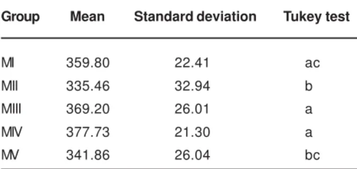
The mean value (KHN) of superficial microhardness of bovine enamel, in the diferent experiment estages, can be seen in Table 1.
There was a statistically significant reduction in the enamel superficial microhardness following the treatment with sodium bicarbonate jet (MII), when compared to the initial superficial microhardness.
Data have shown that the permanency of the specimens in artificial saliva permited that the enamel surface microhardness, which was low after the prophylaxis (MII), has returned to the initial condition, because the values, both from Group MIII, and Group MIV as well as Group MV, did not present statistically significant differences related to the values of initial surface microhadness (MI).
Considering the immersion period of time of the specimens in saliva, there was no statistically significant difference in enamel superficial microhardness in Group MIII (1 hour), compared to Group MIV (24 hours). However, the superficial microhardness of Group MV was lower than those groups.
Table 2 contain the mean values (µm) of the wear observed in bovine enamel, in the different phases of the experiment.
The simulation of prophilaxis (DI) caused a enamel wear of 0.564 µm which was repaired by the treatment with saliva. Within one hour immersion of the specimens in saliva (DII) the wear value had reduced to 0.519 µm, but the difference compared to the wear obtained following the prophylaxis simulation (DI) was not statistically significant.
The treatment with saliva for 24 hours (DIII) and by seven days (DIV) presented better results, because the wear values in these groups were lower than the value found in the specimens immediately after the prophilaxis simulation procedure (DI) at a level statistically significant.
DISCUSSION
This research was carried out with the same 15 bovine enamel specimens which were submitted to sucessive treatments. The employed methodology permitted this procedure. In the wear testing, the rugosimeter was used, which does not alter the tested surface allowing the further performance of other stages of the experiment. In the microhardness test, the KNOOP indenter was used, which neither produces distortions nor damages the enamel structure15. These methods have made possible to evaluate
sucessive alterations occurring in a same specimen. The enamel surface profile was evaluated by a rugosimeter.As the bovine enamel was polished, the surface outline not treated by the sodium bicarbonate jet was similar to a straight line and thus, the alteration of this line observed on the enamel expressed the result of the prophylaxis simulation.
By the literature, it was possible to infer that, even though the enamel abrasion has not been considerable, it has been observed after prophylaxis procedures. Although there is report of wear on the enamel of deciduous tooth with the sodium bicarbonate jet9, it was not verified abrasion in sound
enamel of permanent tooth with this procedure 3,6,13,16. The
wear caused by the sodium bicarbonate jet only occurred in human demineralized enamel3.
As the human enamel is less porous than the bovine enamel 2,14,20, results of wear of both of them must not be
compared without a careful understanding of this fact. In bovine enamel, it was evaluated the wear caused by the sodium bicarbonate. Gerbo7 (1993) had observed no
wear, whereas Honório8 (2003) and Fraga5 (2005), verified
the presence of rugosity, which was higher on enamel previously demineralized than on the sound enamel.
In the present study, it was found a mean wear of 0.564µm, following the simulation of prophylaxy with sodium bicarbonate jet. This value is higher than the one reported by Honório8 (2003), which was of 0.418µm, and by Fraga5
(2005) which verified a wear of 0.319 µm.
It is known that small alterations on the enamel surface can reflect in its physical properties, one of them is the microhardness.
In this study the microhardness mean value of the initial bovine enamel surface, before any treatment, was 359.80 KHN, very close to the value reported by Newbrun;
Group Mean Standard deviation Tukey test
MI 359.80 22.41 ac
MII 335.46 32.94 b
MIII 369.20 26.01 a
MIV 377.73 21.30 a
MV 341.86 26.04 bc
TABLE 1- Mean value of superficial microhardness (KHN) and standard deviation before and after prophylaxis and after immersion in artificial saliva in the different times of treatment
Same letters denote no statistically significant difference (p>0.05) by Tukey test
Group Mean Standard deviation Tukey test
DI 0.564 0.106 a
DII 0.519 0.103 ab
DIII 0.441 0.096 bc
DIV 0.428 0.084 c
TABLE 2- Mean value of wear (µm) and standard deviation after prophylaxis simulation and after immersion in artificial saliva in the different times of treatment
Timberlake; Pigman15 (1959), and higher than the value
reported by Fraga5 (2005) which was of 300.47 KHN and
lower compared to Honório8, wich was 394.0 KHN.
With the treatment of profylaxis simulation with sodium bicarbonate jet, the bovine enamel microhardness has reduced from 359.80 KHN to 335.46 KHN, being the difference statistically significant. There was, therefore a loss of enamel hardness. In a research carried out by Fraga5
(2005) the application of the sodium bicarbonate jet has not altered the superficial microhardness of sound bovine enamel, but its mean value on the enamel presenting artificial carious lesion increased in a significant level, following the simulation of prophylaxis.
It is difficult to explain how, working in similar conditions, there was a discordanceof results as to the sound enamel surface microhardness observed in the present study as well as in Fraga´s study5.
The lack of previous studies in the literature, assessing the effect of sodium bicarbonate on the enamel related to the microhardness, impossibilitates a comparison of the values found in this research. This makes difficult to afirm if the result obtained here was the one expected or not.
Once that, in this in vitro study, alterations on the bovine dental enamel were found by the action of sodium bicarbonate jet, it is important to consider that if those alterations can occur in the clinical practice, although minimally, it can be expected the return to the initial conditions, simply by the fact that the tooth is constantly bathed by saliva.
In the present study, the specimens were treated by artificial saliva by periods of one hour, 24 hours, and 7 days. These periods of time were based on the study carried out by Fraga5 (2005) due to the lack of informations in the
literature about the right moment when the effects of saliva can be detected in the repairing of the alterations resulting from the prophylaxis in sound enamel.
Fraga5 (2005) could not detect any alterations in the
microhardness value in sound teeth subjected to the prophylaxis simulation with sodium bicarbonate jet. Thus it is natural that the further treatment with saliva has not been reflected in this value.
Only with the immersion period in saliva for 28 days Fraga5 (2005) obtained an alteration in wear and
microhardness of enamel.
In the present research, in which there was a reduction in initial microhardness value following the treatment with the sodium bicarbonate jet, when the specimens were immersed in saliva, it could really be verified its effect in the restitution of the tissue integrity.
The specimens immersion in saliva, for just one hour, was enough for the microhardness to reach a value that was not different, statistically significant, from the initial value. The same has occurred with the immersion of the blocks in saliva for 24 hours and for 7 days, permiting to afirm that the repairing of the alterations generated by simulation prophylaxis already occurs, right after the contact with the saliva.
The treatment with saliva also had a repairing effect on
the wear. With one hour of the specimens immersion in saliva, the wear value of 0.564µm caused by the prophylaxis simulation was reduced to 0.519µm, although the difference has not been statistically significant.
However the treatment with saliva by 24 hours, had an expressive reduction of wear, which value was 0.441 with statistically significant difference from the value found after the application of the sodium bicarbonate jet.
There was no higher value in the result of wear with the extended period of immersion of the specimens in saliva for 7 days. It was similar to the one obtained with 24 hours of treatment.
Great discrepancy was found between these results and the Fraga´s results5 which study the immersion period of 4
hours in saliva was not sufficient to alter the microhardness and the wear of hygid bovine enamel. Only with 28 days period of the specimens immersion in saliva, Fraga5 (2005)
could observe an effect statistically significant of wear and microhardness in sound bovine enamel. However, the too long interval between the two observations (4 hours and 28 days) did not permit to know which was the period of time necessary to visualize an effect of treatment.
The results of the current research are in agreement to the Johansson10 (1965). The repairing effect of saliva, in the
present work on the alterations on superficial microhardness and wear enamel was revealed in, at most, 24 hours, and, with the aditional time of 7 days no benefit was noted.
In the study of Johansson10 (1965) using demineralized
human teeth, the remineralizing process by saliva occurred rapidly within the first 24 hours and there was no increment of mineral deposition with the immersion in saliva by a period of up to 3 weeks. A possible explanation for this fact is that, with the passing time, must occur an ionic difusion blocking toward the inner enamel, due to the mineral deposition in the most external layer.
This immediate remineralizing of enamel, when saliva or artificial remineralizing solutions with high calcium concentration are used, was verified in a literature review carried out by Silverstone19 (1977). Also, Ögaard; Ten
Bosch18 (1994) observed in vivo that the remineralization in
the presence of saliva is a relatively rapid process. Although it is not known, in which extension, the obtained results can be transfered to the clinical reality, the indication that the remineralizing process occurs very fast, simply by the action of saliva, this fact gives the dentist reassurement to perform the professional prophylaxis how many times it is necessary.
CONCLUSIONS
1. The application of sodium bicarbonate jet caused a wear and a reduction in the surface microhardness on bovine enamel.
3. The wear resulting from the application of sodium bicarbonate jet were repaired in, at most, 24 hours, following the immersion in artificial saliva.
REFERENCES
1- Amaechi BT, Higham SM. In vitro remineralization of eroded lesions by saliva. J Dent.2001;29(5):371-6.
2- Arends J, Schuthof J, Jongebloed WG. Lesion depth and microhardness indentations on artificial white spot lesions. Caries Res. 1980;14(4):190-5.
3- Boyde A. Airpolishing effects on enamel, dentine, cement and bone. Brit Dent J. 1984;156(8):287-91.
4- Christensen RP, Bangerter VW. Immediate and long-term in vivo effects of polishing on enamel and dentin. J Prosthet Dent. 1987;57(2):150-60.
5- Fraga ACA. Avaliação “in vitro” do efeito da profilaxia com jato de bicarbonato de sódio sobre o esmalte hígido e com lesão de cárie artificial e posterior remineralização. Bauru; 2005. [Dissertação de Mestrado - Faculdade de Odontologia de Bauru da USP].
6- Galloway SE, Pashley DH. Rate of removal of root structure by the use of the Prophy-Jet device. J Periodont. 1987;58(7):464-9. 7- Gerbo LR. Enamel roughness after air powder polishing. Am J Dent. 1993; 6(2):96-8.
8- Honório HM. Avaliação do desgaste de diferentes métodos de profilaxia sobre o esmalte bovino hígido e desmineralizado. Bauru; 2003. [Dissertação de Mestrado – Faculdade de Odontologia de Bauru da USP].
9- Hosoya Y, Johnsto NJW. Evaluation of various cleaning and polishing methods on primary enamel. J Pedod. 1989;13(3):253-69. 10- Johansson B. Remineralization of slightly etched enamel. J Dent Res. 1995;44(1):64-70.
11- Lenz H. Ultrastructure of tooth in respect of mineralization, demineralization and remineralization. Int Dent J. 1967;17(4):693-708.
12 Lutz F, Imfeld T. Advances in abrasiveness: a new technology -prophylaxis pastes. Comp Continuing Educ Dent. 2002;23(1):61-70.
13- Marta SN. Avaliação do efeito do jato de bicarbonato de sódio no esmalte de dentes permanentes jovens. Bauru; 1997. [Dissertação de Mestrado – Faculdade de Odontologia de Bauru da USP].
14- Mellberg JR. Hard-tissue substrates for evaluation of cariogenic and anti cariogenic activity in situ. J Dent Res. 1992;71(sp. Issue):913-9.
15- Newbrun E, Timberlake P, Pigman W. Changes microhardness of enamel following treatment with lactate buffer. J Dent Res. 1959;38(2):293-300.
16- Newman PS, Silverwood RA, Dolby AE. The effects of an air abrasive instrument on dental hard tissues, skin and oral mucosa. Brit Dent J. 1985;159(1):9-12.
17- Oesterle LJ, Shellhart WC, Belanger GK. The use of bovine enamel in bonding studies. Am J Orthod Dentofacial Orthop. 1998;114(5):514-9.
18- Ögaard B, Ten Bosch JJ. Regression of white spot enamel lesions. A new optical method for quantitative longitudinal evaluation in vivo. Am J Dent Orthop. 1994;106(3):238-42.
19- Silverstone LM. Remineralization phenomena. Caries Res. 1977b;11(Supplement 1):59- 84.
20- Wilson M. Bactericidal effect of laiser light and its potential use in the treatment of plaque-related diseases. Int Dent J. 1994;44(2):181-9.