The influence of protective varnish on the integrity
of orthodontic cements
Érika Machado Caldeira1, Antonio de Moraes Izquierdo2, Felipe Giacomet1,
Eduardo Franzotti Sant’Anna3, Antônio Carlos de Oliveira Ruellas4
How to cite this article: Caldeira EM, Izquierdo AM, Giacomet F, Sant’Anna EF, Ruellas ACO. The inluence of protective varnish on the integrity of orth-odontic cements. Dental Press J Orthod. 2013 Nov-Dec;18(6):45-50.
» The authors report no commercial, proprietary or financial interest in the prod-ucts or companies described in this article.
Contact address: Programa de Pós-Graduação em Odontologia (Ortodontia) - Faculdade de Odontologia - Universidade Federal do Rio de Janeiro
Rua Professor Rodolpho Paulo Rocco, 325 – Ilha do Fundão – Rio de Janeiro/ RJ — Brazil.
1 MSc in Orthodontics, Department of Pediatric Dentistry and Orthodontics,
Federal University of Rio de Janeiro (UFRJ).
2 Doctorate Student in Orthodontics, Department of Pediatric Dentistry and
Orthodontics, Federal University of Rio de Janeiro (UFRJ).
3 Adjunct professor, Department of Pediatric Dentistry and Orthodontics,
Federal University of Rio de Janeiro (UFRJ).
4 Adjunct professor, Department of Pediatric Dentistry and Orthodontics,
Federal University of Rio de Janeiro (UFRJ).
Submitted: January 02, 2011 - Revised and accepted: February 19, 2012
Objective:The aim of the present study was to assess the inluence of saliva contamination over the structural strength and integrity of conventional glass-ionomer cements used for cementing orthodontic bands in the absence and pres-ence of a surface-protecting varnish. Method: 48 samples were prepared by inserting 3 types of glass-ionomer ce-ments into standardized metallic matrixes of 10 mm of diameter and 2 mm of depth. The cece-ments used were: Meron (VOCO), Ketac-Cem (3M ESPE) and Vidrion C (DFL), all of which comprised groups A, B and C, respectively. Sub-groups A1, B1 and C1 comprised samples with no surface protection, whereas subSub-groups A2, B2 and C2 comprised samples of which surface was coated with Cavitine varnish (SS White), ater cement manipulation and application, in order to protect the cement applied. All samples were stored in artiicial saliva for 24 hours at 37°C. A Vickers dia-mond micro-durometer was used to produce indentations on the non-treated group (non-varnished) and the treated group (varnished). Results: Varnished materials had signiicantly higher microhardness values in comparison to non-varnished materials. Ketac-Cem had the highest microhardness value among the non-varnished materials. Conclusion:
Varnish application is necessary to preserve the cement and avoid enamel decalciication. Glass-ionomer cements should be protected in order to fully keep their properties, thus, contributing to dental health during orthodontic treatment.
Keywords:Glass-ionomer cements. Artiicial saliva. Microhardness.
Objetivo:avaliar a inluência da contaminação salivar na resistência estrutural e integridade de cimentos de ionômero de vidro convencionais utilizados para cimentação de bandas ortodônticas na ausência e na presença de um verniz protetor de superfície. Métodos: quarenta e oito corpos de prova foram confeccionados a partir de três cimentos or-todônticos, com auxílio de matrizes metálicas padronizadas com 10mm de diâmetro e 2mm de altura. Os cimentos utilizados foram: Meron (Voco), Ketac-Cem (3M ESPE) e Vidrion C (DFL), compondo os grupos A, B e C, respec-tivamente. Metade dessas amostras não recebeu nenhum tipo de proteção supericial, constituindo os subgrupos A1, B1 e C1, enquanto, os subgrupos A2, B2 e C2 tiveram suas superfícies isoladas com verniz Cavitine (SS White) após manipulação e aplicação do cimento, com intuito de proteger a superfície do cimento. As amostras foram armazenadas em saliva artiicial por 24 horas a 37°C. Foi realizado um ensaio de microdureza (Vickers) para avaliação da dureza de superfície do grupo não-tratado (sem isolamento) e do grupo tratado (agente protetor). Resultados: os materiais pre-viamente isolados com o verniz obtiveram valores de microdureza signiicativamente maiores que os não-isolados. O cimento Ketac-Cem apresentou, estatisticamente, a maior microdureza entre os materiais protegidos. Conclusão: o isolamento com verniz mostrou-se necessário para preservação do cimento e, consequentemente, de sua capacidade de evitar possíveis desmineralizações dentárias. Os cimentos de ionômero de vidro devem ser protegidos para manutenção de sua integridade, contribuindo para saúde dental durante o tratamento ortodôntico.
INTRODUCTION
Maintaining an adequate oral health is a constant challenge in Orthodontics, since the high number of retentive surfaces present in the orthodontic appliances hinders bacterial plaque removal. Some issues involving attachment material, such as poor sealing, inadequate structural and adhesive strength as well as cement solubil-ity in oral luids contribute to enamel decalciication.1,2,3
Failure in band cementation usually results in serious problems for orthodontic treatment. If any failure in ce-ment seal occurring between the band and the tooth is not immediately detected, enamel demineralization may oc-cur at the margins of the band.4 In addition to attaching the bands, the cement protects the banded tooth against cavity. The resistance of these dental cements to oral luids can be measured by their solubility and disintegration.5
Glass-ionomer cement is a generic denomination for a group of materials produced by the reaction between sili-cate glass powder and polyacrylic acid.6 The main charac-teristics of this material are luoride release, important for enamel remineralization of carious teeth, biocompatibili-ty, and adhesion to the enamel, which occurs by chemical attraction between the apatite and the polyacrylic acid.7
Due to its capacity of adhering to the dental struc-ture and its ability to release luoride ions, the glass-ion-omer cement is indicated not only for preventive resto-rations, but also as a illing and attaching material.6 In addition, glass-ionomer cement can adhere to stainless steel, which favors its use as an attaching material.8
Bands attached with glass-ionomer cement require less recementations, present less decalciication in the surrounding enamel, and show higher amount of rem-nant cement ater debonding.9 On the other hand, the glass-ionomer cement has the disadvantage of being susceptible to humidity.10
During the initial curing phase of the glass-ionomer cement, any contamination can adversely afect its sur-face hardness, thus, altering its properties.11,12 In clini-cal cases in which contamination control is diicult, its use is contra-indicated.13 For this reason, it seems to be reasonable to isolate the glass-ionomer cement from the oral environment with impermeable materials during initial curing in order to avoid any undesired changes.
There are several studies in the literature relat-ing materials and their adhesive capacity, particu-larly using composites and hybrid ionomers on shear bond tests,14 in addition to those that emphasize
bond strength and ts failures.9,15,16 Other studies have correlated the luoride-releasing ability of certain mate-rials with their cariostatic efects.3,9 However, no studies have been carried out on the need for isolating conven-tional glass-ionomer cements surfaces during cementa-tion of orthodontic bands, even though this accementa-tion is es-sential for the preservation and longevity of this type of cement in restorative Dentistry practice.17
In this study, Cavitine varnish was chosen for such protection. According to the manufacturer, it is a ni-trocellulose-based (8%) varnish with some unique features, including excipients (ethyl acetate, ethanol) which cause it to be volatile and fast-drying. It is used as cavity liner and for protection of silicate restorations, since it prevents salivary action and avoids moisture dur-ing crucial reaction steps.
Microhardness is among the properties which may be affected by contamination during the initial curing phase. Surface microhardness is defined as being the microstructure and texture activities of a given material, which are used for predicting mate-rial strength as well as its ability of abrading opposite structures.18 It is, in fact, one of the most important mechanical properties for comparative studies of den-tal materials.11 Strength, curing time and erosion of the glass-ionomer cement have evolved as new hard-ness tests are performed.19 Knowing the material sur-face microhardness behavior is crucial when choosing the best material, since this property changes when it is exposed to humidity — condition that is similar to that of the oral cavity.18
Therefore, the objective of the present study was to assess the efects of varnish isolation on microhardness of conventional glass-ionomer cements in the presence of salivary contamination during initial curing phase.
MATERIAL AND METHODS
In the present study, conventional glass-ionomer ce-ments were assessed. They were manipulated according to the manufacturer’s instructions and only one investi-gator tested the materials.
batch 0010106 — liquid). Each group was divided into two subgroups containing 8 samples each (Subgroups A1, A2, B1, B2, C1, and C2), in which number 1 refers to those samples without isolation ater manipulation and application of the cement, and number 2 refers to those samples receiving protection against humidity. A total of 48 samples was obtained and humidity protec-tion was achieved by means of applying Cavitine var-nish (SS White, Rio de Janeiro, Brazil, batch 013) onto the cement surfaces.
The samples were obtained by inserting the ma-terials into standardized metallic matrixes of 10 mm in diameter and 2 mm in depth. Insertion was per-formed by using Centrix syringe (DFL) in order to avoid air bubbles. A glass plate was put onto the matrixes so that a plane surface could be obtained, thus, preserving the surface layer as well as enabling the focus during microhardness test.
After the initial 10 minutes, all samples were im-mersed into artificial saliva, including those from subgroups A2, B2, and C2 which had been coated with Cavitine varnish by using microbrushes (Cavi-brush – FGM). The material was kept in a stove at 37oC for 24 hours.17
All samples were kept under moist conditions in order to avoid dehydration and test the varnish ef-ficiency. Artificial saliva was chosen because it in-creases surface hardness of glass-ionomer cements and simulates the conditions found within the oral environment, which would not happen if distilled water was used.19
The microhardness tests were performed by us-ing a Vickers diamond microdurometer (E. LEITZ, Germany) with a 100 gf load being applied during 30 seconds in order to produce indentations, which were measured in Vickers hardness (HV). Five in-dentations were performed in each sample of each group, with a total of 80 indentations — 40 in varnish-coated samples and 40 in non-varnished ones. The microhardness values were obtained by measuring the diagonal of the imprints magnified
by 50 times with ZoomMagic 2.0 software (Peak-Star, USA). The results were calculated with the following formula: HV = 1854.4 P/d2, where P = in-dentation load and d = diagonal obtained.11
Analysis of variance was employed to compare all the subgroups, whereas Tukey’s test was used for mul-tiple comparisons. The signiicance level was set at 1%.
RESULTS
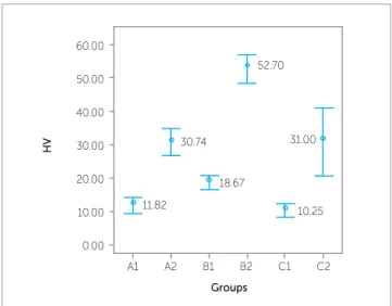
Table 1 and Figure 1 show the microhardness values obtained for all subgroups. They demonstrate that varnish-coated samples had higher microhard-ness values in comparison to the non-varnished ones (P < 0.01). Ketac-Cem glass-ionomer cement (3M ESPE), either varnished or non-varnished, had the highest microhardness value after being stored in artificial saliva for 24 hours.
The varnish-coated samples were found to have smoother and more regular surfaces (Fig 2), as well as the highest microhardness values. On the other hand, cracks and rugosities were observed in the non-varnished samples which also presented inap-propriate microhardness values (Fig 3).
Table 1 - Vickers microhardness values (kg/mm2) for diferent subgroups (mean ± standard deviation).
Figure 1 - Graphic representation of microhardness diferences found for each subgroup.
Group A (Meron, Voco) Group B (Ketac-Cem, 3M ESPE) Group C (Vidrion, DFL)
A1c A2b B1c B2a C1c C2b
HV = 11.82 ± 2.73 HV = 30.74 ± 4.88 HV = 18.67 ± 1.74 HV = 52.69 ± 4.27 HV = 10.24 ± 2.39 HV = 31.00 ± 12.21 Superscribed letters indicate statistically signiicant diferences (P < 0.01) for a>b>c.
10.25 18.67
30.74
11.82
A1 A2 B1 B2 C2
Groups
HV
0.00 10.00 20.00 30.00 40.00 50.00 60.00
on the cement properties.23,25,26 As glass-ionomer ce-ment is susceptible to humidity, it is recommended that its surface be coated during the initial curing phase, that is, soon ater loosing its surface brightness.10,18 However, glass-ionomer cements have a slow curing process and during this phase, they are susceptible to saliva and wa-ter attacks, which dissolve such mawa-terials.27
Water plays a crucial role during the curing phase of glass-ionomer cements. First, it serves as a reaction me-dium and then it slowly hydrates the cross-linked matrix, allowing formation of a stable gel structure which is more resistant and also less susceptible to humidity. If the new-ly-used cement is exposed to the environment with no protective layer, its surface will show cracks and issures caused by dehydration. Both dehydration and excess hu-midity can damage the integrity of the material.26,28
If no protection is provided, the material surface will inevitably become porous and cracked, as ob-served in the non-varnished samples (subgroups A1, B1 and C1). This result can be explained by the nature of the material, because glass-ionomer cements, even when reinforced with composites, react in a more sen-sitive manner to moistness and abrasion.29
Since water balance is crucial to form a stable matrix, and, consequently, to cement maturation, surface pro-tection is extremely important during the initial setting. Thus, due to the fact that glass-ionomer cements exhib-it a high degree of solubilexhib-ity and disintegration in the oral environment, protecting the material surface, characterized by its rugosity, is essential to prevent degradation and avoid S. mutans colonization.17,30 DISCUSSION
Mechanical properties of materials are intimately re-lated to their overall quality and integrity. Material’s sur-face degradation is associated with an increase in rugosity and allows bacteria lodging and, as a consequence, causes undesirable tissue reactions. This explains the need for materials to preserve their mechanical properties, spe-cially hardness.20,21 Materials with better microhardness are more likely to withstand saliva biochemistry, constant pH variations, diferent temperatures and, specially, the resident oral microbiome.22 This is the reason why the present study aimed at verifying surface behavior of 3 types of conventional glass-ionomer cements with regard to humidity and varnish-protection efects. Therefore, the methodology chosen has proved to be adequate.17
In the present study, a 10-minute curing time was adopted before saliva contamination, as stated in previ-ous studies.11,18 The curing time for conventional glass-ionomer cements ranges between 4.4 and 12.2 minutes.23 The irst 10 minutes of chemical reaction are the most important, since acid attack occurs during this period of time, thus, resulting in ion release (luoride, sodium, calcium, aluminum, and phosphate).24 Dehydration was also avoided in the irst 10 minutes of chemical reaction by means of using glass plates on the samples, which pre-vented direct contact between the material and the air, in addition to making the surfaces smooth and uniform.
Total surface hardness of glass-ionomer cements is achieved nearly 24 hours ater application and the testing procedures are usually carried out within this period of time to investigate the efects of storage
Figure 2 - Subgroup B1 sample surface (non-varnished). Presence of cracks, issures, and large indentations . 50 times magniication.
Cem proved to have the highest microhardness values, as previously observed.33 Additionally, no statistically signiicant diferences were observed in the microhard-ness values of the non-varnished samples. Therefore, surface protection of glass-ionomer cements during the initial curing phase was found to be useful.
Considering the complexity of the oral environ-ment, preserving the integrity of the material is neces-sary,30 since failures in cement bands may lead to tooth demineralization and delay treatment due to the need for recementation. Selecting appropriate materials and caring for their preservation is of professional respon-sibility, in view of the longevity of orthodontic treat-ments and commitment to patient health.
CONCLUSION
Protecting the surface of glass-ionomer cements with varnish during the initial curing phase promoted higher microhardness values 24 hours ater its application, cor-roborating the fact that adequate cement preservation is necessary to avoid enamel decalciication. In addition, further clinical comparative studies are required to assess orthodontic bands cemented with either varnished or non-varnished glass-ionomer cements.
Protecting the surface of glass-ionomer cement is a process that lasts for at least 1 hour, although the ideal time required to increase resistance to dis-integration is 24 hours.31 It is known, however, that in the oral environment, protective materials are lost within the first 24 hours because of friction.17 That is the reason why in the methodology used for this study, Cavitine varnish was able to protect the surfaces of the materials within 24 hours.
Although varnishes are indicated as surface protec-tion materials for cements,13,32 there are reports in the literature in which the evaporation of the varnish sol-vent is associated with protective ilms with faulty lines, which does not ensure adequate protection.17 On the other hand, according to the manufacturer’s specii-cations and the results of this study, Cavitine varnish can be used in orthodontic practice. That is because it proved to be eicient with regard to protection, special-ly for cementation, since onspecial-ly a small amount of mate-rial is actually exposed to the oral environment when a correct adaptation of the band is achieved.
Ketak-1. Sadowsky PL, Retief DH. A comparative study of some dental cements used in orthodontics. Angle Orthod. 1976;46(2):171-81.
2. Gameiro GH, Nouer DF, Cenci MS, Cury JA. Enamel demineralization
with two forms of archwire ligation investigated using an in situ caries model: a pilot study. Eur J Orthod. 2009;31(5):542-6.
3. Passalini P, Fidalgo TKS, Caldeira EM, Gleiser R, Nojima MCG, Maia LC.
Preventive efect of luoridated orthodontic resins subjected to high cariogenic challenges. Braz Dent J. 2010;21(3):211-5.
4. Noyes H. Dental caries and the orthodontic patient. J Am Dent Assoc.
1937;24:1243-54.
5. Cameron JC, Charbeneau GT, Craig RG. Some properties of dental
cements of speciic importance in the cementation of orthodontic bands. Angle Orthod. 1963;33(4):233-45.
6. Yli-Urpo H, Vallittu PK, Narhi TO, Forsback AP, Vakiparta M. Release of
silica, calcium, phosphorus, and luoride from glass ionomer cement containing bioactive glass. J Biomater Appl. 2004;19(1):5-20.
7. Vermeersch G, Leloup G, Vreven J. Fluoride release from
glass-ionomer cements, compomers and resin composites. J Oral Rehabil. 2001;28(1):26-32.
8. Coury TL, Willer RD, Miranda FJ, Probst RT. Adhesiveness of
glass-ionomer cement to enamel and dentin: a laboratory study. Oper Dent. 1982;7:2-6.
9. Caves GR, Millett DT, Creanor SL, Foye RH, Gilmour WH. Fluoride release
from orthodontic band cements - a comparison of two in vitro models. J Dent. 2003;31(1):19-24.
10. Pellegrinetti MB, Imparato JCP, Bressan MC, Pinheiro SL, Echeverria S. Avaliação da retenção do cimento de ionômero de vidro. Pesq Bras Odontoped Clin Integr. 2005;5(3):209-13.
11. Ellakuria J, Triana R, Minguez N, Soler I, Ibaseta G, Maza J, et al. Efect of one-year water storage on the surface microhardness of resin-modiied versus conventional glass-ionomer cements. Dent Mater. 2003;19(4):286-90.
12. McLean JW, Wilson AD, Prosser HJ. Development and use of water-hardening glass-ionomer luting cements. J Prosthet Dent. 1984;52(2):175-81.
13. Christensen GJ. Glass ionomer as a luting material. J Am Dent Assoc. 1990;120:59-62.
14. Reicheneder CA, Gedrange T, Lange A, Baumert U, Prof P. Shear and tensile bond strength comparison of various contemporary orthodontic adhesive systems: an in-vitro study. Am J Orthod Dentofacial Orthop. 2009;135(4):422.e1-6.
15. Passalini P, Fidalgo TKS, Caldeira EM, Gleiser R, Nojima MCG, Maia LC. Mechanical properties of one and two step fluoridated orthodontic resins submitted to different pH cycling regimes. Braz Oral Res. 2010;24(2):197-203.
16. Caldeira EM, Fidalgo TKS, Passalini P, Marquezan, M, Nojima MCG, Maia LC. Análise in vitro da inluência da aplicação tópica de luoreto nas propriedades mecânicas de uma resina ortodôntica sob ciclagem de pH. Pesq Bras Odontoped Clin Integr. 2011;11(1):47-52.
REFERENCES
17. Zancopé BR, Novaes TF, Mendes FM, Imparato JCP, Benedetto MS, Raggio DP. Inluência da proteção supericial na rugosidade de cimento de ionômero de vidro. ConScientiae Saúde. 2009;8(4):559-63. 18. Borges AFS, Puppin-Rontani RM, Sinhoreti MAC, Sobrinho LC. Inluência
do tempo de estocagem em meio úmido sobre a microdureza inicial de materiais restauradores estéticos. Ciênc Odontol Bras. 2004;7(4):79-86. 19. Okada K, Tosaki S, Hirota K, Hume WR. Surface hardness change of
restorative illing materials stored in saliva. Dent Mater. 2001;17(1):34-9. 20. Silva KG, Pedrini D, Delbem ACB, Cannon M. Microhardness and luoride
release of restorative materials in diferent storage media. Braz Dent J. 2007;18(4):309-13.
21. Beyth N, Bahir R, Matalon S, Domb AJ, Weiss EI. Streptococcus mutans bioilm changes surface-topography of resin composites. Dent Mater. 2008;24(6):732-6.
22. Silva KG, Pedrini D, Delbem ACB, Cannon M. Efect of pH variations in a cycling model on the properties of restorative materials. Oper Dent. 2007;32(4):328-35.
23. Khouw-Liu VH, Anstice HM, Pearson GJ. An in vitro investigation of a poly (vinyl phosphonic acid) based cement with four conventional glass-ionomer cements. Part 2: Maturation in relation to surface hardness. J Dent. 1999;27(5):359-65.
24. Crisp S, Wilson AD. Reactions in glass ionomer cements: I. Decomposition of the powder. J Dent Res. 1974;53(6):1408-13. 25. Millett DT, Duf S, Morrison L, Cummings A, Gilmour WH. In vitro
comparison of orthodontic band cements. Am J Orthod Dentofacial Orthop. 2003;123(1):15-20.
26. Aguiar DA, Silveira MR, Ritter DE, Locks A, Calvo MCM. Avaliação das propriedades mecânicas de quarto cimentos de ionômero de vidro convencionais utilizados na cimentação de bandas ortodônticas. Rev Dental Press Ortod Ortop Facial. 2008;13(3):104-11.
27. Algera TJ, Kleverlaan CJ, Prahl-Andersen B, Feilzer AJ. The inluence of environmental conditions on the material properties of setting glass-ionomer cements. Dent Mater. 2006;22(9):852-6
28. Feilzer AJ, Kakaboura AI, De Gee AJ, Davidson CL. The inluence of water sorption on the development of setting shrinkage stress in traditional and resin-modiied glass ionomer cements. Dent Mater. 1995;11(3):186-90. 29. Cefaly DF, Wang L, Mello LLCP, Santos JL, Santos JR, Lauris, JRP. Water
sorption of resin-modiied glass-ionomer cements photoactivated with LED. Braz Oral Res. 2006;20(4):342-6.
30. Caldeira EM, Osório A, Oberosler ELC, Vaitsman DS, Alviano DS, Nojima MCG. Antimicrobial and luoride release capacity of orthodontic bonding materials. J Appl Oral Sci. 2013;21(4):327-34.
31. Earl MSA, Ibbetson RJ. The clinical disintegration of a glass ionomer cement. Br Dent J. 1986;161(8):287-91.