101
Jacob Jr C et al. Thoracolumbar burst fracture
Radiol Bras. 2012 Mar/Abr;45(2):101–104
Thoracolumbar burst fracture: what the radiologist should
know
*
Fratura toracolombar do tipo explosão: o que o radiologista deve conhecer
Chárbel Jacob Junior1, Diogo Miranda Barbosa2, Priscila Rossi de Batista3, Dimitri Mori Vieira4,
Igor Cardoso Machado5, Rodrigo Rezende6
Thoracolumbar burst fractures are defined as those fractures involving compromise of the anterior, middle and posterior vertebral columns. The treatment for such vertebral fractures still remains undefined, raising questions about the best form of intervention in these cases. Because of these doubts, imaging methods play a key role in the preoperative workup. However, several tests and measurements are performed by spine surgeons before deciding on the best approach to be adopted, with no standardization and neither consensus. The present review was aimed at standardizing and describing the main techniques and radiological findings on the basis of instability criteria adopted by surgeons in the assessment of thoracolumbar burst fractures, namely extent of height loss of the anterior wall of the fractured vertebra, level of spinal canal compromise by bone fragments and degree of widening of interspinous and interpedicular distance. It is the authors’ opinion that the standardization of the main measurements in the evaluation of thoracolumbar burst fractures by radiological methods will provide the information required for a better interpretation of tests results and consequently aiding in the decision making about the most appropriate treatment.
Keywords: Radiology; Fracture; Spine.
As fraturas vertebrais do tipo explosão são definidas como fraturas nas quais ocorre comprometimento da coluna anterior, média e posterior da vértebra. O tratamento destas fraturas vertebrais persiste indefinido, gerando questionamentos quanto à melhor forma de intervenção destes pacientes. Devido a estas dúvidas, os métodos de imagem apresentam papel fundamental na propedêutica pré-operatória. No entanto, diversas análises e mensurações são realizadas pelos cirurgiões de coluna sem padronização e consenso antes de se decidir sobre a melhor abordagem destes casos. Nesta revisão temos como objetivo padronizar e descrever as principais técnicas e achados radiológicos, com base nos prin-cipais critérios de instabilidade utilizados pelos cirurgiões na avaliação da fratura toracolombar tipo explosão, sendo eles, a medida da perda da altura da parede anterior da vértebra fraturada, a porcentagem de fragmento intracanal e o grau de abertura da distância interespinhosa e interpedicular. Acreditamos que, ao padronizar as principais mensu-rações realizadas para avaliação das fraturas toracolombares do tipo explosão por meio dos métodos radiológicos, estaremos fornecendo informações necessárias para a melhor interpretação dos resultados dos exames e, consequen-temente, para uma tomada de decisão mais adequada acerca do tratamento.
Unitermos: Radiologia; Fratura; Coluna vertebral. Abstract
Resumo
* Study developed at Hospital Universitário da Santa Casa de Misericórdia de Vitória, Vitória, ES, Brazil.
1. Orthopedist and Traumatologist, Specialist in Vertebral Spine Surgery, Physician Assistant, Group of Vertebral Spine – Hospi-tal Vila Velha and Santa Casa de Misericórdia de Vitória, Vitória, ES, Brazil.
2. Radiologist, Musculoskeletal System Specialist, Chief Phy-sician, Centro de Diagnóstico por Imagem (Center of Imaging Diagnosis), Vitória, ES, Brazil.
3. Master, Fellow PhD degree in Physiological Sciences, Phys-iotherapist, Group of Vertebral Spine – Santa Casa de Misericórdia de Vitória, Vitória, ES, Brazil.
4. Trainee of Musculoskeletal Radiology, Centro de Diagnós-tico por Imagem, Vitória, ES, Brazil.
5. Orthopedist and Traumatologist, Specialist in Vertebral Spine Surgery, Physician Assistant, Group of Vertebral Spine – Hospi-tal Meridional and Santa Casa de Misericórdia de Vitória, Vitó-ria, ES, Brazil.
6. PhD of Health Sciences, Orthopedist, Specialist in Verte-bral Spine Surgery, Chief Physician, Group of VerteVerte-bral Spine –
Jacob Jr C, Barbosa DM, Batista PR, Vieira DM, Machado IC, Rezende R. Thoracolumbar burst fracture: what the radiologist should know. Radiol Bras. 2012 Mar/Abr;45(2):101–104.
0100-3984 © Colégio Brasileiro de Radiologia e Diagnóstico por Imagem
REVIEW ARTICLE
bral columns(2). Most burst fractures occur
at the level of the thoracolumbar junction whose vulnerability is partially explained by the anatomic and biomechanical fea-tures of the region. Such an explanation is due to the radial shape of the thoracic cage and stability provided by the costotrans-verse ligaments in the thoracic spine which give a higher resistance to load in the coro-nal and sagittal planes, and to axial rotation. Such a protection degree and the relatively rigid shape contrast with the underlying lumbar spine – more flexible and less pro-tected than the thoracic spine –, resulting in a fragile segment that is named thora-INTRODUCTION
Thoracolumbar burst fracture was first described by Sir Frank Holdsworth, in 1963(1), as an injury typically resulting from
fall from height or motor vehicle accidents, causing a significant axial load on the spine, leading to a failure by compression of the anterior, middle and posterior
verte-Santa Casa de Misericórdia de Vitória, Hospital Meridional and Hospital Vila Velha, Vitória, ES, Brazil.
Mailing Address: Dr. Rodrigo Rezende. Rua Desembargador Augusto Botelho, 209/801, Praia da Costa. Vila Velha, ES, Bra-zil, 29101-110. E-mail: [email protected]
102
Jacob Jr C et al. Thoracolumbar burst fracture
Radiol Bras. 2012 Mar/Abr;45(2):101–104 columbar transition (T11-L2)(3). In a
spe-cific study developed by Avanzi et al.(2),
83% of injuries have occurred between T12 and L2, the first lumbar vertebra being the most affected, likewise in studies devel-oped by other authors(4–7).
Currently, there is a consensus in the medical literature about the best approach to be adopted in the treatment of vertebral fractures types such as luxation, flexion/ distraction and wedge fractures, but not on the treatment of burst fractures, since most patients neither present neurological defi-cit nor meet direct instability criteria(2,3,8– 10). So, the decision making process about
the best treatment to be adopted for thora-columbar burst fractures is based on indi-rect vertebral instability criteria(2,5,11–13).
The main instability criteria adopted by spine surgeons to evaluate burst fractures are the following: extent of height loss of the anterior wall of the fractured vertebra, level of spinal canal compromise by bone fragments, degree of kyphosis, and degree of widening of interspinous and inter-pedicular spaces(2,11–14).
Based on such criteria which have al-ready been defined by the literature, sur-geons can determine the likelihood of ver-tebral collapse and progression to late neu-rological deficit, besides estimating the post-treatment residual pain, and also to change de fracture classification, consider-ing that for each criterion there is a ques-tion to be answered before the definiques-tion of the treatment.
The motivation for developing the present study is the necessity of standard-izing and diffusing the main treatment tech-niques and radiological findings, with ba-sis on the main spinal instability criteria utilized by surgeons in the assessment of thoracolumbar burst fractures, with the objective of helping them to define the best form of management for these patients
MATERIALS AND METHODS
Radiological routine
In a systematic fashion, the authors started the radiological workup to evaluate patients with suspected thoracolumbar burst fracture utilizing posteroanterior (PA) and lateral views of spine radiography. Pos-teroanterior (PA) view allows the
measure-ment of widening of interspinous and interpedicular spaces, according to the Neumann’s method(13).
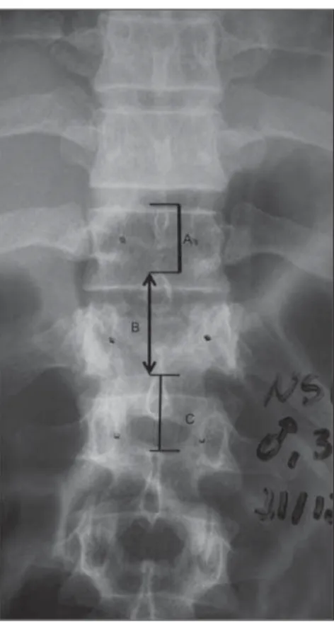
The calculation of the degree of widen-ing of interpedicular space of the injured vertebra is based on the average of interpedicular spaces between the fractured vertebra and the immediately superior and immediately inferior vertebrae(2,12),
corre-sponding to the rate of widening of the interpedicular space (Figure 1). The mea-surement of the interpedicular space was performed with a transparent ruler with a millimeter scale.
Also on the PA view, the Neumann’s method(13) allows the measurement of the
variation of the distance between the spinous processes which constitutes an in-direct sign of injury to structures of the posterior spinal column. The method
con-sists in marking the distance from the up-per border of the spinous processes pro-jected on PA radiographs. Variations of up to 7 mm in the distance is considered as normal, also allowing the calculation of the percentage of spinous processes widening, with 20% being considered as unstable posterior ligamentous complex, requiring surgical treatment (Figure 2).
Continuing the radiological evaluation, the authors utilized the lateral view of the spine which allows the evaluation of the rate of height loss of the anterior wall of the vertebra affected by compression fracture. For this purpose, the authors utilized the method developed by Willén et al.(11),
uti-lizing the average between the heights of the vertebral bodies immediately superior
Figure 1. Calculation of interpedicular distance. Postero-anterior radiography of thoracolumbar spine demonstrating interpedicular distances at the level of the fracture (B) and between the injured vertebra and the immediately superior and imme-diately inferior vertebrae (A and C, respectively). The mean interpedicular distance is calculated by sub-traction of the interpedicular distance at the level of the fractured vertebra on the average between the interpedicular distances of the vertebrae imme-diately superior and immeimme-diately inferior to the frac-tured vertebra, multiplied by 100, according to the following formula: {(A+C)/2 – B}/(A+C)/2 × 100. Example: A = 2.0 cm, B = 3.0 cm, C = 2 cm; so, {(2+2)/2} – 3/(2+2)/2 = –1/2 = 50%.
103
Jacob Jr C et al. Thoracolumbar burst fracture
Radiol Bras. 2012 Mar/Abr;45(2):101–104
vide data regarding the fracture trace analy-sis and evaluation of the involvement of the vertebra components, with possibility of utilizing sagittal and coronal image recon-structions. Such method allows a highly accurate assessment of the vertebral canal compromise by bone fragments (retropul-sion of the posterior wall of the vertebral body) by measuring the mid-sagittal diam-eter, as proposed by Trafton and Boyd, in 1984(12), and of the presence of lamina
frac-ture.
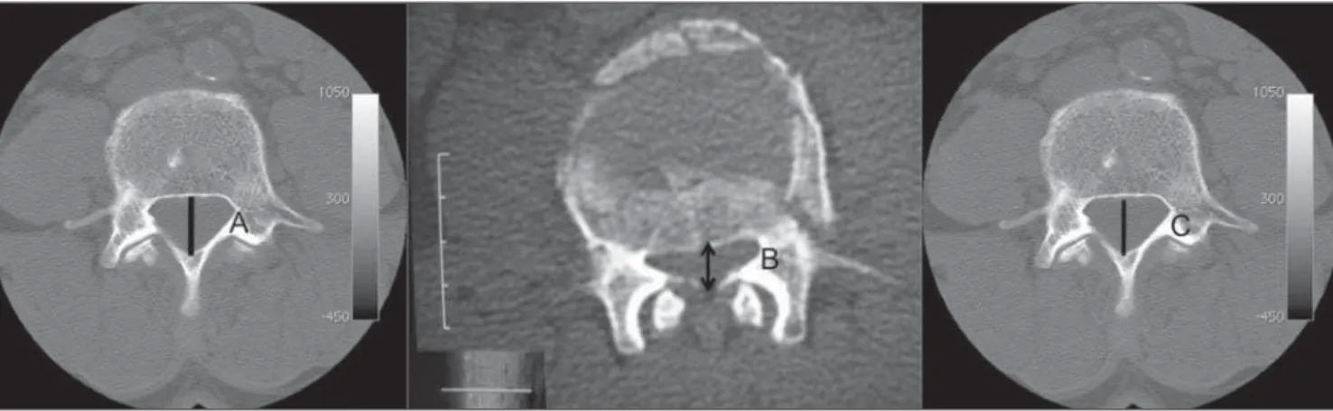
The evaluation of the percentage of ver-tebral canal stenosis as proposed by Trafton and Boyd is based on the average between the values found on axial CT sections of the superior and inferior vertebrae adjacent to the affected vertebra. The evaluation of the neural canal narrowing produced by burst fracture was performed by measuring the mid-sagittal diameter of the canal on CT sections at the level of the affected verte-bra, expressed as a percentage in compari-son with the adjacent levels (Figure 4).
At CT, it is also possible to evaluate the presence or not of laminar fracture, which may be related to the severity of the trauma and associated with a higher risk for neu-rological involvement (Figure 5)(10).
DISCUSSION
There remains considerable difficulty in determining the best treatment for thora-columbar burst fractures, generating debate in several medical schools. Because of such uncertainty, some authors have created in-and inferior to the fractured vertebrae to
obtain the rate of height loss of the anterior wall of the fractured vertebra (Figure 3).
Continuing the imaging workup, the uti-lization of computed tomography may
pro-stability criteria based on objective mea-surements to support the indication of the most appropriate treatment.
In the present article, the authors de-scribe the main radiological methods and findings both at radiography and computed tomography, justifying the relevance of the utilization of objective measurements for both medical first responders and spine surgery specialists. Based on such stan-dardization, the radiologists will be able to adopt such criteria in their reports, in prac-tice, contributing to the indication of the treatment for these patients, since lesions considered as stable are likely to be conser-vatively treated with the use thoraco-lumbosacral orthoses, and fractures consid-ered as unstable require surgery for stabi-lization with pedicle screws.
The measurement of the degree of inter-pedicular widening on PA radiographs is Figure 5. Lamina fracture. Axial computed tomog-raphy section demonstrating burst fracture of the last dorsal vertebra. Lamina fracture is highlighted at left (arrow).
Figure 4. Calculation of anteroposterior distance of the vertebral canal. Computed tomography of vertebral spine demonstrating the anteroposterior diameter of the vertebral canal at the level of the fractured segment (B) and of the vertebral canal at the superior and inferior levels (A and C, respectively). The mean diameter of the vertebral canal at the level of the immediately superior and immediately inferior vertebras was calculated by subtraction from the diameter of the vertebral canal at the level of the fracture, on the average between the vertebral canal diameters of the superior and inferior segments, multiplied by 100, according to the following formula: {(A+C)/2 – B}/(A+C)/2 × 100. Example: A = 4 cm, B = 2 cm, C = 4 cm; so, {(4+4)/2 – 2}/(4+4)/2 = 2/4 = 50%. Figure 3. Calculation of vertebral bodies height.
104
Jacob Jr C et al. Thoracolumbar burst fracture
Radiol Bras. 2012 Mar/Abr;45(2):101–104 extremely relevant for allowing a correct
classification of burst fractures (Denis clas-sification for involvement of middle and anterior columns) which require greatest care and enhanced radiological investiga-tion by means of computed tomography. In 1992, Mumford et al. described a direct relation between percentage of vertebral canal compromise and interpedicular wid-ening visualized at plain anteroposterior radiography, suggesting that the inter-pedicular widening occurs in burst frac-tures. Such data is extremely relevant for the medical first responder because, after the identification of interpedicular widen-ing in the fracture, its classification changes from wedge fracture to burst fracture, re-quiring enhanced care with the patient(7).
On the other hand, the degree of inters-pinous widening, also measured at PA ra-diography, indirectly evaluates the risk for severe posterior ligament injury, which might change the classification of the frac-ture from burst fracfrac-ture to flexion/distrac-tion fracture, determining the type of treat-ment to be adopted. The most utilized method for measuring interspinous widen-ing is the one proposed by Neumann(13),
considering a widening > 7 mm as patho-logical. Also, an interpedicular widening > 20% can be utilized as a reference value, as already reported in the literature(13).
Magnetic resonance imaging is the gold standard for the diagnosis of posterior liga-ment injury, but this method is not widely available in many hospitals.
The evaluation of the intracanal frag-ment percentage is extremely important, since the degree of compromise may lead or not neurological involvement, directly depending on the percentage of decrease in the vertebral canal width. According to
Meves & Avanzi, the neurological condi-tions of patients with burst fracture were directly associated with vertebral canal nar-rowing(15). Trafton & Boyd(12) have
re-ported that thoracolumbar spine fractures with narrowing of the middle-sagittal diam-eter = 50% resulting from retropulsed frag-ments associated with lamina fracture present a significant risk for neurological deficit.
Lamina fracture is other finding evalu-ated at computed tomography. Such a find-ing suggests that the impact of the trauma was distributed among the three columns, so increasing the probability of neurologi-cal involvement(5). Tisot & Avanzi(10) have
observed that the mean percentage of ver-tebral canal narrowing caused by the pres-ence of bone fragments was significantly higher in cases of association with lamina fracture. Such finding has represented an important data to be investigated at com-puted tomography (Figure 5) because of the association between lamina fracture and dural tears.
CONCLUSION
The authors highlight the extreme rel-evance of the radiologists’ knowledge of the main criteria e measurements utilized by spine surgeons, as well as on the adop-tion of such data in the determinaadop-tion of treatment for thoracolumbar burst frac-tures, allowing their inclusion in the radio-logical workup and routine radioradio-logical reports.
REFERENCES
1. Holdsworth F. Fractures, dislocations and frac-ture-dislocations of spine. J Bone Joint Surg Br. 1963;45:6–20.
2. Avanzi O, Meves R, Caffaro MFS, et al.
Correla-ção entre a abertura interpedicular e o comprome-timento do canal vertebral na fratura toracolom-bar em explosão. Coluna. 2008;7:361–6. 3. Vaccaro AR, Kim DH, Brodke DS, et al.
Diagno-sis and management of thoracolumbar spine frac-tures. J Bone Joint Surg. 2003;85:2456–70. 4. Kim NH, Lee HM, Chun IM. Neurologic injury
and recovery in patients with burst fracture of the thoracolumbar spine. Spine. 1999;24:290–3. 5. Vaccaro AR, Nachwalter RS, Klein GR, et al. The
significance of thoracolumbar spinal canal size in spinal cord injury patients. Spine. 2001;26:371– 6.
6. McDonough PW, Davis R, Tribus C, et al. The management of acute thoracolumbar burst frac-tures with anterior corpectomy and Z-plate fixa-tion. Spine. 2004;29:1901–8.
7. Mumford J, Weinstein JN, Spratt KF, et al. Tho-racolumbar burst fractures. The clinical efficacy and outcome of nonoperative management. Spine. 1993;18:955–70.
8. Wood K, Buttermann G, Mehbod A, et al. Opera-tive compared with nonoperaOpera-tive treatment of a thoracolumbar burst fracture without neurologi-cal deficit. A prospective, randomized study. J Bone Joint Surg Am. 2003;85-A:773–81. 9. McCormack T, Karaikovic E, Gaines RW. The
load sharing classification of spine fractures. Spine. 1994;19:1741–4.
10. Tisot RA, Avanzi O. Fratura da coluna vertebral tipo explosão na área da cauda eqüina: correla-ção entre funcorrela-ção neurológica e alterações estru-turais no canal vertebral. Acta Ortop Bras. 2008; 16:85–8.
11. Willén J, Lindahl S, Nordwall A. Unstable thora-columbar fractures. A comparative clinical study of conservative treatment and Harrington instru-mentation. Spine.1985;10:111–22.
12. Trafton PG, Boyd CA Jr. Computed tomography of thoracic and lumbar spine injuries. J Trauma. 1984;24:506–15.
13. Neumann P, Wang Y, Kärrholm J, et al. Determi-nation of inter-spinous process distance in the lumbar spine. Evaluation of reference population to facilitate detection of severe trauma. Eur Spine J. 1999;8:272–8.
14. Whitesides TE Jr. Traumatic kyphosis of the tho-racolumbar spine. Clin Orthop Relat Res. 1977; (128):78–92.