online | memorias.ioc.fiocruz.br
Viability study of a multiplex diagnostic platform for Chagas disease
Leonardo Foti1, Bruna de Paula Fonseca e Fonseca2, Lilian Dias Nascimento2,
Christiane de Fatima Silva Marques2, Edmilson Domingos da Silva2, Cesar Augusto Barros Duarte1,
Christian M Probst1, Samuel Goldenberg1, Antônio Gomes Pinto2, Marco Aurélio Krieger1/+
1Instituto Carlos Chagas-Fiocruz, R. Prof. Algacyr Munhoz Mader 3775, 81350-010 Curitiba, PR, Brasil 2Laboratório de Tecnologia
Diagnóstica, Biomanguinhos-Fiocruz, Rio de Janeiro, RJ, Brasil
A new multiplex assay platform was evaluated to detect Trypanosoma cruzi infection using the recombinant an-tigens CRA, FRA, CRAFRA fusion and parasite lysate. The anan-tigens presented different sensitivity and specificity in a singleplex test when compared to a serial dilution of two pools comprising 10 positive serum samples and one pool of 10 negative samples. The recombinant protein CRA presented lower sensitivity (55%) in contrast to the 100% spe-cificity and sensitivity of FRA, CRAFRA and T. cruzi lysate. These antigens also showed good results in a duplex test and the duplex test with CRAFRA/T. cruzi lysate showed better performance with 100% specificity and sensitivity, as well as a lower cut-off value in comparison to the other duplex test, FRA/T. cruzi lysate. Hence, when the antigens were used in duplex format, both tests showed decreased cut-off values and no interference between different bead sets, resulting in increasing sensitivity and specificity. The results of these multiplex tests show that they could be an alternative to singleplex detection for Chagas disease, and also indicate the necessity of using multiplex diagnostic tools to increase the sensitivity and specificity for diagnostic tests. Emerging data from the T. cruzi genome and from its ORFeome project will also allow the identification of new antigens for this disease detection application.
Key words: Chagas disease - diagnostic - multiplex - bead-based assay - Trypanosoma cruzi
Over one billion people worldwide (1/6 of the world’s population) suffer from one or more neglected diseases (O’Conell 2007). Beyond mortality, neglected diseases cause severe and permanent disabilities and deformities, hampering human development and resulting in signifi-cant socio-economic losses (King et al. 2006, Hopkins et al. 2007).
Chagas disease is one of the world’s leading causes of heart disease, representing the second highest illness burden among tropical diseases in the Americas (TDR 2007). The protozoan Trypanosoma cruzi is the etiologic agent of the disease in mammals; the parasite is transmit-ted by blood-sucking triatomine bugs, blood transfusion or across the placenta (Chagas 1909, Brener et al. 2000).
Since 1990, a series of multinational initiatives based on vector control, systematic screening of blood donors in all endemic countries and detection and treatment of congenital transmission have been launched for control and elimination of Chagas disease. These strategies have led to significant reductions in the number of infected people worldwide. However, approximately eight mil-lion people are still infected with T. cruzi; thus Chagas disease continues to represent a health threat for an esti-mated 28 million people (TDR 2007).
+ Corresponding author: [email protected] Received 19 May 2009
Accepted 16 June 2009
Accurate and early diagnosis is essential for cor-rect therapeutic treatment of T. cruzi infected patients. However, Chagas disease is often not diagnosed suffi-ciently early in most asymptomatic or oligosymptomatic infected individuals (MSF 2008). In several cases, by the time patients presented clinical manifestations of advanced chronic Chagas disease, specific treatment with currently available drugs had limited effectiveness (Coura 2007). T. cruzi diagnosis is frequently reached after multiple serological tests, in combination with epi-demiological data and clinical symptoms (Cooley et al. 2008). Thus, mechanisms for epidemiologic surveillance and accurate diagnosis are necessary to compensate for the lack of inexpensive and effective diagnostic tools and therapies that meet the requirements of health needs in endemic countries (Burgess et al. 2006).
Despite this recommendation, there is still a great de-gree of discordance among results obtained in different laboratories (Schumunis & Cruz 2005). These tests also have other important drawbacks, including low process-ing speed, high labour time, low throughput capacity, limited multiplex capability and high cost (Joos et al. 2002, Yan et al. 2005). These points and the fact that Chagas disease treatment is monitored on a serodiagnos-tic basis prompts the use of antigens that allow the detec-tion of specific antibodies in acute and chronic infecdetec-tions (Villa et al. 2007). The use of defined antigens together with parasite lysates in a multiplexed assay would be a significant improvement for serodiagnostic testing and immunopathologic studies of Chagas disease.
In the past decade, several technologies have emerged as diagnostic tools capable of improving diagnosis by us-ing multiplex principles. The diagnostic process becomes faster, less expensive, and the hands-on time in laborato-ries decreases substantially since these platforms can be fully automated (Jani et al. 2002). One of the most prom-ising multiplex techniques consists of a suspension array created by immobilising capture reagents on the surface of encoded microspheres in conjunction with flow cy-tometry analysis (Vignali 2000, Jani et al. 2002, Joos et al. 2002). Briefly, serum antibodies are first bound to antigens chemically coupled to the surface of micro-spheres, and then fluorochrome-conjugated reporter antibodies are bound onto the serum antibodies. As the microspheres pass sequentially through the microfluidic system, fluorescence signals from each microsphere and the reporter antibodies are measured to reveal the iden-tity of bead set and the presence of the captured antibod-ies, respectively (Joos et al. 2002).
Microsphere-based immunoassays allow quantita-tive and qualitaquantita-tive analysis of multiple targets with a unique combination of features, including high through-put capacity, small sample volume, high reproducibility, high sensitivity (especially with efficient fluorochromes such as phycoerythrin) and a wide dynamic range (Yan et al. 2005). This system is an open platform for analysis of different molecular interactions and has been used for detection of several molecules, from cytokines (Prab-hakar et al. 2002) to single-nucleotide polymorphisms (Lee et al. 2004). It has also been used as a serology tool for screening of antibodies against viruses (Dias et al. 2005) and bacteria (Lal et al. 2004), as well as autoanti-bodies (Martins et al. 2004).
In this study, we developed a microsphere-based im-munoassay for T. cruzi infection diagnosis. By using different types of parasite antigens (crude extracts and recombinant proteins) we describe a multiplex test capa-ble of detecting T. cruzi infection in serum samples from patients, demonstrating that this platform can be used to develop new and more informative diagnostic tests for neglected diseases.
MATERIALS AND METHODS
Human sera -Negative and positive sera for Cha-gas disease were obtained from the National Panel for Blood Screening Quality Control (AEQ); sera were elaborated and tested by the Technology Institute for
Immunobiologicals (Bio-Manguinhos/Fiocruz) and the National Institute for Health Quality Control (Fiocruz). All sera samples were analysed using conventional sero-logic tests (e.g., IFI, IHA and ELISA) to detect specific IgG performed at Bio-Manguinhos. Three serum pools were constructed: one ‘‘negative’’ pool, consisting of 10 sera that were negative in all assays, and two ‘‘positive’’ pools, containing 10 sera each that provided unequivo-cally positive results on all tests. These samples were also individually used throughout the experiments, with addition of 10 other negative sera.
Antigens -The T. cruzi recombinant antigens CRA and FRA, their recombinant fusion protein designated CRA-FRA (Krieger et al. 1992) and T. cruzi lysate (Leon et al. 2001) were produced by Bio-Manguinhos/FIOCRUZ.
Microsphere coupling to T. cruzi antigens -Coupling of recombinant antigens or T. cruzi lysate to paramag-netic carboxylated microspheres (Luminex Corp, Aus-tin-TX, USA) was performed according to the manufac-turer’s instruction. Briefly, 2 x 10⁶ microspheres were
vortexed and sonicated to assure homogeneous distribu-tion. The microsphere suspension was then washed twice with double-distilled water (dH2O) and suspended in 80 µL of activation buffer (100 mM sodium phosphate, pH 6.2). Solutions (10 µL of each) of N-hydroxysulfo-succinimide (Pierce, Rockford-IL, USA) and 1-ethyl-3-(3-dimethylaminopropyl)-carbodiimide hydrochloride (Pierce), both diluted to 50 mg/mL in dH2O, were added to stabilise the reaction and activate the microspheres. After mixing, the microspheres were incubated for 20 min in the dark at RT. The activated microspheres were subsequently washed with coupling buffer (50 mM MES, pH 5.0 and PBS 1 x, pH 7.0), after which 200 µL of an-tigen solution was added. Microspheres were incubated with rotation for 2 h. After incubation, the microspheres were washed with washing buffer (PBS, containing 1% bovine serum albumin (BSA), 0.05% Tween 20) and sus-pended in 500 µL of blocking/storage buffer (PBS con-taining 1% BSA and 0.02% sodium azide). The micro-spheres were counted with a haemocytometer, adjusted to a concentration of 4 x 10⁴ microspheres/mL with
stor-age buffer and stored protected from light at 2-8°C.
in assay buffer was added and microspheres were in-cubated for 30 min in the dark. The microspheres were washed twice with 100 µL of wash buffer and reporter fluorescence, expressed as median fluorescence inten-sity (MFI), was determined with a LabScan 100 device (ONE LAMBDA, USA).
Cut-off determination -Twenty negative serum sam-ples were assayed in duplicate and the obtained MFI was considered valid when the bead count reached a mini-mum of 100 beads per bead set per well. The net MFI for each serum and antigen in the assay was obtained by subtracting the MFI from background wells (no serum added) and then calculating the mean of the replicates. The cut-off values were determined from the mean MFI of the duplicates added to four times the standard de-viation of the net MFI of the negative samples: cut-off= mean of net MFIs + (4 x STD of net MFIs).
Δsep_pop determination -This value represents the fold difference between the positive and negative serum
population. Δsep_popwas calculated as the least positive
serum net MFI value minus the highest negative serum net MFI value. The obtained value was divided by the standard deviation of the negative samples [Δsep_pop = (least positive net MFI - highest negative net MFI)/ standard deviation of negative samples].
RESULTS
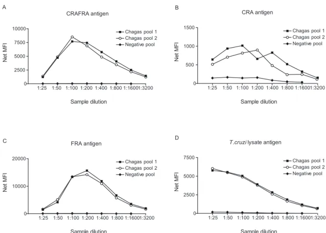
Singleplex assay for Chagas disease diagnosis - De-velopment of a microsphere-based immunoassay for Chagas disease required an initial assessment of the ef-ficiency of antigen coupling to the microspheres and an analysis of the best sample dilution to be used for the assay. For this purpose, we analysed three serum sample pools derived from the AEQ panel: two positive for Cha-gas disease and one negative.
Each Chagas antigen was conjugated to an individual microsphere set. Serial dilutions of pools ranging from 1:25-1:3200 were made and assayed with the standard assay protocol. All analyses were made singly in order to assess the range of fluorescence generated by each individual microsphere set.
Despite the different profiles of each dilution curve, a 1:200 dilution gave optimal signal results for all antigens (Fig. 1). This dilution also allowed a greater degree of discrimination between negative and positive samples, thus it was used in all subsequent experiments.
Singleplex specificity analysis - Once the sample dilution was established, experiments were performed to determine whether the assays were indeed measur-ing antibodies raised against T. cruzi; 20 positive and 20 negative sera for Chagas disease were individually assayed against all antigens.
CRAFRA antigen
1:25 1:50 1:100 1:200 1:400 1:800 1:16001:3200
0 2500 5000 7500 10000
Chagas pool 1 Chagas pool 2 Negative pool
Sample dilution
N
e
t
M
F
I
CRA antigen
1:25 1:50 1:100 1:200 1:400 1:800 1:16001:3200
0 500 1000 1500
Chagas pool 1 Chagas pool 2 Negative pool
Sample dilution
N
e
t
M
F
I
FRA antigen
1:25 1:50 1:100 1:200 1:400 1:800 1:16001:3200
0 10000 20000
Chagas pool 1 Chagas pool 2 Negative pool
Sample dilution
N
e
t
M
F
I
T.cruzi lysate antigen
1:25 1:50 1:100 1:200 1:400 1:800 1:16001:3200
0 2500 5000 7500
Chagas pool 1 Chagas pool 2 Negative pool
Sample dilution
N
e
t
M
F
I
A
C
B
D
Fig. 2 demonstrates a clear separation of negative and positive samples for three of the antigens analysed. The Δsep_pop presented a 50.7, 21.8 and 15.9-fold difference between positive and negative sera populations for CRA-FRA, lysate and FRA antigens, respectively. No false pos-itives were detected for these antigens (100% specificity) at their respective cut-off lines. The CRA antigen, howev-er, presented a lower sensitivity (55%) as it was unable to detect all positive samples; this antigen was consequently excluded from all subsequent tests.
Duplex immunoassay - Two duplexes were tested: one containing two microsphere sets each coupled with CRAFRA or lysate antigens and another duplex con-taining two microsphere sets each coupled with FRA or lysate antigens.
All three antigens in the duplex assays showed com-parable results for positive and negative samples when compared to their singleplex performance (Fig. 3). In-terference between the different microsphere sets was investigated by comparing the net MFIs generated by the singleplex assay with those generated by the duplex assay. Fig. 3 shows that no interference was observed be-tween bead sets. Intensity dispersion of the samples was also maintained in the duplex assays with the exception of the CRAFRA antigen, which presented slightly higher net MFI signals in the duplex assay format (Fig. 3C).
DISCUSSION
Diagnostic tools are important for identification of diseases and can determine their correct treatment. However, current diagnostic tools are costly and not sensitive or specific enough to be widely used in public health programs in developing countries. Efficient di-agnostic tools for such countries must be able to detect a large number of diseases in an inexpensive, fast and precise way (Urdea et al. 2006, O’Meara et al. 2007). A multiplexing tool that can quickly and accurately identify specific antibodies against a certain pathogen is critically needed for both public health improvement and epidemiological surveillance.
Chagas Negative Chagas Negative Chagas Negative Chagas Negative 0
200 400 600 800 5000 10000 15000 20000
CRAFRA CRA FRA Lysate
cut-off
N
e
t
M
F
I
∆sep_pop = 50.7 ∆sep_pop = -2.5 ∆sep_pop = 15.9 ∆sep_pop = 21.8
Fig. 2: specificity analysis for Chagas individual antigens. Net me-dian fluorescence intensity (MFI) obtained by the 1:200 dilution of 20 Chagas positive serum samples and 20 negative sera evaluated in singleplex assays against the antigens CRAFRA, CRA, FRA and
Trypanosoma cruzi lysate.
Chagas Negative Chagas Negative Chagas Negative 0
100 200 300
Singleplex lysate
Duplex lysate + FRA 1000
2000 3000 4000 5000
Duplex lysate + CRAFRA
cut-off sep_pop = 21.8 sep_pop = 63.3 sep_pop = 69.0
N
e
t
M
F
I
Chagas Negative Chagas Negative
0 100 200 800 4000 7200 10400 13600 16800 20000
Single plex FRA
Duplex lysate + FRA
cut-off sep_pop = 15.9 sep_pop = 31.0
N
e
t
M
F
I
Duple x lysate + CRAFRA
Chagas Negative Chagas Negative
0 100 200 300 800 7200 13600 20000 26400
cut-off
Singleplex CRAFRA
N
e
t
M
F
I
sep_pop = 50.7 sep_pop = 46.0
A
B
C
In this study we developed a microsphere-based as-say for Chagas disease using the antigenic properties of CRAFRA and FRA recombinant proteins (Krieger et al. 1992), which are different T. cruzi antigens, as well as a
T. cruzi lysate (Leon et al. 2001).
As the sensitivity of each antigen affects the resulting output, our first approach was to identify the response obtained by each antigen upon comparison with positive and negative serum pools. All antigens presented high net MFI values for the positive pools and lower net MFI values for the negative pool. However, the CRA antigen showed high reactivity with the negative serum pool and low reactivity with the positive serum pool at the given detection threshold. This result, and the fact that the CRAFRA antigen showed better distinction between positive and negative serum pools, reinforces the impor-tance of combining distinct antigens for better detection.
When the CRAFRA, FRA and lysate antigens were compared with individual samples in singleplex format, they were able to clearly differentiate the negative from the positive sera samples, whereas the CRA antigen failed to do so. This was not an unexpected result since CRA is part of the fusion protein CRAFRA, which pre-sented high MFI values and the lowest cut-off even when compared to the results obtained with FRA alone. This result shows that CRA used together with FRA does not interfere with CRAFRA sensitivity. For this reason, CRA antigen was excluded from the subsequent tests.
In order to evaluate multiplex capability, duplex as-says utilising beads coupled with lysate/FRA and lysate/ CRAFRA antigens were performed. Interference be-tween microsphere sets is an important consideration for multiplex assays, but comparison of MFIs generated in the singleplex and duplex assays showed no interference between microsphere sets.
The duplex of lysate/CRAFRA showed the low-est cut-off value and was able to differentiate between negative and positive samples, with only one negative serum sample within the cut-off limit. The lysate/FRA duplex was also able to differentiate between negative and positive samples while maintaining the same profile of sample dispersion, but the same negative sample was detected as a false positive
The duplex assays developed in the present study are sensitive, rapid and show good specificity. Thus, the duplex microsphere assay has the potential to become a viable alternative to standard ELISA, as it requires much smaller sample volumes as well as greatly reduced time and labour spent when assaying a large number of samples. Diagnosis of T. cruzi infection using a mul-tiple antigen strategy is therefore feasible, as mulmul-tiplex technology can be used to diagnose multiple diseases in one single reaction. Also, it allows the screening of dif-ferent physiological manifestations of the host during infection evolution.
Although this was a preliminary study, the pres-ent work showed robust results, especially in the ability of this test to separate the positive and negative sample populations by the obtained Δseq_pop values. The use of a larger sample size with different characteristics such as
low positives as well as different disease samples to ad-dress cross reactivity is important for realisation of the true capability of this multiplex test as a diagnostic tool.
It is important to note that, after publication of the
T. cruzi reference genome (El-Sayed et al. 2005), a wealth of information became available, enabling new approaches for Chagas disease diagnosis. During the search for new markers, all coding sequences identified in the T. cruzi genome were cloned into an appropri-ate vector, resulting in the construction of a collection named ORFeome (Rual et al. 2004). These coding se-quences can be expressed in a high-throughput manner (Luan et al. 2004, Chen et al. 2008) and used for screen-ing patient sera, enablscreen-ing the potential identification, at unprecedented speed, of new markers for diagnosis of Chagas disease. This strategy could also be used to find more specific markers for clinic subtypes, disease evolu-tion and for the evaluaevolu-tion of Chagas disease treatments. The T. cruzi ORFeome is currently under construction in our laboratory and, in the near future, will be used for the abovementioned screenings, as well as for many oth-er applications in diffoth-erent research fields involving T. cruzi biology and pathogenesis. This resource will pres-ent an invaluable toolset for researchers working with T. cruzi and Chagas Disease.
ACKNOWLEDGEMENTS
To Alexandre Dias Tavares, from the Institute of Molec-ular Biology of Paraná, Brazil, for the critical review of the manuscript.
REFERENCES
Brener Z, Andrade ZA, Barral-Neto M 2000. Trypanossoma cruzi e doença de Chagas. Guanabara Koogan, Rio de Janeiro, 431 pp. Burgess DCH, Wasserman J, Dahl CA 2006. Global health
diagnos-tics. Nature 444 (Suppl. 1): 1-2.
Chagas C 1909. Nova tripanozomiase humana. Estudos sobre a mor-folojía e o ciclo evolutivo de Schizotrypanum cruzi n.gen., n.sp., ajente etiolójico de nova entidade morbida do homen. Mem Inst Oswaldo Cruz 1: 159-218.
Chen Y, Qiu S, Luan CH, Luo M 2008. A high throughput platform for eukaryotic genes. Methods Mol Biol426: 209-220.
Cooley G, Etheridge RD, Boehlke C, Bundy B, Weatherly DB, Min-ning T, Haney M, Postan M, Laucella S, Tarleton RL 2008. High throughput selection of effective serodiagnostics for Trypano-soma cruzi infection. PLoS Med2: e316.
Coura JR 2007. Chagas disease: what is known and what is needed - a background article. Mem Inst Oswaldo Cruz 102 (Suppl. I): 113-122.
Dias D, Van Doren J, Schlottmann S, Kelly S, Puchalski D, Ruiz W, Boerckel P, Kessler J, Antonello JM, Green T, Brown M, Smith J, Chirmule N, Barr E, Jansen KU, Esser MT 2005. Optimization and validation of a multiplexed luminex assay to quantify anti-bodies to neutralizing epitopes on human papillomaviruses 6, 11, 16 and 18. Clin Diagn Lab Immunol 12: 959-969.
Darban H, da Silveira JF, de Jong P, Edwards K, Englund PT, Fazelina G, Feldblyum T, Ferella M, Frasch AC, Gull K, Horn D, Hou L, Huang Y, Kindlund E, Klingbeil M, Kluge S, Koo H, Lac-erda D, Levin MJ, Lorenzi H, Louie T, Machado CR, McCulloch R,McKenna A, Mizuno Y, Mottram JC, Nelson S, Ochaya S, Os-oegawa K, Pai G, Parsons M, Pentony M, Pettersson U, Pop M, Ramirez JL, Rinta J, Robertson L, Salzberg SL, Sanchez DO, Seyler A, Sharma R, Shetty J, Simpson AJ, Sisk E, Tammi MT, Tarleton R, Teixeira S, Van Aken S, Vogt C, Ward PN, Wick-stead B, Wortman J, White O, Fraser CM, Stuart KD, Andersson B 2005. The genome sequence of Trypanosoma cruzi, etiologic agent of Chagas disease. Science 309: 409-415.
Hopkins AL, Witty MJ, Nwaka S 2007. Mission possible. Nature 449: 166-169.
Jani IV, Janossy G, Brown DW, Mandy F 2002. Multiplexed immu-noassays by flow cytometry for diagnosis and surveillance of infectious diseases in resource-poor settings. Lancet Infect Dis 2: 243-250.
Joos TO, Stoll D, Templin MF 2002. Miniaturised multiplexed im-munoassays. Curr Opin Chem Biol 6: 76-80.
King DA, Peckham C, Waage JK, Brownlie J, Woolhouse ME 2006. Epidemiology. Infectious diseases: preparing for the future.
Science 313: 1392-1393.
Krieger MA, Almeida E, Oelemann W, Lafaille JJ, Pereira JB, Krieg-er H, Carvalho MR, GoldenbKrieg-erg S 1992. Use of recombinant an-tigens for the accurate immunodiagnosis of Chagas’ disease. Am J Trop Med Hyg 46: 427-434.
Lal G, Balmer P, Joseph H, Dawson M, Borrow R 2004. Development and evaluation of a tetraplex flow cytometric assay for quantita-tion of serum antibodies to Neisseria meningitidis serogroups A, C, Y and W-135. Clin Diagn Lab Immunol 11: 272-279.
Lee SH, Walker DR, Cregan PB, Boerma HR 2004. Comparison of four flow cytometric SNP detection assays and their use in plant improvement. Theor Appl Genet 110: 167-174.
Leon JS, Godsel LM, Wang K, Engman DM 2001. Cardiac myosin autoimmunity in acute Chagas’ heart disease. Infect Immun 69: 5643-5649.
López-Antuñano FJ 1997. Quimioterapia de las infecciones produci-das por Trypanosoma cruzi. Salud Publica Mex 39: 463-471.
Luan CH, Qiu S, Finley JB, Carson M, Gray RJ, Huang W, John-son D, Tsao J, Reboul J, Vaglio P, Hill DE, Vidal M, Delucas LJ, Luo M 2004. High-throughput expression of C. elegans proteins.
Genome Res 14: 2102-2110.
Martins TB, Burlingame R, von Mühlen CA, Jaskowski TD, Litwin CM, Hill HR 2004. Evaluation of multiplexed fluorescent micro-sphere immunoassay for detection of autoantibodies to nuclear antigens. Clin Diagn Lab Immunol 11: 1054-1059.
MSF - Médecins Sans Frontières 2008. Campaign for access to essen-tial medicines. International meeting. New diagnostic tests are urgently needed to treat patients with Chagas disease. Rev Soc Bras Med Trop 41: 315-319.
O’Connell D 2007. Neglected diseases. Nature 449: 157-182. O’Meara WP, Hall BF, McKenzie FE 2007. Malaria vaccine efficacy:
the difficulty of detecting and diagnosing malaria. Malar J 6: 36. Prabhakar U, Eirikis E, Davis HM 2002. Simultaneous
quantifica-tion of proinflammatory cytokines in human plasma using the LabMAP assay. J Immunol Methods 260: 207-218.
Rual JF, Hill DE, Vidal M 2004. ORFeome projects: gateway between genomics and omics. Curr Opin Chem Biol 8: 20-25.
Schmunis GA, Cruz JR 2005. Safety of the blood supply in Latin America. Clin Microbiol Rev 18: 12-29.
TDR 2007. Reporte del grupo de trabajo científico sobre la enferme-dad de Chagas, 2005. Availble from: http://apps.who.int/tdr/pub-Availble from: http://apps.who.int/tdr/pub-lications/tdr-research-publications/reporte-enfermedad-chagas/
pdf/swg_chagas.pdf. [Accessed January, 5th, 2009].
Urdea M, Penny LA, Olmsted SS, Giovanni MY, Kaspar P, Shepherd A, Wilson P, Dahl CA, Buchsbaum S, Moeller G, Hay Burgess DC 2006. Requirements for high impact diagnostics in the devel-oping world. Nature 444 (Suppl. 1): 73-79.
Vignali DAA 2000. Multiplexed particle-based flow cytometric as-says. J Immunol Methods 243: 243-255.
Villa L, Morote S, Bernal O, Bulla D, Albajar-Vinas P 2007. Access to diagnosis and treatment of Chagas disease/infection in en-demic and non-enen-demic countries in the XXI century. Mem Inst Oswaldo Cruz 102 (Suppl. I): 87-94.