Expression of
DLK1
and
MEG3
genes in porcine tissues
during postnatal development
Maria Oczkowicz
1, Agata Piestrzyska-Kajtoch
2, Katarzyna Piórkowska
1, Barbara Rejduch
2and Marian Rózycki
11
Department of Genetics and Animal Breeding, National Research Institute of Animal Production,
Balice, Poland.
2
Department of Immuno and Cytogenetics, National Research Institute of Animal Production, Balice,
Poland.
Abstract
TheDrosophila-like homolog 1 (DLK1), a transmembrane signal protein similar to other members of the Notch/Del-ta/Serrate family, regulates the differentiation process in many types of mammalian cells. Callipyge sheep andDLK1 knockout mice are excellent examples of a fundamental role of the gene encoding DLK1 in muscle growth and fat de-position.DLK1 is located within co-regulated imprinted clusters (the DLK1/DIO3 domain), along with other imprinted genes. Some of these,e.g. the RNA coding MEG3 gene, presumedly interfere with DLK1 transcription. The aim of our study was to analyzeDLK1 and MEG3 gene expression in porcine tissues (muscle, liver, kidney, heart, brain stem) during postnatal development. The highest expression of bothDLK1 and MEG3 variant 1 (MEG3 var.1) was observed in the brain-stem and muscles, whereas that ofMEG3 variant 2 (MEG3var.2) was the most abundant in muscles and the heart. During development (between 60 and 210 days of age) expression of analyzed genes was down-regulated in all the tissues. An exception was the brain- stem, where there was no significant change inMEG3 (both variants) mRNA level, and relatively little decline (2-fold) in that ofDLK1 transcription. This may indicate a dis-tinct function of theDLK1 gene in the brain-stem, when compared with other tissues.
Key words: DLK1, imprinting,MEG3, pigs, polar overdominance.
Received: September 25, 2009; Accepted: June 11, 2010.
TheDrosophila-like homolog 1 (DLK1) is a trans-membrane signal protein similar to other members of the Notch/Delta/Serrate family (Jensenet al., 1994). This pro-tein controls several cell-differentiation processes through-out embryonic and adult life. A role of DLK1 in adi-pogenesis has been well documented (Smas and Sul, 1996; Sul , 2009; Nuedaet al., 2007). Recently, DLK1 was shown to regulate fate of myogenic cells (Andersenet al., 2009) and human skeletal stem cells (Abdallahet al., 2004). A significant role ofDLK1in maintaining proper organism function was demonstrated by generating DLK1 knock-out-mice, which exhibited accelerated obesity, growth dis-orders and skeletal malformation (Moonet al., 2002). Nev-ertheless, knowledge about exact function of DLK1 in particular tissues and organs remains fragmentary.
A gene forDLK1is located in imprinted gene clusters on chromosomes 12, 14 and 7 within the so-called
DLK1/DIO3 domain of mice, humans and pigs,
respec-tively. There are few imprinted genes in the neighborhood ofDLK1, among which the best studied are paternally ex-pressedPEG11/RTL1, maternally expressedMEG3/GTL2
andMEG8. The full-length PEG11 protein was recently identified in callipyge sheep muscle (Byrneet al., 2010). On the other hand,MEG3andMEG8genes produce non-coding RNAs (ncRNA). These play an essential role in growth and differentiation, as the deletion ofMEG3/GTL2, together with its differentially methylated region, induces lethal parent-origin-dependent defects in mice (Takahashi
et al., 2009). The whole domain has been intensively stud-ied in reference to molecular mechanism of imprinting con-trol, but to date no satisfactory model has been proposed for this cluster.
Among farm animals, the most extensive studies of theDLK1/PEG11domain have been undertaken in sheep, mainly in an attempt to identify the Single Nucleotide Poly-morphism (SNPCLPG) responsible for the so called “calli-pyge phenotype” in this region. The calli“calli-pyge phenotype (muscle hypertrophy of the hindquarters) is inherited in a non-Mendelian manner (the so-called polar overdominan-ce), and appears in the offspring only when the mutated
www.sbg.org.br
Send correspondence to Maria Oczkowicz. National Research In-stitute of Animal Production, Department of Genetics and Animal Breeding, Ul. Krakowska 1, 32-083 Balice, Poland. E-mail: [email protected].
allele comes from the father and the wild one from the mother (Cpat/Nmatgenotype) (Cockettet al., 1996). Animals with other genotypes (Cpat/CmatNpat/Nmat, Npat/Cmat) do not exhibit the characteristic phenotype. There is a theory ex-plaining the molecular mechanism underlying the unusual inheritance of callipyge phenotype: CLPG mutation en-hances the level ofDLK1,MEG3,PEG11andMEG8gene expression incis, probably by modifying the activity of a common regulatory element. The callipyge phenotype re-sults from aDLK1and/orPEG11overexpression in skele-tal muscle with simultaneous underexpression ofMEG3
and MEG8. In Cmat/Cpat individuals, overexpression of
MEG8andMEG3or other maternally expressed genes in-terfere in trans withDLK1andPEG11and inhibit expres-sion ofDLK1andPEG11(Charlieret al., 2001; Georgeset al., 2003).
The considerable effect of SNPCLPGon sheep muscu-lature has made DLK1an interesting candidate gene for marker-assisted selection in other farm animals. Unfortu-nately, SNPCLPG appeared to be a private allele, encoun-tered exclusively in callipyge flocks (Smit et al., 2003). Nevertheless, Kim et al. (2004), on identifying a DLK1
polymorphism (silent SNP mutation) associated with growth, fatness and body composition in pigs, proved its polar-overdominant inheritance. These results have been confirmed through QTL (Quantitative Trait Locus) analy-sis, although the molecular mechanism involved has, as yet, not been stated (Liet al., 2008). Recently,gDLK1has been proposed as a novel selection-marker for high muscle growth in chickens, since observedDLK1mRNA expres-sion was greater in the muscles of broilers than in layers (Shinet al., 2009).
In pigs, four differentDLK1and twoMEG3transcripts have been identified, so far (Deiuliiset al., 2006; Liet al., 2008; Samulin et al., 2009). In the pigs, the short form (DLK1C2) is the most abundant transcript in all analyzed tissues (Deiuliiset al., 2006, Samulinet al., 2009).MEG3
expression has recently been observed in the liver, heart, spleen, fat, kidney and skeletal muscle of two-month old pigs. Nevertheless, relative abundance of differentMEG3
variants has not been studied so far (Jiang and Yang, 2009). Since DLK1appears to be an interesting candidate gene for marker-assisted selection, and as little is known about postnatal gene expression within theDLK1/DIO3 do-main in pigs, we decided to analyze relative mRNA abun-dance ofDLK1andMEG3genes in various porcine tissues (muscle, liver, brain, kidney, heart), as well as changes in the expression of these genes during postnatal develop-ment.
Animals for the study were kept in the Pilot Plant of the National Research Institute of Animal Production in Pawlowice under identical housing and feeding conditions. They were divided into 6 age-groups, according to the day of slaughter (60-, 90-, 120-, 150-, 180- and 210-days-old pigs). In the first part of the experiment, two batches each of
the breeds Large White and Duroc, with 4 to 6 pigs of both in each age group, were analyzed separately. As no signifi-cant differences between breeds were found, we decided to create only age groups consisting of 8 to12 animals (4 to 6 Large White and 4-6 Duroc). Animals were related – all pigs within the breed had the same father, and their mothers were sisters. Fragments of muscle(longissimus dorsi)and the liver were collected from animals of all the age groups, whereas fragments of the brain-stem, heart and kidney were collected only from the 60 and 210 days groups. Tissues were collected immediately after slaughter, and kept in liq-uid nitrogen during transportation. All animals were stress-resistant (RYR1 C/C).
Total RNA was extracted using TRI-Reagent (Sigma) and a Silent Crusher S homogenizer (Heidolph), according to the method described by Chomczyski (1993). RNA from the brain-stem was isolated by using the SV Total RNA System (Promega). The amount of extracted RNA was esti-mated by BioPhotometer (Eppendorf), and its quality eval-uated by gel electrophoresis.
The RNA (1mg) was reverse transcribed into cDNA at 37 °C by using a High Capacity cDNA Reverse Tran-scription Kit with random primers (Applied Biosystems), according to the manufacturer’s protocol.
Primers and probes for all the genes were designed with Primer Express software (Applied Biosystems).
MEG3 primers were designed to distinguish alternative isoforms, whereasDLK1primers covered exon-exon junc-tion and amplified all transcripts. The sequences of probes and primers were as follows:DLK1for-5’AGGACGGCT GGGATGGA, rev-5’CGAGGTTCGCGCAGGTT, pro-be-5’TCTGTGACCTAGACATCMEG3 var. 1for-5’GG AAGGGACCTCAGACATGTG, rev-5’CCTAGCCTGC CGATTTCAGA, probe-5’CTCCTGCACCCTCC,MEG3 var. 2 for- 5’TGAGGCTGGAGGAGCGTTAG, rev-5’CCAACTTGGACCCCTTCTCTT, probe-5’ATCTTGT CGCATTGCT.
Relative quantification of expression was performed on a 7500 Real-Time PCR System using labeled TaqMan® Tamra and MGB probes and TaqMan® Universal PCR Master Mix with UNG AmpErase (Applied Biosystems). Reactions, in a total volume of 25mL were done in triplicate and according to the TaqMan® Universal PCR Master Mix protocol. GAPDHwas used as endogenous control to com-pare expression changes ofDLK1andMEG3during devel-opment in muscle and liver.RPL27was used for between-tissue comparison, due to variations inGAPDHexpression among the analyzed tissues. The results were analyzed us-ing Sequence Detection System software v. 2.0 (Applied Biosystems). Statistical analysis was performed using One Way Anova, Tukey test (SAS Institute).
and 210 days) were taken into consideration, whereas for the brain-stem, heart and kidney only two were (60 and 210 days).
The lowest expression ofDLK1andMEG3 var. 1was noted in the liver, and the highest in the brain-stem (DLK1– 3.8-fold higher than in liver,MEG3 var.1- 26-fold higher). An exception was theMEG3 var.2 transcript, which was the most abundant in the muscle (23-fold higher than in liver). In the muscle, abundance of other transcripts was also relatively high (DLK1- 3-fold higher than in liver,
MEG3 var. 1– 14-fold higher). The level of expression was intermediate in the heart and kidneys, when compared with other tissues, althoughMEG3transcripts were more abun-dant in the heart than in the kidney, whereas inDLK1 tran-scripts this was to the contrary (Figure 1).
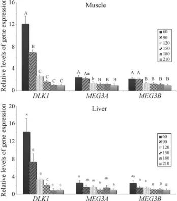
We also analyzed alteration in the expression of these genes during postnatal development. In the brain stem, kid-ney and heart DLK1 andMEG3 mRNA abundance was compared between 60 and 210 days of age, while in the muscle and liver additional developmental periods were analyzed (90, 120, 150 and 180 days of age). The expres-sion level of all transcripts decreased with age in all the tis-sues investigated (Figures 1 and 2), with the exception of
MEG3(both variants) in the brain, where there was no sig-nificant change. DLK1 gene expression declined much more intensively (2-fold in the brain between 60 and 210 days, 8-fold in the heart, 14-fold in the liver and 12-fold in the muscle) thanMEG3(both transcripts, 1.4-, 2.6-, 2,2-fold higher in the respective tissues). In the brain-stem, ex-pression of the analyzed genes seemed to be more stable during aging, when compared to the other analyzed tissues (Figure 1). In the muscle, where more developmental stages were analyzed, no fluctuation during the 90-180-days interval was observed.DLK1 expression decreased gradually, whereas that ofMEG3remained similar at 60 and 90 days to afterwards enter in sharp decline (Figure 2a). The pattern of DLK1 expression in the liver was similar to
that observed in the muscle, whereas that ofMEG3 was more pronounced only at 60 days (Figure 2b).
Previous studies have shown that in humans,DLK1
gene expression is high during prenatal development, whereas after birth this is restricted to hormone-secreting cells and monoaminergic neurons in the central nervous system (Abdallahet al., 2007). In contrast, postnatalDLK1
expression in pigs was observed in the kidneys, heart, spleen, fat and muscles (Deiuliiset al., 2006), as confirmed herein. Moreover, high mRNA abundance ofDLK1 and
MEG3 in the brain-stem was detected, with the MEG3 var.1being the predominant variant of the latter. To date,
DLK1expression has not been studied in the brain-stem of farm animals, although in adult mice, high DLK1 and
MEG3expression has been observed in certain regions of the brain,e.g.hypothalamus, medulla and cerebral cortex (www.brain-map.org) (Labialle et al., 2008). A semi-quantitative analysis of tissue-specific expression of por-cineMEG3 gene has been performed previously by RT-PCR (Jiang and Yang, 2009).The authors noted the highest expression of this gene in the brain and lungs, followed by the tongue, spleen, stomach, fat, kidneys, liver, skeletal muscles, heart and small intestine. Contrarily, our results suggest a much higher expression ofMEG3(both variants) in skeletal muscles than in the liver or kidneys. However, the results are difficult to compare due to the multiple isoforms of theMEG3gene.
Figure 1-DLK1,MEG3var.1 andMEG3var. 2 gene expession in various porcine tissues at 60 and 210 days after birth. Values are presented as mean±SEM. Asterisks indicate significant differences between 60 and 210 days for each gene. *p < 0.05, **p < 0.01.
Figure 2-DLK1,MEG3var.1 andMEG3var. 2 gene expression in the
Declining expression of eleven imprinted genes (in-cludingDLK1andMEG3) during postnatal development in multiple tissues (heart, lung and kidney) of mice at 1, 4 and 8 weeks of age has been previously reported (Luiet al., 2008). The authors inferred that the down-regulation of im-printed genes contributes to a deceleration in organ growth. Recently DLK1has been shown to regulate growth hor-mone (GH)expression (Ansellet al., 2007). This, as well as our results, strongly supports the hypothesis for the kidney, liver and heart. In the present study, the decrease inDLK1
gene expression was also very pronounced in muscles. This may reflect a deceleration in muscle-mass growth. In con-trast, the decline ofDLK1transcript abundance in the brain was relatively low (2-fold), with no significant change in
MEG3 transcripts, thereby implying the maintenance of high expression levels even at 210 days. This may indicate a distinct function of theDLK1gene in the brain-stem.
Although the biological consequences remain un-known, it is evident that expression of theDLK1/DIO3 do-main in pigs, as in mice, is regulated in a tissue-specific manner. A full understanding of the complex mechanism involved in gene expression control in theDLK1/DIO3 do-main, requires an analysis of the imprinting status of genes in various tissues during development.DLK1is generally considered as a paternally expressed gene andMEG3as a maternally expressed one. An increasing number of experi-ments, however, imply that the imprinting status of many genes, besides changing during development, differs among tissues (Khatib, 2007). Further studies of expression and the methylation status of this important domain in a wider range of tissues and developmental stages are plan-ned in the future.
Acknowledgments
This work was supported by the National Research Institute for Animal Production. Statutory activity No. 1130.1.
References
Abdallah BM, Ding M, Jensen CH, Ditzel N, Flyvbjerg A, Jensen T, Dagnaes-Hansen F, Gasser JA and Kassem M (2007) Dlk1/FA1 is a novel endocrine regulator of bone and fat mass and its serum level is modulated by growth hormone. Endocrinology 148:3111-3121.
Abdallah BM, Jensen CH, Gutierrez G, Leslie RG, Jensen TG and Kassem M (2004) Regulation of human skeletal stem cells differentiation by Dlk1/Pref-1. J Bone Miner Res 19:841-852.
Andersen DC, Petersson SJ, Jørgensen LH, Bollen P, Jensen PB, Teisner BD, Schroeder H and Jensen CH (2009) Character-ization of DLK1+ cells emerging during skeletal muscle re-modeling in response to myositis, myopathies, and acute in-jury. Stem Cells 27:898-908.
Ansell PJ, Zhou Y, Schjeide BM, Kerner A, Zhao J, Zhang X and Klibanski A (2007) Regulation of growth hormone
expres-sion by Delta-like protein 1 (Dlk1). Mol Cell Endocr 271:55-63.
Byrne K, Colgrave ML, Vuocolo T, Pearson R, Bidwell CA, Cockett NE, Lynn DJ, Fleming-Waddell JN and Tellam RL (2010) The imprinted retrotransposon-like gene PEG11 (RTL1) is expressed as a full-length protein in skeletal mus-cle from Callipyge sheep. PLoS One 5:e8638.
Charlier C, Segers K, Karim L, Shay T, Gyapay G, Cockett N and Georges M (2001) The callipyge mutation enhances the ex-pression of coregulated imprinted genes in cis without af-fecting their imprinting status. Nat Genet 27:367-369. Chomczynski P (1993) A reagent for the single-step simultaneous
isolation of RNA, DNA and proteins from cell and tissue samples. Biotechniques 15:532-537.
Cockett NE, Jackson SP, Shay TL, Farnir F, Berghmans S, Snow-der GD, Nielsen DM and Georges M (1996) Polar overdo-minance at the ovine callipyge locus. Science 273:236-238. Deiuliis JA, Li B, Lyvers-Peffer PA, Moeller SJ and Lee K (2006)
Alternative splicing of delta-like 1 homolog (DLK1) in the pig and human. Comp Biochem Physiol B 145:50-59. Georges M, Charlier C and Cockett N (2003) The callipyge locus:
Evidence for the trans interaction of reciprocally imprinted genes. Trends Genet 19:248-252.
Jensen CH, Krogh TN, Højrup P, Clausen PP, Skjødt K, Larsson LI, Enghild JJ and Teisner B (1994) Protein structure of fetal antigen 1 (FA1). A novel circulating human epidermal-growth-factor-like protein expressed in neuroendocrine tu-mors and its relation to the gene products of dlk and pG2. Eur J Biochem 225:83-92.
Jiang C and Yang Z (2009) Characterization, imprinting status and tissue distribution of porcine GTL2 gene. Agric Sci China 8:216-222.
Khatib H (2007) Is it genomic imprinting or preferential expres-sion? Bioessays 29:1022-1028.
Kim KS, Kim JJ, Dekkers JC and Rothschild MF (2004) Polar overdominant inheritance of a DLK1 polymorphism is asso-ciated with growth and fatness in pigs. Mamm Genome 15:552-559.
Labialle S, Yang L, Ruan X, Villemain A, Schmidt JV, Hernandez A, Wiltshire T, Cermakian N and Naumova AK (2008) Co-ordinated diurnal regulation of genes from the Dlk1-Dio3 imprinted domain: Implications for regulation of clusters of non-paralogous genes. Hum Mol Genet 17:15-26.
Li XP, Do KT, Kim JJ, Huang J, Zhao SH, Lee Y, Rothschild MF, Lee CK and Kim KS (2008) Molecular characteristics of the porcine DLK1 and MEG3 genes. Anim Genet 39:189-192. Lui JC, Finkielstain GP, Barnes KM and Baron J (2008) An
im-printed gene network that controls mammalian somatic growth is down-regulated during postnatal growth decelera-tion in multiple organs. Am J Physiol – Regul Integr Comp Physiol 295:R189-196.
Moon YS, Smas CM, Lee K, Villena JA, Kim KH, Yun EJ and Sul HS (2002) Mice lacking paternally expressed Pref-1/Dlk1 display growth retardation and accelerated adiposity. Mol Cell Biol 22:5585-5592.
Nueda ML, Baladrón V, Sánchez-Solana B, Ballesteros MA and Laborda J (2007) The EGF-like protein dlk1 inhibits notch signaling and potentiates adipogenesis of mesenchymal cells. J Mol Biol 367:1281-1293.
adi-pocyte development in vitro and in vivo. Anim Genet 40:239-241.
Shin J, Velleman SG, Latshaw JD, Wick MP, Suh Y and Lee K (2009) The ontogeny of delta-like protein 1 messenger ribo-nucleic acid expression during muscle development and re-generation: Comparison of broiler and Leghorn chickens. Poultry Sci 88:1427-1437.
Smas CM and Sul HS (1996) Characterization of Pref-1 and its in-hibitory role in adipocyte differentiation. Int J Obesity Rel Metabol Disorders 20(Suppl 3):65-72.
Smit M, Segers K, Carrascosa LG, Shay T, Baraldi F, Gyapay G, Snowder G, Georges M, Cockett N and Charlier C (2003) Mosaicism of solid gold supports the causality of a
non-coding A-to-G transition in the determinism of the callipyge phenotype. Genetics 163:453-456.
Sul HS (2009) Minireview: Pref-1: Role in adipogenesis and mesenchymal cell fate. Mol Endocrinol 11:1717-1725. Takahashi N, Okamoto A, Kobayashi R, Shirai M, Obata Y,
Ogawa H, Sotomaru Y and KonoT (2009) Deletion of Gtl2, imprinted non-coding RNA, with its differentially methyl-ated region induces lethal parent-origin-dependent defects in mice. Hum Mol Genet 18:1879-1888.
Associate Editor: Ricardo Guelerman P. Ramos