Genotoxicity of
Nicotiana tabacum
leaves on
Helix aspersa
Fernanda R. da Silva
1, Bernardo Erdtmann
1, Tiago Dalpiaz
2, Emilene Nunes
2, Alexandre Ferraz
2,
Tales L.C. Martins
3, Johny F. Dias
4, Darlan P. da Rosa
5, Marilene Porawskie
5,6, Silvia Bona
5and Juliana da Silva
21
Programa de Pós-Graduação em Genética e Biologia Molecular,
Universidade Federal do Rio Grande do Sul, Porto Alegre, RS, Brazil.
2Laboratory of Genetic Toxicology, Programa de Pós-graduação em Biologia Molecular
e Celular Aplicada à Saúde, Universidade Luterana do Brasil, Canoas, RS, Brazil.
3Universidade Federal do Pampa, Campus Bagé, Bagé, RS, Brazil.
4
School of Physics, Universidade Federal do Rio Grande do Sul, Porto Alegre, RS, Brazil.
5Laboratory of Experimental Hepatology, Teaching Hospital of Porto Alegre, Porto Alegre, RS, Brazil.
6Faculty of Biosciences, Pontificial Catholic University of Rio Grande do Sul, Porto Alegre, RS, Brazil.
Abstract
Tobacco farmers are routinely exposed to complex mixtures of inorganic and organic chemicals present in tobacco leaves. In this study, we examined the genotoxicity of tobacco leaves in the snailHelix aspersa as a measure of the risk to human health. DNA damage was evaluated using the micronucleus test and the Comet assay and the concen-tration of cytochrome P450 enzymes was estimated. Two groups of snails were studied: one fed on tobacco leaves and one fed on lettuce (Lactuca sativa L) leaves (control group). All of the snails received leaves (tobacco and lettuce leaves were the only food provided) and waterad libitum. Hemolymph cells were collected after 0, 24, 48 and 72 h. The Comet assay and micronucleus test showed that exposure to tobacco leaves for different periods of time caused significant DNA damage. Inhibition of cytochrome P450 enzymes occurred only in the tobacco group. Chemical anal-ysis indicated the presence of the alkaloid nicotine, coumarins, saponins, flavonoids and various metals. These re-sults show that tobacco leaves are genotoxic inH. aspersa and inhibit cytochrome P450 activity, probably through the action of the complex chemical mixture present in the plant.
Keywords: comet assay, genotoxicity, micronucleus test,Helix aspersa,Nicotiana tabacum, tobacco leaves.
Received: November 30, 2012; Accepted: March 12, 2013.
Introduction
Brazil is the world’s second largest producer of to-bacco (Nicotiana tabacum) leaves after China and accounts for 14.1% of the global production. The southern Brazilian state of Rio Grande do Sul (RS) is the largest tobacco pro-ducer in the country. More than 320,000 tons are produced annually, with over 906,000 workers employed in direct farming and 40,000 jobs in cigarette factories (AFUBRA, 2012).
Tobacco farmers are routinely exposed to the com-plex mixture of chemicals present in tobacco leaves, in ad-dition to organic and inorganic pesticides used to treat to-bacco plants. Toto-bacco leaves contain an unusually high number of different chemical compounds, such as nicotine,
nicotianine and malic acid (Hinds, 2012). These farmers are at risk of Green Tobacco Sickness (GTS), a disease caused by the dermal absorption of nicotine from wet to-bacco leaves. The signs and symptoms of GTS include nau-sea, pallor, chills, vomiting, headache, difficulty in breath-ing, abdominal pain, diarrhea, loss of appetite, runny eyes, blurred vision, weakness, prostration and dizziness, and, occasionally, fluctuations in blood pressure or heart rate (Onukiet al., 2003; Parikhet al., 2005).
Nicotine is the most important alkaloid present in to-bacco. The quantity of nicotine in dried tobacco leaves var-ies from 4% to 8% (Hinds, 2012; IPCS-INCHEM, 2012). During harvest time, farm workers may be exposed to up to 600 mL of dew or rain on the tobacco plants, which roughly corresponds to the nicotine content of 36 cigarettes (NIOSH, 2012). There have been few studies on the genotoxicity of nicotine and the findings are contradictory. Some authors have reported that nicotine and its four major metabolites are not genotoxic when assayed in the Ames www.sbg.org.br
Send correspondence to Juliana da Silva. Programa de Pós-Graduação em Biologia Molecular e Celular Aplicada à Saúde, Universidade Luterana do Brasil, Av. Farroupilha 8001, Prédio 22, Quarto andar, 92425-900 Canoas, RS, Brazil. E-mail: juliana.silva@ulbra.br.
test or other bacterial indicator assays (Doolittle et al., 1995), whereas others have observed that tobacco induces DNA alterations in mammalian cells (Munzner and Ren-ner, 1989; Trivediet al., 1990).
In contrast to other techniques, the study of bioindicator organisms can reveal the biologic impact of xenobiotics. Among terrestrial invertebrates, the snail He-lix aspersacan accumulate different classes of chemicals and therefore serve as a pertinent species for monitoring trace metals and agrochemicals (Snyman et al., 2000; Beeby and Richmond, 2002) as well as plant extracts (Pereira et al., 2009). Xenobiotics accumulated through different routes are transported by blood cells to the diges-tive gland, which is also the main target organ for metabolic and detoxification processes (Beeby and Richmond, 2002).
In this study, we adapted the micronucleus test to in-vestigate the response ofH. aspersato exposure to tobacco leaves as an indicator of the risk to human health. DNA damage was assessed with the micronucleus test (MN) and the Comet assay in hemolymph cells, and the inhibition of cytochrome P450 enzyme activity was also examined. In addition, trace element analysis, phytochemical screening and nicotine quantification were also done.
Materials and Methods
Plant material
Nicotiana tabacumleaves were collected on a farm in the south-central region of Rio Grande do Sul State, Brazil. Only tobacco leaves without pesticides were sampled. The leaves were packed and stored in a freezer at -30 °C.
Snails
Adult individuals (n = 20) of the land snailH. aspersa
(8.96 + 1.62 g; mean+ SD) were obtained from a snail
breeder at the Lutheran University of Brazil, Canoas, RS, Brazil. The snails were acclimatized in a cage at 22+3 °C for seven days, during which period they received leaves from organically grown lettuce (Lactuca sativaL.) and wa-terad libitum. After acclimatization, the snails were as-signed to control and test groups.
Treatments and hemolymph sampling
The study involved two groups of snails: the first was fed tobacco leaves and the second was fed lettuce leaves (control group) (n = 10 each). All snails received tobacco or lettuce leaves (the only food provided) and waterad libi-tum. Hemolymph samples for the Comet assay were ob-tained from the snails 0 h, 24 h, 48 h and 72 h after the be-ginning of feeding each type of leaf, and after 72 h for the micronucleus test. The snails were weighed every day throughout the experiment. The hemolymph samples were collected using a syringe containing heparin and processed within 30 min of collection. At the end of the 72 h period, all snails were killed and stored in a freezer at -30 °C for
trace element analysis and determination of cytochrome P450 activity.
Comet assay
The alkaline Comet assay was done as described by Singh et al. (1998) with several modifications for hemolymph (Ianistcki et al., 2009). Images of 100 ran-domly selected cells (50 cells from each of two replicate slides) were analyzed for each individual. To calculate a damage index (DI), cells were visually classified into one of five classes based on tail size (0 = no tails and 4 = maxi-mum-length tails) as assessed by light microscopy. This classification resulted in a single DNA damage score for each individual and consequently for each group studied. The DI for each individual could range from 0 (completely undamaged = 100 cells x 0) to 400 (maximum damage = 100 cells x 4). The damage frequency (DF, in %) was calcu-lated for each sample based on the number of cells with tails
vs.those without tails (Heuseret al., 2002). All sides were coded for blind analysis.
Micronucleus test (MN)
The MN test was done on hemolymph as a second monitoring system for comparison with the alkaline Comet assay. The MN test was adapted to the characteristics of the test organism based on a protocol described for
Limnoperna fortunei, a mussel species (Villela et al., 2007). A syringe containing 0.5 mL of freshly prepared methanol:acetic acid (1:1, v/v) was used to collect the same volume (0.5 mL) of hemolymph. The syringe was briefly placed on ice (7-10 min) and the hemolymph samples then smeared onto microscope slides, fixed in methanol for 10 min, air-dried and stained for 7 min with a mixture of 10 mL Giemsa and 90 mL 0.2 M phosphate buffer (pH 5.8) prior to examination by light microscopy. Two thousand cells in two hemolymph smears from each snail were exam-ined for the presence or absence of micronuclei. All sides were coded for blind analysis.
Enzyme analysis
For microsome preparation the tissue (digestive gland) was homogenized in an Ultra-Turrax homogenizer for 15 s in a tissue:buffer (0.1 M potassium phosphate, pH 7.5, 0.15 M KCl, 10 mM EDTA and 0.1 mM phenylmethylsufonyl fluoride) ratio of 1:5 (w/v). The ho-mogenate was centrifuged at 12,000 xgfor 20 min and the supernatant then centrifuged twice at 105,000 xg(60 min each). The resulting microsome fraction was resuspended in buffer (0.2 M potassium phosphate, pH 7.5, 0.15 M KCl, 10 mM EDTA, 0.1 mM phenylmethylsufonyl fluoride and 20% glycerol) at a tissue:buffer ratio of 1:3 and then stored at -70 °C (Vrolijket al., 1994).
The microsomal protein concentrations (expressed in mg/mL) were determined as described by Lowry et al.
samples were assayed in duplicate and the final absorbance was read at 625 nm in a spectrophotometer.
For the spectral characterization of cytochrome b5,
P450 and P420: the microsomal suspension was diluted to 1 mg/mL and sodium dithionite crystals were added for analysis of cytochrome b5. The samples were read against a
suspension of microsomes. Cytochromes P450 and P420 were identified based on the compound formed by the inter-action of reduced microsomes with sodium dithionite and 80 mL of carbon monoxide. This mixture was analyzed
against a microsomal suspension reduced with sodium di-thionite (Omura and Sato, 1964). The measurements were done in a Beckman DU70 spectrophotometer at 400-700 nm.
Chemical analysis of tobacco leaves
Tobacco leaves were oven-dried for 4 h at 60 °C and then were ground to a fine powder. The tobacco leaf pow-der was used to prepare pellets for trace element analysis using the PIXE (Particle-Induced X-ray Emission) tech-nique and phytochemical screening. Nicotine was quanti-fied by HLPC.
For the PIXE analysis, the pellets were placed in the target holder inside the reaction chamber. During the exper-iments, the pressure inside the reaction chamber was ~10-5mbar. The experiments were carried out at the Ion Im-plantation Laboratory of the Institute of Physics, Federal University of Rio Grande do Sul (IF-UFRGS). A 3 MV Tandetron accelerator provided a 2 MeV proton beam with an average current of 5 nA at the target. The X-rays pro-duced in the samples were detected by a germanium (Ge) detector with an energy resolution of about 180 eV in 5.9 keV with high efficiency between 3 and 100 keV. The spectra were analyzed with the GUPIX software package and the amounts were expressed in ppm (Campbellet al., 2000).
The phytochemical profile ofN. tabacumleaves was determined as described by Harborne (1998). The method consists of several procedures for the detection of flavonoids, tannins, anthraquinones, alkaloids, saponins, coumarins and cardiac glycosides. The confirmatory thin layer chromatography analysis was done using the systems and developers indicated by Wagner and Bladt (1996).
The nicotine dosage in tobacco leaves extracted with water was determined by HPLC. Standard nicotine was purchased from Sigma (lot no. 093K4121). Analysis was done on a C18 reverse-phase column (cartridge: 5 mm,
250 x 4.6 mm). The separation was done at 35 °C. The isocratic mobile phase used was a mixture of aqueous phase (phosphate buffer, pH 6.8, and methanol at a ratio of 35:65, v/v). The solution was sonicated before use and the flow rate was 1.2 mL/min. The HPLC method used was based on work by Tambwekaret al.(2003). A stock solution of nico-tine (1 mg/mL) was prepared by dissolving 100 mg in 100 mL of phosphate buffer (pH 6.8). Various dilutions
(2.5-50mg/mL; five points per curve) were prepared the
ar-eas under the peaks were determined for each concentration (assayed in quadruplicate) and used to construct the stan-dard curve. The retention time of nicotine was ~3.93 min. The assay was linear within the expected concentration range, indicating its suitability for this analysis. The equa-tion of the regression curve was y = 0.02475 + 0.00005x and r2was 0.996. Tobacco leaves (65 g) were washed with 70 mL of distilled water and the resulting solution was lyophilized and yielded 705 mg of residue. The crude ex-tract (100 mg; labeled as solution A) was dissolved in 100 mL of phosphate buffer (pH 6.8) and a 20mL aliquot
was injected (in triplicate) and eluted with the mobile phase (10 mM phosphate buffer (pH 6.8):methanol, 35:65 v/v). No interfering substances were detected in the chroma-togram.
Statistical analysis
The normality of the data was evaluated using the Kolmogorov-Smirnov test. Statistical comparisons be-tween the groups were done using Student’s two-tailed
t-test. Differences between exposure times were analyzed using the non-parametric two-tailed Kruskal-Wallis test followed by Dunns test for multiple comparisons. The criti-cal level for rejection of the null hypothesis was p < 0.05.
Results
Table 1 summarizes the Comet assay data expressed as Damage index and Damage frequency for H. aspersa
hemolymph cells exposed to tobacco leaves compared to cells from control snails. Control snails (fed lettuce leaves) showed no significant variation throughout the experiment, although there was a slight increase in DNA damage after 48 h and 72 h. There was also no difference between the groups (control and tobacco leaves) at 0 h. However, snails fed tobacco leaves had higher values than the control group at all subsequent times. When analyzed by the Kruskal-Wallis test, the mean DI and DF for snail hemolymph cells after 24, 48 and 72 h of exposure were significantly higher than those of the corresponding controls and the values at 0 h (see Table 1 for p values).
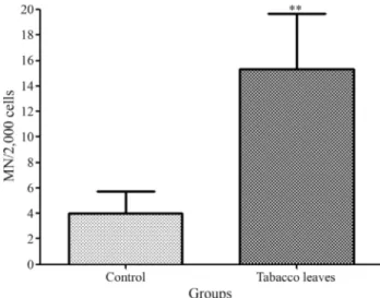
Figure 1 shows the micronucleus test (MN) results for hemolymph cells after 72 h. Cells from snails fed tobacco leaves had a significantly higher MN value than the control group (Student’st-test). Signs of toxicity such as swelling and death were observed in some snails at the end of the 72 h treatment period.
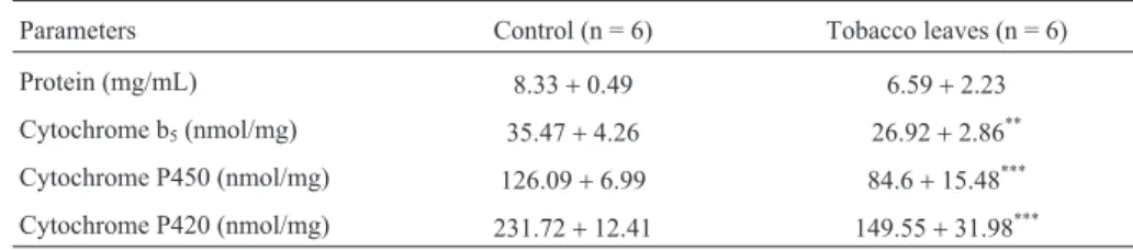
Table 2 shows that 11 trace elements were detected in the tobacco leaf samples analyzed using PIXE. There was no significant difference in the protein content of the diges-tive glands of snails in the two groups, but there was a sig-nificant reduction in the cytochrome b5, P450 and P420
The nicotine concentration in the extract was 22.36 mg/mL (solution A). The solution obtained from
washed tobacco leaves was lyophilized and yielded 705 mg of residue. Extraction of 65 g of this residue with 70 mL of water yielded 15.76 mg of nicotine, which corresponded to ~0.02% of nicotine per leaf. Phytochemical analysis of to-bacco leaves revealed the presence of coumarins, saponins traces, alkaloids and flavonoids.
Discussion
Substances present in tobacco form a complex mix-ture of organic and inorganic compounds that may interact to produce additive, synergistic or antagonistic effects (Fent, 2003). In the present study, the results of the Comet assay and MN test (Table 1 and Figure 1) of hemolymph from snails (H. aspersa) fed tobacco leaves were signifi-cantly higher than those of the control group (fed lettuce). The application of the Comet assay in these snails has been shown to be an inexpensive, effective and sensitive method for assessing the genotoxicity of chemical mixtures
(Ianistckiet al., 2009). The Comet assay detects recent le-sions that can be repaired, such as breaks and alkali-labile sites, while the MN test detects non-repairable damage, such as clastogenic and aneugenic lesions (Villelaet al., 2007). Our findings show that the compounds present in to-bacco leaves were genotoxic and mutagenic inH. aspersa. The slight time-dependent increase in DNA damage (seen in the Comet assay) in untreated snails can be explained by animal stress. Other reports have described similar results for controls (Villelaet al., 2007). Since the frequency of MN seen here in the control group was within an acceptable range, our results can be considered reliable.
The phytochemical analysis revealed the presence of alkaloids in tobacco leaves. Hinds (2012) stated that nico-tine is the main tobacco alkaloid. Using HPLC, we found a nicotine content of ~0.02% per leaf. This lower value of nicotine compared to that reported by others
(IPCS-Table 1- Damage index and damage frequency (Comet assay) in hemolymph of snails exposed to tobacco leaves or lettuce leaves (control group).
Exposure time (h) Exposure group
Control Tobacco leaves
Damage index Damage frequency Damage index Damage frequency 0 56.80+34.53 29.90+14.16 60.00+22.98 29.00+10.88 24 53.60+36.76 28.30+17.34 166.80+45.29**, a 73.90
+14.22**, a
48 96.60+41.18 44.50+18.11 207.60+48.52**, a 83.00+16.05**, a 72 97.50+38.55 45.60+17.78 224.10+42.95***, a 88.40+7.32***, a
The values are the mean+SD of 10 snails per group.**p < 0.01 and *p < 0.001 compared to the corresponding control parameter (Studentst-test). a
p < 0.05 (24 h) and p < 0.001 (48 h and 72 h) compared to 0 h (Kruskal-Wallis test).
Figure 1- Detection of micronuclei (MN) in hemolymph cells ofHelix aspersaexposed to tobacco leaves. The columns represent the mean+SD
determined in 10 snails of each group. *p < 0.01 compared to snails fed let-tuce leaves (control) (Studentst-test).
Table 2- Content of trace elements (ppm) in tobacco leaves.
Trace element Tobacco leaves Lettuce leavesa Na 458965+38828 995.70
K 33449+508 0.11
Mg 4138+224 15.31
P 1659+94 0.03
S 1822+75 0.01
Cl 3425+112 0.01
Al 1272+342 0.90
Si 1470+90 1.95
Ti 140+10 2.47x10⁻⁴
Ca ND 0.07
Fe ND 1.32x10⁻³
Cu ND 7.25x10⁻⁵
Mn 85+5 4.60x10⁻⁵
Zn 31+4 1.35x10⁻⁴
Values are the mean+SD of 3 determinations.aData from Leffaet al.
INCHEM, 2012) can be explained by the extraction method used. The highest frequency of damage was observed after 72 h, although a similar extent of damage was observed af-ter 24 h. Argentin and Cichetti (2004) applied the MN test to cultured mammalian cells treated with nicotine and ob-served rapid induction of DNA damage in samples treated for up to 72 h, with maximum levels observed after 24 h of exposure. Nicotine caused a concentration-dependent in-crease in DNA fragmentation, as assessed by the Comet as-say, in mini-organ cultures of human nasal epithelium, indicating a direct genotoxic effect (Sassenet al., 2005); similar findings have been reported for lymphatic tissue of palatine tonsils and peripheral lymphocytes (Kleinsasseret al., 2005). Nicotine has also been implicated in free radical generation in various types of rodent and human cells, with a direct relationship between reactive oxygen species (ROS) induction and DNA damage (Yildizet al., 1999; Argentin and Cichetti, 2004; Da Silvaet al., 2010).
Another chemical group present in tobacco leaves is coumarins. Gasparotto Jr et al. (2005) showed that coumarins have significant molluscicidal activity against
Biomphalaria glabrata. Our results confirmed the presence of coumarins and we believe that this chemical group ac-counts for the toxicity inH. aspersaafter 72 h, when 50% of the snails exposed to tobacco leaves died (data not shown). Overall, coumarins are not genotoxic, and both negative and positive responses have been reported for these compounds in the Ames test, sister chromatid ex-change (SCE), MN and chromosome aberration tests in mammalian cells (Lake, 1999).
Flavonoids are phenolic compounds that are widely distributed in all foods of plant origin. Several beneficial properties have been attributed to these compounds, includ-ing antioxidant, anti-inflammatory and anticarcinogenic ef-fects (Da Silva et al., 2002; Galati and O’Brien, 2004; Nuneset al., 2011). Flavonoids can be potentially harmful since some are mutagenic in bacterial and mammalian test systems (MacGregor, 1986; Skibola and Smith, 2000; Da Silva et al., 2002). Nevertheless, the data on flavonoid genotoxicity are incomplete and misleading results have been obtained with respect to mutagenicity in tests using mammalian cells (Skibola and Smith, 2000).
Our phytochemical analysis revealed only traces of saponins in tobacco leaves. This chemical group has
anti-genotoxic and anti-mutagenic (Scarpatoet al., 1998), anti-tumoral (Leeet al., 1999) and anti-inflammatory (Navarro
et al., 2001) activities, and probably did not increase the DNA damage observed in snails fed tobacco leaves.
PIXE analysis revealed different levels of inorganic elements in tobacco leaves when compared with lettuce leaves (Table 2). The levels of trace elements in lettuce were obtained in a previous study by our group. All of the inorganic elements detected were present in much higher levels in tobacco leaves than in lettuce leaves. This differ-ence reflects the fact that the two plants are different spe-cies. Studies in humans and animals have demonstrated that a wide variety of metals act as mutagenic and carcinogenic agents (Leonard et al., 2004). In general, metal geno-toxicity is caused by indirect mechanisms, a major one of which involves interference with cellular redox regulation and the induction of oxidative stress (ROS generation), which may cause oxidative DNA damage (Beyersmann and Hartwig, 2008).
When digestive gland proteins and enzymes were an-alyzed (Table 2), the cytochrome P450 and P420 activities were lower in snails fed tobacco leaves compared with the control group. This decrease probably reflected the fact that nicotine and flavonoids inhibit cytochrome P450 activity, as observed by Shaoet al.(2009) and Galati and O’Brien (2004). Galati and O’Brien (2004) also demonstrated that flavonoids inhibit drug-metabolizing enzymes. In addition, P450 isoforms are responsive to induction and inhibition by xenobiotics, including plant constituents (Ioannides, 2002; Ueng and Chen, 2004). Thus, inhibition of cytochrome en-zymes inH. aspersafed tobacco leaves may indicate that these snails can accumulate genotoxic agents capable of causing DNA damage. As indicated by Kimet al.(2002), the inhibition of cytochrome activity may disturb endocrine systems and lead to adverse effects such as carcinogenicity, immunological dysfunction and reproductive abnormality. In conclusion, our results demonstrate that tobacco leaves are genotoxic in H. aspersa. The genotoxicity, mutagenicity and enzymatic inhibition caused by exposure to tobacco leaves was probably mediated by the complex mixture of substances (nicotine, coumarins, traces of sapo-nins, flavonoids and different inorganic elements) present in these leaves. Our findings confirm the sensitivity of the Comet assay and MN test in detecting damage caused by
Table 3- Protein content and cytochrome activities of digestive glands fromHelix aspersaindividuals exposed to lettuce leaves (control) or tobacco leaves.
Parameters Control (n = 6) Tobacco leaves (n = 6) Protein (mg/mL) 8.33+0.49 6.59+2.23 Cytochrome b5(nmol/mg) 35.47+4.26 26.92+2.86** Cytochrome P450 (nmol/mg) 126.09+6.99 84.6+15.48*** Cytochrome P420 (nmol/mg) 231.72+12.41 149.55+31.98***
complex mixtures such as that present in tobacco leaves. Several biological markers have been proposed for assess-ing exposure to tobacco or tobacco smoke in order to eluci-date the mechanisms of DNA damage. In this work, we attempted to interpret the biological action of complex mix-tures of components present in tobacco leaves. This ap-proach may be useful for studying complex mixtures in other plant species.
Acknowledgments
The authors thank the farmer who granted us permis-sion to collect tobacco leaves on his farm. This work was supported by grants from the Coordenadoria de Aperfei-çoamento de Pessoal de Nível Superior (CAPES), Conse-lho Nacional de Desenvolvimento Científico e Tecnológico (CNPq) and Fundação de Amparo à Pesquisa do Estado do Rio Grande do Sul (FAPERGS).
References
Argentin G and Cichetti R (2004) Genotoxic and antiapoptotic ef-fect of nicotine on human gingival fibroblasts. Toxicol Sci 79:75-81.
Beeby A and Richmond L (2002) EvaluatingHelix aspersaas a sentinel for mapping metal pollution. Ecol Indic 1:261-270. Beyersmann D and Hartwig A (2008) Carcinogenic metal
com-pounds: Recent insight into molecular and cellular mecha-nisms. Arch Toxicol 82:493-512.
Campbell JL, Hopman TL, Maxwell JA and Nejedly Z (2000) The Guelph PIXE software package III: Alternative proton data-base. Nucl Instr Meth Phys Res 170:193-204.
Da Silva F, Erdtmann B, Dalpiaz T, Nunes E, Rosa D, Porawski M, Bona S, Simon C, Allgayer M and Da Silva J (2010) Ef-fects of dermal exposure toNicotiana tabacum(Jean Nicot, 1560) leaves in mouse evaluated by multiple methods and tissues. J Agric Food Chem 58:9868-9874.
Da Silva J, Herrmann SM, Heuser V, Peres W, Marroni NP, Gon-zalez-Gallego J and Erdtmann B (2002) Evaluation of the genotoxic effect of rutin and quercetin by comet assay and micronucleus test. Food Chem Toxicol 40:941-947. Doolittle DJ, Winegar R and Lee CK (1995) The genotoxic
poten-tial of nicotine and its major metabolites. Mutat Res 344:95-102.
Fent K (2003) Ecotoxicological problems associated with con-taminated sites. Toxicol Lett 141:353-365.
Galati G and O’Brien PJ (2004) Potential toxicity of flavonoids and other dietary phenolics: Significance for their chemo-preventive and anticancer properties. Free Radic Biol Med 37:287-303.
Gasparotto Jr A, Brenzan MA, Piloto IC, Cortez DGA, Nakamura CV, Filho BP, Filho ER and Ferreira AG (2005) Phyto-chemical study and evaluation of the molluscicidal activity ofCalophyllum brasilienseCamb (Clusiaceae). Quim Nova 28:575-578.
Harborne JB (1998) Phytochemical Methods, A Guide to Modern Techniques of Plant Analysis. Chapman & Hall Co., Lon-don, 320 pp.
Heuser V, Da Silva J, Moriske H, Dias J, Yoneama ML and Freitas T (2002) Genotoxicity biomonitoring in regions
ex-posed to vehicle emissions using the comet assay and the micronucleus test in native rodentCtenomys minutus. Envi-ron Mol Mutagen 40:227-235.
Ianistcki M, Dallarosa J, Sauer C, Teixeira CE and Da Silva J (2009) Genotoxic effect of polycyclic aromatic hydrocar-bons in the metropolitan area of Porto Alegre, Brazil, evalu-ated by H. aspersa (Müller, 1774). Environ Pollut 157:2037-2042.
Ioannides C (2002). Pharmacokinetic interactions between herbal remedies and medicinal drugs. Xenobiotica 32:451-478. Kim JS, Ahn T, Yim S and Yun CH (2002) Differential effect of
copper (II) on the cytochrome p450 enzymes and NADPH-cytochrome p450 reductase: Inhibition of NADPH-cytochrome p450-catalyzed reactions by copper (II) ion. Biochemistry 41:9438-9447.
Kleinsasser NH, Sassen AW, Semmler MP, Harréus UA, Licht AK and Richter E (2005) The tobacco alkaloid nicotine demonstrates genotoxicity in human tonsillar tissue and lymphocytes. Toxicol Sci 86:309-317.
Lake BG (1999) Coumarin metabolism, toxicity and carcinoge-nicity: Relevance for human risk assessment. Food Chem Toxicol 37:423-453.
Lee SJ, Sung JH, Leeb SJ, Moon CK and Lee B (1999) Antitumor activity of a novel ginseng saponin metabolite in human pul-monary adenocarcinoma cells resistant to cisplatin. Cancer Lett 144:39-43.
Leffa DD, Damiani AP, Da Silva J, Zocche JJ, dos Santos CE, Boufleur LA, Dias JF and De Andrade VM (2010) Evalua-tion of the genotoxic potential of the mineral coal tailings through the Helix aspersa (Müller, 1774). Arch Environ Contam Toxicol 59:614-621.
Leonard S, Bower J and Shi X (2004) Metal-induced toxicity, carcinogenesis, mechanisms and cellular responses. Mol Cell Biochem 255:3-10.
Lowry OH, Rosebrough NJ, Farr AL and Randall RJ (1951) Pro-tein measurement with the Folin phenol reagent. J Biol Chem 193:265-275.
MacGregor JT (1986) Mutagenic and carcinogenic effects of flavonoids. In: Cody V, Middlenton E and Harbone JB (eds) Plant Flavonoids in Biology and Medicine, Biochemical Pharmacological and Structure-Activity Relationships. John Wiley & Sons Inc., New York, pp 411-424.
Munzner R and Renner HW (1989) Genotoxic investigations of tobacco protein using microbial and mammalian test sys-tems. Z Ernährungswiss 28:300-309.
Navarro P, Giner RM, Recio MC, Máñez S, Cerdá-Nicolás M and Rios JL (2001)In vivoanti-inflammatory activity of sapo-nins fromBupleurum rotundifolium. Life Sci 68:1199-1206. Nunes R, Kahl V, Sarmento M, Richter M, Costa-Lotufo L,
Abin-Carriquiry J, Martinez M, Ferronatto S, Ferraz A and Da Silva J (2011) Antioxidant and antigenotoxicity activity of acerola fruit (Malpighia glabra l.) at two stages of ripe-ness. Plant Foods Hum Nutr 66:129-135.
Omura T and Sato R (1964) The carbon monoxide-binding pig-ment of liver microsomes. II - Solubilization, purification and properties. J Biol Chem 239:2379-2385.
Parikh JR, Gokani VN, Kulkani PK, Shah AR and Saiyed HN (2005) Acute and chronic health effects due to green tobacco exposure in agricultural workers. Am J Ind Med 47:494-499.
Pereira B, Rosa R, Da Silva J, Guecheva T, Oliveira I, Ianistcki M, Benvegnú V, Furtado G, Ferraz A, Richter M,et al.(2009) Protective effects of three extracts from Antarctic plants against ultraviolet radiation in several biological models. J Photochem Photobiol 96:117-129.
Sassen AW, Richter E, Semmler M, Harréus U, Gamarra F and Kleinsasser NH (2005) Genotoxicity of nicotine in mini-organ cultures of human upper aerodigestive tract epithelia. Toxicol Sci 88:134-141.
Scarpato R, Bertoli A, Naccarati A, Migliore L, Cocchi L, Barale R and Pistelli L (1998) Different effects of newly isolated saponins on the mutagenicity and cytotoxicity of the anti-cancer drugs mitomycin C and bleomycin in human lym-phocytes. Mutat Res 420:49-54.
Shao T, Yuan H, Yan B, Lu Z and Min H (2009) Antioxidant en-zyme activity in bacterial resistance to nicotine toxicity by reactive oxygen species. Arch Environ Contam Toxicol 57:456-462.
Singh NP, McCoy MT, Tice RR and Schneider E (1998) A simple technique for quantification of low levels of DNA damage in individual cells. Exp Cell Res 175:184-191.
Skibola CF and Smith MT (2000) Potential health impacts of ex-cessive flavonoid intake. Free Radic Biol Med 29:375-383. Snyman R, Reinecke S and Reinecke A (2000) Hemocytic lyso-some response in the snailHelix aspersaafter exposure to the fungicide copper oxychloride. Arch Environ Contam Toxicol 39:480-485.
Tambwekar KR, Kakariya RB and Garg S (2003) A validated high performance liquid chromatographic method for analy-sis of nicotine in pure form and from formulations. J Pharm Biomed Anal 32:441-450.
Trivedi A, Dave B and Adhvaryu S (1990) Assessment of geno-toxicity of nicotine employingin vitromammalian test sys-tems. Cancer Lett 54:89-94.
Ueng Y and Chen R (2004) The role of cytochrome P450 in herb-drug interaction. Curr Pharmacogenom 2:209-218. Villela I, Oliveira I, Silveira J, Dias J, Henriques J and Da Silva J
(2007) Assessment of environmental stress by the micro-nucleus and comet assays onLimnoperna fortuneiexposed to Guaíba hydrographic region samples (Brazil) under labo-ratory conditions. Mutat Res 628:76-86.
Vrolijk N, Targett N, Woodin B and Stegeman J (1994) Toxico-logical and ecoToxico-logical implication of biotransformation en-zymes in tropical teleost Chaetodon capistratus. Marine Biol 119:151-158.
Wagner H and Bladt S (1996) Plant Drug Analysis: A Thin Layer Chromatography Atlas. Springer Verlag, Berlin, 369 pp. Yildiz D, Liu Y, Ercal N and Armstrong D (1999) Comparison of
pure nicotine and smokeless tobacco extract-induced toxici-ties and oxidative stress. Arch Environ Contam Toxicol 37:434-439.
Internet Resources
AFUBRA Associação dos Fumicultores do Brasil (2012) Fumicultura no Brasil, http://www.afubra.com.br (accessed May 15, 2012).
Hinds J (2012) The use of tobacco. Cumberland Presbyterian Publishing House.
http://medicolegal.tri-pod.com/hinds1882.htm (accessed May 23, 2012). IPCS-INCHEM International Programme on Chemical Safety
(2012). Nicotine. http://www.inchem.org/docu-ments/pims/chemical/nicotine.htm (accessed May 22, 2012).
NIOSH Agricultural Health & Safety Center News (2012) Southeast Center Studies Ways to Prevent Green Tobacco Sickness.
http://www.cdc.gov/niosh/nora/symp06/pdfs/NORASymp osium2006Book.pdf (accessed December 5, 2012).
Associate Editor: Daisy Maria Fávero Salvadori