Antiproliferative effects of Tubi-bee propolis in glioblastoma cell lines
Kleiton Silva Borges
1, María Sol Brassesco
2, Carlos Alberto Scrideli
2, Ademilson Espencer Egea Soares
1and Luiz Gonzaga Tone
1,21
Departamento de Genética, Faculdade de Medicina de Ribeirão Preto, Universidade de São Paulo,
Ribeirão Preto, SP, Brazil.
2
Divisão de Pediatria Oncológica, Departamento de Pediatria, Escola de Medicina de Ribeirão Preto,
Universidade de São Paulo, Ribeirão Preto, SP, Brazil.
Abstract
Propolis is a resin formed by a complex chemical composition of substances that bees collect from plants. Since an-cient times, propolis has been used in folk medicine, due to its biological properties, that include antimicrobial, anti-inflammatory, antitumoral and immunomodulatory activities. Glioblastoma is the most common human brain tu-mor. Despite the improvements in GBM standard treatment, patients' prognosis is still very poor. The aim of this work
was to evaluatein vitro the Tubi-bee propolis effects on human glioblastoma (U251 and U343) and fibroblast
(MRC-5) cell lines. Proliferation, clonogenic capacity and apoptosis were analyzed after treatment with 1 mg/mL and 2 mg/mL propolis concentrations for different time periods. Additionally, glioblastoma cell lines were submitted to treatment with propolis combined with temozolomide (TMZ). Data showed an antiproliferative effect of tubi-bee propolis against glioblastoma and fibroblast cell lines. Combination of propolis with TMZ had a synergic anti-proliferative effect. Moreover, propolis caused decrease in colony formation in glioblastoma cell lines. Propolis treat-ment had no effects on apoptosis, demonstrating a cytostatic action. Further investigations are needed to elucidate the molecular mechanism of the antitumor effect of propolis, and the study of its individual components may reveal specific molecules with antiproliferative capacity.
Key words:glioblastoma, propolis, temozolomide, U251, U343.
Received: August 6, 2010; Accepted: February 4, 2011.
Propolis is a resinous product composed of various botanical exudates, collected from plants and used by bees as a protective hive barrier against different pathogens. Its chemical composition includes flavonoids, aromatic acids, esters, aldehydes, ketones, fatty acids, terpenes, steroids, amino acids, polysaccharides, hydrocarbons, alcohols, hy-droxybenzene, and several other compounds in trace amounts (Bankovaet al., 1983; Marcucci, 1995). Its com-position varies according to the resin sources in the hive specific region. Propolis has been widely used in folk medi-cine, with therapeutic or preventive effects against inflam-mation, heart disease, diabetes mellitus, microbial hepa-totoxicity and cancer (Bankova et al., 1998; Burdock, 1998).
Glioblastoma is the most frequent and aggressive pri-mary brain tumor in adults. Despite standard treatments, consisting of surgery and postoperative radiotherapy, pa-tient survival remains poor, mainly attributed to
tumor-inherent radio- and chemoresistance (Estelleret al., 2000; Scrideliet al., 2008). In recent years, the combination of the alkylating agent temozolomide (TMZ) with standard daily fractionated irradiation therapy followed by adjuvant TMZ have shown to improve prognosis, therefore becoming a part of the standard therapy for patients with newly diag-nosed glioblastoma (van Nifteriket al.2007; Brandeset al., 2010). This chemotherapeutic pro-drug is transformed un-der physiological conditions into its active unstable methy-lating metabolite, 5-(3-methyl-1-triazeno)imidazole-4-car-boxamide (MTIC). Methylation of the DNA by MTIC results in O6-methylguanine adducts, which are considered to be responsible for the cytotoxic effect of TMZ (Esteller
et al., 2000; Brandeset al., 2010). O6-methylguanine ad-ducts can result in futile attempts of the mismatch repair system, leading to DNA double-strand breakage and even-tually cell death (van Nifteriket al., 2007)
Several reports have shown the antiproliferative ef-fects of propolis from different origins and their fractions in several cancer cell lines (Grunbergeret al., 1988; Khalil, 2006). In the present study, we describe the anti-cancer ef-fects of ethanolic propolis extract produced by
Scaptotrigona sp(Tubi-bee propolis) in glioblastoma cell
www.sbg.org.br
Send correspondence to Kleiton Silva Borges. Laboratório de Pe-diatria, Faculdade de Medicina de Ribeirão Preto, Hospital das Clínicas, Universidade de São Paulo, Bloco G, Av. Bandeirantes 3900, 14048-900 Ribeirão Preto, SP, Brazil. E-mail: ksborges@usp.br.
lines, associated or not with TMZ, and in one non-neo-plastic fibroblast cell line.
Propolis samples obtained from the stingless “Tubi” beehives (Scaptotrigona sp) were collected in the Serra do Corda region (Maranhão State, Brazil). Tubi-bee propolis extracts were obtained as previously described (Farnesiet al., 2009). Briefly, propolis was ground and an ethanol ex-tract was prepared, as follows: 30 g of propolis/100 mL eth-anol (70%). The solution was kept at room temperature for 20 days and shaken once a day. After filtration, the solvent was totally evaporated in a water bath, at temperatures not exceeding 50 °C. For cell assays the crude extract was di-luted in dimethylsulphoxide (DMSO, Sigma-Aldrich).
TMZ, known commercially as Temodal®, was ac-quired from Schering-Plough Brazil and diluted according to the manufacturer's instructions. The capsule content was diluted in water at a ratio of 22 mg drug/100 mL water. This solution was placed in a shaker for 30 min at 37 °C and then filtered through a Millipore®filter (0.5mm). By this proce-dure, 85% of dissolved active principle was obtained. Aliquots of the drug were stored at -20 °C. TMZ concentra-tions of 20, 50 and 100mM were used in the experiments.
Human adult glioblastoma cell lines U251 and U343 and the human fibroblast cell line MRC5 were purchased from the American Type Culture Collection. Cells were cultured in HAM F10 medium (Gibco BRL, Life Technol-ogies®, Carlsbad, CA, USA) supplemented with 10% fetal bovine serum, penicillin (100 U/mL) and streptomycin (100mg/mL), at 37 °C in a humidified 5% CO2incubator.
The effects of propolis on clonogenic capacity were evaluated by a clonogenic assay (Frankenet al., 2006). Af-ter trypsinization, single cell suspensions of 300 cells were seeded into 6-well plates and treated with propolis extract at the concentrations of 1 and 2 mg/mL for 48 h. After this treatment, the culture medium was removed and replaced with extract-free medium. The cell cultures were then incu-bated for 7-10 days and thereafter the colonies were rinsed with PBS, fixed with methanol and stained with Giemsa. All colonies with > 50 cells were counted. Assays were per-formed in triplicate.
For the proliferation assay, cells were seeded on 96-well plates (1x10³cells/well). After 24 h, the medium was replaced with fresh media containing the treatment (pro-polis, TMZ or DMSO at 0,5%) and then cultured for 24, 48 and 72 h. After the treatment, the culture medium was re-moved and replaced with medium containing 10mL of XTT dye (3 mg/mL) (XTT II; Roche Molecular Biochemicals, Indianapolis, USA) in each well. The plates were incubated for 2 h at 37 °C, and the formazan product was measured at 450 nm in an iMark microplate reader (Bio-Rad Labora-tories). All experiments were performed in triplicate. Va-lues are shown as mean±SD.
For apoptosis assessment, a total of 3x10⁵cells were seeded in 25 cm2tissue culture flasks containing 5 mL of culture medium. After 24 h, the medium was replaced,
propolis and DMSO were added, and then the cells were cultured for additional 48 h. Apoptotic cells were recog-nized by nuclear condensation and fragmentation, accord-ing to Lee and Shacter (1999). Treated cells were centri-fuged and incubated for 5 min at 37 °C with bisbenzimide (Hoechst 33342), propidium iodide and fluorescein dia-cetate (Sigma Chemical Co., St. Louis, USA). Then, sam-ples were mounted on slides, coverslipped and analyzed by fluorescence microscopy with a triple filter. Cells were scored and categorized according to differential staining: (1) normal: blue nucleus and green cytoplasm, (2) apo-ptotic: fragmented blue nucleus and green cytoplasm, and (3) necrotic: spherical red nucleus. 500 nuclei were ana-lyzed per treatment.
One-way or two-way ANOVA followed by the ap-propriatepost-hoctest (Bonferroni) were used to check for significant differences between groups (differences be-tween doses or times). Differences were considered signifi-cant at p < 0.05.
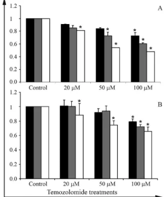
To determine the TMZ concentration for combination with propolis, U343 and U251 cells were submitted to dif-ferent concentrations of TMZ for 24, 48 and 72 h. The TMZ concentration chosen for combined treatment was 50mM, which reduced proliferation at 48 h for U251 and U343 cell lines (Figure 2).
U251, U343 and MCR-5 cell lines were treated with propolis extract at the concentrations of 1 and 2 mg/mL for 24, 48 and 72 h. The glioblastoma cell lines were also treated with a propolis concentration of 2 mg/mL associ-ated with 50mM of temozolomide. Cell viability was deter-mined by an XTT assay, as described above.
Propolis extract concentrations inhibited growth of the three cell lines when compared with DMSO (0.5%) (p < 0.05) (Figure 1). For cell line U251, proliferation inhi-bition was observed at 24, 48 and 72 h corresponding to a 10, 24 and 46% decrease with the 1 mg/mL dose, and a 15, 32 and 59% decrease with the 2 mg/mL dose, respectively. However, there was no statistically significant difference between the concentrations for this cell line at the times studied (Figure 1A).
In cell line U343, a decrease in proliferation was ob-served at 24, 48 and 72 h for both dosages, corresponding to a 14, 21 and 30% decrease for the 1 mg/mL, and a 30, 42 and 48% decrease for the 2 mg/mL dose. For this cell line, all treatments except the 72 h treatment showed statistically significant differences between the concentrations (Fig-ure 1B).
In both the U251 and U343 cell lines treated with 1 and 2 mg/mL propolis extract for 48 h, a decrease in colony formation capacity was observed; however, there was no difference between the two treatments (data not shown).
To determine the occurrence of apoptosis in GBM cells treated with the propolis extract, the cells were differ-entially stained. Apoptosis was not observed after the treat-ment with propolis at neither of the concentrations tested. The methodology applied also allowed the detection of ne-crotic cells, observed at a low number and without differ-ences between treatments (data not shown).
Propolis, a complex mixture of plant metabolites, shows a broad spectrum of biological activities including antibiotic, antioxidative, anti-inflammatory and anticancer effects (Bankovaet al., 1983; Marcucci, 1995; Banskotaet al., 2001). Its cytotoxicity in cultures of human and animal tumor cells, including breast carcinoma, melanoma, colon and renal carcinoma cell lines, has been frequently reported in the literature (Khalil, 2006).
The present study showed that propolis extract inhib-ited proliferation in glioblastoma and fibroblast cell lines, as already demonstrated by previous studies. Propolis ex-tract from the Netherlands showed an interesting antiproli-ferative activity against highly metastatic liver murine
colon 26-L5 carcinoma cells (Banskota et al., 2000). A butanolic Greek propolis extract was also found to be cyto-toxic in two malignant human cell lines (HT-1080 fibro-sarcoma and HT-29 colon adenocarcinoma), whereas it was not equally toxic when tested in normal human skin fibroblasts (Pratsiniset al., 2010).
In cell line U343, the antiproliferative effect of pro-polis was dose- and time-dependent, suggesting that this cell line is more sensitive than U251 that did not present the same effect. These differences could be associated withp53
status (mutant or wild type). Cell line U343, but not U251, carries the wild type gene (Ishiiet al., 1999) and the anti-proliferative effects of propolis may be p53-dependent. Other studies have shown increased expression ofp53after treatment with different propolis extracts (Weng et al., 2007; Ishiharaet al., 2009; Xuanet al., 2010). Several func-tions and activities are attributed to p53, and it also acts in different cellular metabolism processes, such as cell cycle, apoptosis, senescence and DNA repair (Joerger and Fersht, 2008).
The combination of propolis with temozolomide, a chemoterapeutic drug used in the treatment of glioblastoma which produces DNA alkylation (Estelleret al., 2000), evi-denced synergistic antiproliferative effects, demonstrating the ability of propolis to predispose cells to the action of chemotherapy (Figures 1A and B). However, this effect should be further investigated.
Figure 2- Proliferation assay of cell lines treated with TMZ at the concen-trations of 20, 50 and 100mM, for 24, 48 and 72 h. Asterisks indicates a statistically significant difference between the TMZ-treated and the con-trol group.
The effects observed in this work can be related with the chemical composition of propolis, which is highly de-pendent on the flora of the region where it is collected. Sawayaet al.(2009) studied the same Tubi-bee propolis used in this work and showed that its composition varied seasonally. The mass spectra ions found in this extract were m/z 371, 373, 401 and 471. This latter was the more impor-tant one; its formula C30H47O4suggests that it has at least
one acid function. Electrospray Ionization – Mass Spec-trometry (ESI-MS) of these ions showed to be compatible with terpenes and with acid groups. All are marker ions of
Schinus terebenthifolius, also known in Brazil as “aroeira mansa”, a preferred source of resins in stingless bee pro-polis in many regions of Brazil.
Propolis from temperate zones predominantly con-tains phenolic compounds, including flavonoids and cin-namic acid derivatives (Marcucci, 1995). On the other hand, diterpenes and prenylated compounds, which are vir-tually absent in propolis from temperate zones, have been reported to be the major constituents of propolis from tropi-cal South America, along with lignans, flavonoids and other classes of compounds (Sawayaet al., 2009).
Several reports have shown apoptosis induction cau-sed by propolis extracts (Wenget al., 2007; Szliszkaet al., 2009; Xuanet al., 2010). However, in the present study, this effect was not observed, suggesting that in the concen-trations used this type of propolis presents only cytostatic effects.
In summary, this investigation of the potential anti-proliferative effects of propolis in human glioblastoma and normal fibroblast cell lines showed a strong inhibitory ef-fect on the proliferation of all cell lines tested. Dose and time dependence were only observed for cell line U343. Moreover, the association of propolis with temozolomide produced synergistic antiproliferative effects. Propolis treatment also inhibited the clonogenic capacity in GBM cell lines, but the antitumor effects observed here were not caused by apoptosis. Further investigations are needed to elucidate the molecular mechanism of the antitumor effect of propolis, and the study of its individual constituents may reveal specific molecules with antiproliferative capacity.
Acknowledgments
Financial Support by Fundação de Amparo a Pesqui-sa do Estado de São Paulo (FAPESP, process nº 2009/50118-2), and FAPESP fellowships to KSB (2010/08699-5) and MSB (2006/04827-3) are acknowl-edged
References
Bankova VS, Popov SS and Marekov NL (1983) A study on flavonoids of propolis. J Nat Prod 46:471-474.
Bankova V, Boudourova-Krasteva G, Popov S, Sforcin JM and Funari SRC (1998) Seasonal variations of the chemical composition of Brazilian propolis. Apidologie 29: 361-36.
Banskota AH, Tezuka Y, Adnyana IK, Midorikawa K, Matsu-shige K, Message D, Huertas AA and Kadota S (2000) Cytotoxic, hepatoprotective and free radical scavenging ef-fects of propolis from Brazil, Peru, the Netherlands and China. J Ethnopharmacol 72:239-246.
Banskota AH, Tezuka Y and Kadota S (2001) Recent progress in pharmacological research of propolis. Phytother Res 15:561-571.
Brandes AA, Franceschi E, Tosoni A, Bartolini S, Bacci A, Agati R, Ghimenton C, Turazzi S, Talacchi A, Skrap M,et al. (2010) O(6)-methylguanine DNA-methyltransferase me-thylation status can change between first surgery for newly diagnosed glioblastoma and second surgery for recurrence: clinical implications. Neuro Oncol 12:283-288.
Burdock GA (1998) Review of the biological properties and tox-icity of bee propolis (propolis). Food Chem Toxicol 36:347-363.
Esteller M, Garcia-Foncillas J, Andion E, Goodman SN, Hidalgo OF, Vanaclocha V, Baylin SB and Herman JG (2000) Inacti-vation of the DNA-repair gene MGMT and the clinical re-sponse of gliomas to alkylating agents. N Engl J Med 343:1350-1354.
Farnesi AP, Aquino-Ferreira R, De Jong D, Bastos JK and Soares AEE (2009) Effects of stingless bee and honey bee propolis on four species of bacteria. Genet Mol Res 8:635-640. Franken NA, Rodermond HM, Stap J, Haveman J and van Bree C
(2006) Clonogenic assay of cells in vitro. Nat Protoc 1:2315-2139.
Grunberger D, Banerjee R, Eisinger K, Oltz EM, Efros L, Cald-well M, Estevez V and Nakanishi K (1988) Preferential cytotoxicity on tumor cells by caffeic acid phenethyl ester isolated from propolis. Experientia 44:230-232.
Ishihara M, Naoi K, Hashita M, Itoh Y and Suzui M (2009) Growth inhibitory activity of ethanol extract of Chinese and Brazilian propolis in four human colon carcinoma cell lines. Oncol Rep 22:349-354.
Ishii N, Maier D, Merlo A, Tada M, Sawamura Y, Diserens AC and Van Meir EG (1999) Frequent co-alterations of TP53, p16/CDKN2A, p14ARF, PTEN tumor suppressor genes in human glioma cell lines. Brain Pathol 9:469-479.
Joerger AC and Fersht AR (2008) Structural biology of the tumor suppressor p53. Annu Rev Biochem 77:557-582.
Khalil ML (2006) Biological activity of bee propolis in health and disease. Asian Pac J Cancer Prev 7:22-31.
Lee Y and Shacter E (1999) Oxidative stress inhibits apoptosis in human lymphoma cells. J Biol Chem 274:19792-19798. Marcucci MC (1995) Propolis: Chemical composition, biological
properties and therapeutic activity. Apidologie 26:83-99. Pratsinis H, Kletsas D, Melliou E and Chinou I (2010)
Antipro-liferative activity of Greek propolis. J Med Food 13:286-290.
Sawaya ACHF, Calado JCP, Santos LC, Marcucci MC, Akatsu IP, Soares AEE, Abdelnur PV, Cunha IBSC and Eberlin MN (2009) Composition and antioxidant activity of propolis from three species of Scaptotrigona stingless bees. J Api-prod ApiMed Sci 1:37-42.
microarray and real-time quantitative PCR. J Neurooncol 88:281-91.
Szliszka E, Czuba ZP, Bronikowska J, Mertas A, Paradysz A and Krol W (2009) Ethanolic extract of propolis augments TRAIL-induced apoptotic death in prostate cancer cells. Evid Based Complement Alternat Med, Ahead-of-Print, doi:10.1093/ecam/nep180.
van Nifterik KA, van den Berg J, Stalpers LJ, Lafleur MV, Leenstra S, Slotman BJ, Hulsebos TJ and Sminia P (2007) Differential radiosensitizing potential of temozolomide in MGMT promoter methylated glioblastoma multiform cell lines. Int J Radiat Oncol Biol Phys 69:1246-1253.
Weng MS, Liao CH, Chen CN, Wu CL and Lin JK (2007) Pro-polin H from Taiwanese propolis induces G1 arrest in hu-man lung carcinoma cells. J Agric Food Chem 55:5289-5298.
Xuan H, Zhao J, Miao J, Li Y, Chu Y and Hu F (2010) Effect of Brazilian propolis on human umbilical vein endothelial cell apoptosis. Food Chem Toxicol 49:78-85.
Associate Editor: Emmanuel Dias Neto