Fluo re sce nt ligands fo r studying
ne uro pe ptide re ce pto rs by co nfo cal
m icro sco py
1Montreal Neurological Institute, McGill University, Montreal, Q uebec, Canada 2Institut de Pharmacologie Moléculaire et Cellulaire du CNRS,
Université de Nice-Sophia Antipolis, Valbonne, France A. Beaudet1, D. Nouel1,
T. Stroh1, F. Vandenbulcke1,
C. Dal-Farra2 and
J.-P. Vincent2
Abstract
This paper reviews the use of confocal microscopy as it pertains to the identification of G-protein coupled receptors and the study of their dynamic properties in cell cultures and in mammalian brain following their tagging with specific fluorescent ligands. Principles that should guide the choice of suitable ligands and fluorophores are discussed. Examples are provided from the work carried out in the authors laboratory using custom synthetized fluoresceinylated or BODIPY-tagged bioactive peptides. The results show that confocal microscopic detection of specifically bound fluorescent ligands permits high reso-lution appraisal of neuropeptide receptor distribution both in cell culture and in brain sections. Within the framework of time course experiments, it also allows for a dynamic assessment of the internal-ization and subsequent intracellular trafficking of bound fluorescent molecules. Thus, it was found that neurotensin, somatostatin and mu-and delta-selective opioid peptides are internalized in a receptor-dependent fashion and according to receptor-specific patterns into their target cells. In the case of neurotensin, this internalization process was found to be clathrin-mediated, to proceed through classi-cal endosomal pathways and, in neurons, to result in a mobilization of newly formed endosomes from neural processes to nerve cell bodies and from the periphery of cell bodies towards the perinuclear zone. These mechanisms are likely to play an important role for ligand inactivation, receptor regulation and perhaps also transmembrane signaling.
Co rre spo nde nce
A. Beaudet
Montreal Neurological Institute 3801 University Street Montreal, Q uebec Canada H3A 2B4 Fax: + 1-514-398-5871 E-mail: mcin@ musica.mcgill.ca
Presented at the International Symposium: Biological Applications of Confocal Microscopy, Belo Horizonte, MG, Brasil, April 6-8, 1998.
Personal work referred to in the text was made possible by a grant from the Medical Research Council of Canada, and by fellowships from the Fonds de la Recherche en Santé du Q uébec, the Deutsche Forschungsgemeinschaft and the Fondation pour la Recherche Médicale, and by a France-Q uebec International exchange program.
Received May 14, 1998 Accepted June 3, 1998
Ke y wo rds
•Neurotensin •Somatostatin •O pioids •Internalization •Endocytosis
•Confocal microscopy
Intro ductio n
There has been a long history of attempts at conjugating fluorescent molecules to re-ceptor ligands with the aim of identifying specific neurotransmitter binding sites (for a review, see 1). However, many of these at-tempts were defeated by the lack of suitable probes as well as by the insensitivity of
provided us with powerful and resolving approaches for studying receptors and re-ceptor-mediated mechanisms in living tis-sue.
Applicatio ns o f fluo re sce nt ligands to the study o f re ce pto r-ligand inte ractio ns
There are several ways in which fluores-cent ligands may be applied to the study of neurotransmitter receptors. These include: 1) classical binding studies in which radio-active probes are replaced with fluorescent ones and specifically bound ligand is de-tected using fluorescence polarization as-says, 2) flow cytometric studies in which fluorescence-activated cell sorters (FACS) are used for assessing specific cell surface ligand binding in dissociated cells (at 4o
C) or for demonstrating ligand internalization within transfected cells (at 37o
C), and 3) photobleaching experiments in which the bleaching of certain ligand molecules (which are only fluorescent when bound) is used to study the rate of recovery of fluorescence after bleaching and hence to provide infor-mation on the association of the fluorescent ligand. In the present review, we will focus on the use of confocal microscopy for: 1) assessing the binding of fluorescent ligands to their receptors in isolated cells in culture or in whole brain sections, 2) visualizing ligand internalization and trafficking in trans-fected cells and live brain slices, and 3) identifying intra-cellular compartments of ligand sequestration.
Se le ctio n o f a fluo re sce nt ligand
The choice of a fluorescent ligand should take into account the metabolic and phar-macological properties of the compound to be tagged as well as the biophysical proper-ties of the fluorophore.
The ligand to be tagged may be an ago-nist or an antagoago-nist. The latter usually
of-fers a higher affinity, and therefore provides for a better signal to noise ratio, than ago-nists. However, antagonists are ill suited for the study of receptor dynamics as they usu-ally do not internalize (3). They do, how-ever, reportedly induce receptor clustering (3). Metabolically stable analogs may prove more advantageous than native ligands for studying ligand-receptor interactions in vivo.
Thus, the use of the stable somatostatin (SRIF) analog, BODIPY-D-Trp8-SRIF, has allowed us to study SRIF binding and inter-nalization both in transfected cells (4) and in neurons (our unpublished data) without hav-ing to resort to catabolic enzyme inhibitors. The downside of these compounds, how-ever, is that their intracellular fate may not faithfully mimic that of the native peptide.
Ideally, a fluorescent ligand should re-tain as much as possible of the affinity and selectivity of the untagged agonist. In prac-tice, however, the addition of a fluorescent moiety will often affect the binding affinity of the ligand. This loss of affinity will usu-ally depend on the site at which the fluoro-phore is tagged to the fluorescent probe (5). It will also vary according to the fluorophore chosen. Thus, whereas the addition of fluo-rescein (FITC) or of BODIPY moieties to D-Trp8-somatostatin (D-Trp8-SRIF) only slightly reduces the affinity of the ligand in brain membrane preparations (4), the tag-ging of a Cy 3.5 fluorophore to the same molecule reduces its binding affinity by al-most an order of magnitude (our unpub-lished data). This loss in affinity, however, is also a function of the intrinsic properties of the ligand and of its pharmacophore, as the neuropeptide neurotensin (NT) may be tagged with any of the FITC, BODIPY or Cy 3.5 moieties without losing its affinity for the NT receptor (our unpublished data).
re-ceptor or transfected with the recombinant receptor of interest (4-6). These binding experiments should be complemented by imaging studies on whole cells that either naturally express the receptor under study or are transfected with the appropriate recombinant DNA to measure concentra-tion-dependent binding in the presence and absence of nonlabeled competitors. When-ever possible, the biological relevance of the fluorescent ligand should also be tested, ei-ther in cell culture or in slice preparations (7).
For the reasons stated above, the choice of a fluorophore will be predicated on its compatibility with the preservation of the pharmacological properties of the ligand. Within this frame of reference, preference should be given to fluorophores with high fluorescent yield and resistance to photo-bleaching. Both BODIPY and Cy compounds are particularly interesting in this regard. One should also ensure that the fluorescent ligand remains hydrophilic, which is critical for the localization of cell surface receptors. Fluorescent ligands have so far been de-veloped against a variety of neuroreceptors, including α-adrenoceptors (1), ß-adrenocep-tors (8,9), benzodiazepine recepß-adrenocep-tors (10), nicotinic (11) and muscarinic (12) acetyl-choline receptors, and D1 (13) and D2 (13,14) dopamine receptors. In the present review, we will focus on the use of peptidic probes as developed by our group for studying neu-ropeptide-receptor interactions in cell cul-tures and mammalian brain sections. Several other studies have made use of fluorescent probes for studying neuropeptide-receptor interactions. These include studies on sub-stance P (15,16), angiotensin II (17), thy-rotropin-releasing hormone (18), opioid (19,20), cholecystokinin (21), gonadotropin-releasing hormone (22), vasopressin (23) and gastrin-releasing peptide (24) receptors. The reader is referred to these publications for further information.
Fluo re sce nt pe ptide binding to ce lls in culture and to who le brain slice s
Confocal microscopic analysis of specif-ic fluorescent peptide binding may be used for static assessment of the cellular distribu-tion of neuropeptide receptor distribudistribu-tion in whole cells in culture or in frozen brain sections.
Binding to culture d ce lls
The binding of FITC- and BODIPY-la-beled neurotensin (fluo-NT), of FITC- and BODIPY-labeled D-Trp8-somatostatin SRIF), of BODIPY-labeled dermorphin (fluo-dermorphin) and of BODIPY-labeled del-torphin (fluo-deldel-torphin) was examined in COS-7 cells transfected with recombinant DNA encoding high- or low-affinity NT re-ceptors (fluo-NT), sst1, sst2A or sst5 SRIF receptors (fluo-SRIF), and mu (fluo-dermor-phin) or delta (fluo-deltor(fluo-dermor-phin) opioid re-ceptors (4,5,25; our unpublished data). Bind-ing of fluo-NT was also assessed in cells naturally expressing the high-affinity neuro-tensin receptor (NT-1; SN17 neuron-neuro-blastoma hybrid cells (26); primary neurons in culture (27)) or the low-affinity NT recep-tor (NT-2; astrocytes in culture (27)). Bind-ing was carried out at 4o
C or at 37o
In all transfected cell systems, fluores-cent peptides bound at 4oC formed at
equi-librium a more or less homogenous pericel-lular ring (Figure 1). This labeling was spe-cific in that it was no longer apparent when the incubations were carried out in the pres-ence of an excess of nonlabeled peptide. It was also exclusively surface-bound, as de-termined by serial confocal sectioning through the cell and confirmed by its com-plete disappearance following a hypertonic
acid wash. In COS-7 cells incubated at 37o
C in the presence of an endocytosis inhibitor, specific fluorescent labeling was also con-fined to the cell surface, but was usually more prevalent at one pole of the cell (Figure 2). This capping of bound fluorescence was taken to reflect cell surface clustering of receptor complexes under conditions of im-peded internalization (5,26).
When the same experiments were carried out in cells naturally expressing the receptor, cell surface binding at 4o
C took the form of small fluorescent hot spots haphazardly dis-tributed upon the surface of the cells. The size and shape of these fluorescent hot spots, corresponding to ligand-receptor clusters, varied according to the type of cell and/or receptor expressed. Thus, in SN-17 cells, which express the NT-1 receptor, these clus-ters were small and rounded (26). By con-trast, in astrocytes, which express the NT-2 receptor, these surface clusters were larger and more geometric in shape (27; Figure 3). However, when incubated at 37o
C in the presence of PAO, naturally expressing cells exhibited the same capping phenomenon as observed in transfected cells.
The differences between patterns gener-ated in naturally expressing versus trans-fected cells at 4o
C probably reflect dispari-ties in receptor densidispari-ties (the concentration of receptors in transfected cells being too high for one to resolve individual clusters). They could also reflect differences in clus-tering properties, but this appears unlikely given the similarity of the patterns observed at 37oC in the presence of PAO. In any event,
the present results suggest that even at 4o
C, the membrane retains a fluidity compatible with cell surface clustering of receptor-ligand complexes.
Binding to fro ze n brain se ctio ns
We have recently resorted to this ap-proach for localizing SRIF binding sites at the cellular level in the rat mediobasal
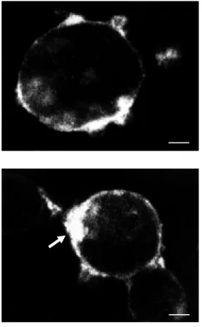
hypo-Figure 1 - Binding of fluo-NT to COS-7 cells transfected w ith c-DNA encoding the low -affinity NT receptor (NT-2). Cells w ere incubated for 30 min, at 4oC, in
the presence of 10 nM fluo-NT. Labeling is strictly membrane-bound and forms a more or less homogeneous pericellular ring. Scale bar: 5 µm.
Figure 2 - Effect of the endocy-tocis inhibitor, phenylarsine ox-ide (PAO), on the cell surface distribution of bound fluo-der-m orphin. COS-7 cells t rans-fected w ith cDNA encoding the mu opioid receptor w ere incu-bated for 45 min, at 37oC, w ith
10 nM fluo-dermorphin in the presence of 10 µM PAO. Note how the bound fluorescent mol-ecules form a single large clus-ter at one pole of the cell (ar-row ). Scale bar: 5 µm.
Figure 3 - Distribution of fluo-NT-labeled NT-2 receptors on the surface of an astrocyte in culture follow ing 60-min incubation at 37oC w ith 20 nM fluo-NT. In
these cells, w hich express but do not internalize NT-2 recep-tors, surface-bound molecules f orm num erous polym orphic clusters. This labeling is entirely suppressed by a hypertonic acid w ash. Scale bar: 20 µm.
thalamus. The labeling technique is the same as those currently used for assessing recep-tor distributions by aurecep-toradiography (29). The advantage of using fluorescent over ra-dioactive ligands is that they provide higher resolution and speedier results. They are also considerably less sensitive, however, and ligand concentrations may have to be increased accordingly.
Briefly, 20 µm-thick sections from the mediobasal hypothalamus were incubated for 45 min at 4o
C with 40 nM fluo-SRIF, in the presence (nonspecific binding) or the absence (specific binding) of a hundred-fold excess of nonfluorescent ligand. In sections incubated with fluo-SRIF alone, both con-ventional epifluorescence and confocal mi-croscopy revealed selective binding of the fluorescent compound in the arcuate nucleus. This binding was specific, as demonstrated by the fact that sections incubated in the presence of a hundred-fold excess of nonla-beled ligand showed only background fluorescence. At the regional level, the la-beled pattern was comparable to that previ-ously observed by autoradiography using
125
I-SRIF (30). However, confocal micros-copy allowed for a more clear-cut localiza-tion of the ligand to small neuronal perikarya distributed throughout the structure. Further-more, serial optical sectioning of the labeled sections revealed that the label was not con-fined to the cell surface but was present throughout the cytoplasm of the labeled per-ikarya.
Inte rnalizatio n and intrace llular trafficking o f re ce pto r-ligand co m ple xe s
The application of confocal microscopy to the visualization of specifically bound fluorescent peptides has proven most pow-erful for studying internalization and traf-ficking of receptor-ligand complexes into their target cells. These processes may be studied both in live cells in culture and in
whole brain slices maintained in a superfu-sion system.
Ce ll culture studie s
We compared the internalization and in-tracellular trafficking of fluo-NT via high-affinity NT receptors (NT-1), of fluo-SRIF via various SRIF receptor subtypes, and of fluo-dermorphin and fluo-deltorphin via µ and δ opioid receptors, respectively, in COS-7 cells transfected with cDNA encoding the appropriate receptor. Internalization was demonstrated by incubation with the appro-priate fluorescent ligand at 37oC, followed
by a series of hypertonic acid washes to strip off surface-bound ligand and visualization of acid-wash resistant labeling by confocal microscopy. Incubations with 125
I-labeled peptide analogues were carried out in paral-lel to provide a quantitative measure of ligand binding and of receptor-mediated endocyto-sis. Confirmation that the internalization was endocytic in nature was obtained by carrying out the incubations in the presence of the endocytosis inhibitor, PAO.
Incubation of COS-7 cells transfected with cDNA encoding the NT-1 receptor for 0-60 min with 10 nM fluo-NT resulted in a rapid build-up of acid-resistant intracellular fluorescence. Parallel experiments using 125
I-NT confirmed that this internalization pro-cess was both rapid (t1/2 = 10 min) and efficient (80% of total surface binding inter-nalized by one hour). At short time intervals (between 0 and 20 min), the label formed small hot spots distributed throughout the cytoplasm of the cells, but sparing the nucleus (Figure 4). At later time points (>20 min), these endosome-like particles de-creased in number and progressively clus-tered towards the center of the cells, next to the nucleus.
sst1, sst2A or sst5. Biochemical experiments using the radioactive SRIF analogue 125
I-Tyr0
-[D-Trp8
]SRIF showed that internaliza-tion of SRIF was dependent on the receptor subtype, with sst2A mediating the internal-ization of radioiodinated Tyr0
-[D-Trp8
]SRIF as effectively as did NT-1 for fluo-NT (ap-proximately 80% of total radioactivity bound), whereas sst1 internalized only very little (<10% of total radioactivity bound; 4).
The capacity of internalization via sst5 was intermediate between sst1 and sst2A recep-tor subtypes (our unpublished data).
By confocal microscopy, incubation of sst1-transfected cells at 37oC with fluo-SRIF
for 0-60 min gave rise to a pericellular ring-like pattern of labeling. This labeling pattern remained remarkably constant with time, closely resembling the one obtained after incubation of peptide-receptor-transfected cells at 4oC (see above). However, it was
entirely preserved after a hypertonic acid wash indicating that it was not surface bound but sub-plasmalemmal (4).
COS cells transfected with cDNA encod-ing sst2A, on the other hand, exhibited fluo-rescent labeling throughout their cytoplasm, as observed after incubation with fluo-NT. As with fluo-NT, this labeling was highly punctate, suggesting that the label had been sequestered in small membrane-bound com-partments (Figure 5). The size of these par-ticles varied significantly with time as their mean surface progressively increased in the course of the experiment. At the same time, they redistributed from a peripheral distribu-tion at short incubadistribu-tion times (1-5 min) by invading the whole cytoplasm and converg-ing towards the nucleus at later stages of the experiment (4).
The intracellular labeling observed in COS cells transfected with cDNA encoding sst5 more closely resembled the eccentric pattern of labeling observed in sst1-trans-fected cells than in cells transsst1-trans-fected with sst2A, at least at early time points (0-20 min; Figure 6). However, at later time intervals (>20 min) the ligand started to pervade the cytoplasm within small endosome-like par-ticles as seen with the sst2A sub-type.
The intracellular distribution of fluo-dermorphin and fluo-deltorphin within COS-7 cells transfected with µ and δ opioid recep-tors, respectively, also varied markedly with time. After short durations of incubation, both fluorescent markers were clustered at one pole of the cell in the immediate vicinity
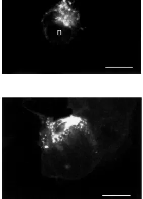
Figure 4 - Distribution of inter-nalized fluo-NT in COS-7 cells transfected w ith cDNA encoding the high-affinity NT receptor (NT-1). Cells w ere incubated for 20 min w ith 20 nM fluo-NT and sub-jected to a hypertonic acid w ash. Internalized ligand forms small, rounded hot spots that pervade the cytoplasm but spare the nucleus (n). Scale bar: 10 µm.
Figure 5 - COS-1 cells trans-fected w ith cDNA encoding the sst2A somatostatin (SRIF) recep-tor and incubated for 30 min w ith 20 nM fluo-SRIF at 37oC. The
int racellular labeling pat t ern, seen here after w ash-out of sur-face-bound ligand w ith hyper-tonic acid buffer, is similar to that observed w ith fluo-NT in NT-1-transfected COS-7 cells (see Fig-ure 4). Small, intensely labeled endosome-like particles pervade the cytoplasm of the cells. n, Nucleus. Scale bar: 10 µm.
Figure 6 - Internalization of fluo-SRIF in COS-7 cells transfected w ith cDNA encoding the sst5 SRIF receptor. After 20 min of incubation, the bulk of the ligand is still confined to a sub-plasma-lemmal zone. Compare the dis-tribution of the acid-w ashable fraction of the ligand, as seen here, w ith that evident at the same time point in sst2A-trans-fected cells. Scale bar: 10 µm.
n
of the plasma membrane. After 30 min, the label had taken on the shape of small fluores-cent particles in the cytoplasm. These were, however, still excentrically clustered. After 60 min the label finally pervaded the entire cytoplasm sparing the nucleus but remaining highly punctate. By this time, differences between fluo-deltorphin and fluo-dermor-phin-labeled endosome-like particles became apparent in that the latter were on average larger and less numerous (5).
In order to directly assess possible differ-ences in the pathways through which µ and
δ opioid receptors had internalized their re-spective ligands, COS cells were co-trans-fected with cDNA encoding µ and δ opioid receptors. Subsequently, the cells express-ing both types of opioid receptors were si-multaneously incubated with BODIPY Green-deltorphin and BODIPY Red-dermor-phin for 90 min at 37o
C. Images were then acquired through the distinct red and green excitation/emission channels of the confocal microscope. The single images showed pat-terns of labeling equivalent to those found in singly transfected cells. Superimposition of the images, however, showed only partial overlap of red and green fluorescent par-ticles indicating that the µ and δ fluorescent ligands were partly sequestered in distinct compartments. Interestingly, the bulk of dis-tinct green and red particles were small and predominantly located in peripheral zones of the cytoplasm whereas double-labeled endosome-like particles were concentrated in the center of the cell, suggesting that dermorphin and deltorphin were internal-ized initially via separate compartments that eventually joined in the course of intracellu-lar trafficking.
These findings indicate that neuropep-tide receptors are internalized following ligand binding when ectopically expressed in transfected epithelial cells. More impor-tantly, however, they demonstrate that the internalization of individual ligands is dif-ferentially regulated according to receptor
subtypes and that their trafficking may pro-ceed along different endocytic pathways.
Inte rnalizatio n in live brain slice s
In order to determine whether neuropep-tides were internalized into live neuronal cells with the same efficiency and according to comparable sequestration patterns as they did in heterologous transfection systems, we examined the binding and internalization of fluo-NT in live brain slices (300 µm thick) superfused in vitro in a recording chamber
system. We selected brain areas (basal fore-brain (31); ventral midfore-brain (32)) in which the transmitter build-up of the cells express-ing the NT-1 receptor had previously been identified (cholinergic in the former, dopa-minergic in the latter), providing a means to assess, using immunohistochemistry for choline acetyltransferase and tyrosine hy-droxylase, respectively, the selectivity of fluo-NT labeling patterns for cells expressing the NT-1.
In both regions, the distributional pattern of fluo-NT labeling varied according to the duration of the wash-out after ligand appli-cation. Between 5 and 10 min after applica-tion of fluo-NT, the labeling was intense and distributed throughout the neuropil over neu-ronal perikarya and processes alike (Figure 7). Thirty and 60 min after application of ligand, the labeling had markedly increased over neuronal perikarya but had almost com-pletely disappeared from the surrounding
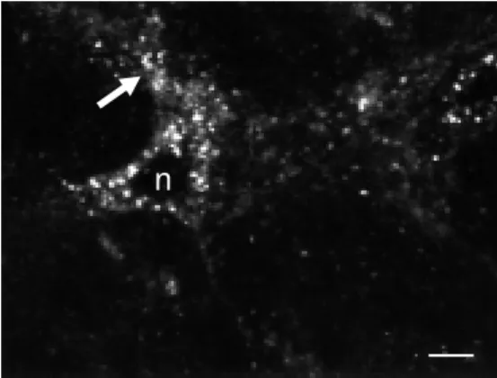
Figure 7 - Fluo-NT-labeled neu-ron in the ventral tegmental area of an adult rat brain. Sections from the ventral midbrain w ere superfused during 3 min w ith 20 nM fluo-NT at 37oC, rinsed for
10 min in buffer, fixed w ith 4% paraformaldehyde and optically sectioned. Internalized ligand molecules are evident w ithin en-dosome-like organelles through-out the perikaryon sparing the nucleus (n). Labeled endosomes are also evident w ithin the proxi-mal processes of the cell (arrow ). No labeling w as apparent w hen the incubation w as carried out at 4oC or at 37oC in the presence
of an excess of nonfluorescent NT. Scale bar: 15 µm.
!
Ide ntificatio n o f e ndo cytic co mpartme nts
One of the advantages of fluorescent pep-tides is that they may be used in conjunction with either other fluorescent markers or with immunofluorescence for double localization studies. We have resorted to both of these approaches to determine whether the inter-nalization of fluorescent peptides witnessed in both transfected epithelial cells and rat brain slices proceeded through the classical, clathrin-mediated endocytic pathway.
We first examined the pH of the internal-ization compartments, as classical endosomes are known to be more acidic than the sur-rounding cytoplasm (33). For this purpose, we resorted to acridine orange, a vital dye documented to accumulate selectively in compartments with low internal pH values (34). COS-7 cells transfected with the NT-1 receptor were first incubated for 30 min at 37o
C in the presence of 10 nM fluo-NT. Cells were then rinsed, rapidly stained with acridine orange (20 µM, 1 min at room tem-perature), rinsed again and observed under the confocal microscope. After 30 min of incubation, fluo-NT formed, as described above, numerous intracellular hot spots (Fig-ure 8A). All of fluo-NT-labeled struct(Fig-ures also exhibited strong acridine orange stain-ing (Figure 8B) indicatstain-ing that they were highly acidic and that they could therefore correspond to early endosomes, late endo-somes or lysoendo-somes.
To further distinguish between these dif-ferent possibilities, and also to monitor the migration of the ligand within endocytic com-partments with time, we combined in the same cell system the visualization of fluo-NT with the immunohistochemical detec-tion of marker proteins which may be used to distinguish between different endocytic com-partments. Thus, at short time intervals (0-20 min) following incubation of the trans-fected cells with fluo-NT, most of the intra-cellular ligand was detected in compartments processes. By single optical sectioning,
in-tracellular labeling was detected in the form of small, intensely fluorescent particles which increased in size but decreased in number with time (Figure 7). These hot spots also got closer with time to the nucleus of the cell as seen in transfected cells. In slices incu-bated with an excess of native NT, no label-ing was observed over either region except for a diffuse reactivity of intraparenchymal capillaries. Similarly, no tissue labeling was observed after incubation with fluorescein isothiocyanate alone.
The concomitant decrease in neuropil labeling and increase in specific fluo-NT observed over time suggest that internaliza-tion occurring at the level of the dendritic tree is rapidly followed by an intracellular mobilization of internalized molecules. The migration and remodeling of intracellular internalization compartments with time fur-ther suggest that, as in transfected cells, NT is internalized via classical internalization pathways.
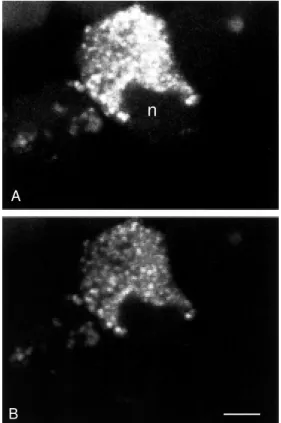
Figure 8 - Double staining of COS-7 cells transfected w ith cDNA encoding the NT-1 recep-tor w ith fluo-NT (A) and acridine orange (B). Fluo-NT labeling w as carried out for 30 min at 37oC.
Cells w ere then rinsed and fur-ther incubated for 1 min w ith acridine orange. Note that both labels are co-localized w ithin the same intracellular organelles, in-dicating that fluo-NT is internal-ized w ithin acidic compartments. n, Nucleus. Scale bar: 10 µm.
n
A
that immunostained positively for Rab 5, a small GTP-binding protein known to be as-sociated with early endosomes (35,36). These results are congruent with those of our acri-dine orange experiments and indicate that fluo-NT is internalized via clathrin-coated pits and that it subsequently proceeds along the endosomal pathway.
Because of the acidic environment of endosomes, this process is known to result in a dissociation of receptor-ligand complexes. Following dissociation, internalized recep-tors may be either targeted to lysosomes for degradation or recycled back to the plasma membrane (37). To confirm that receptor-ligand complexes were indeed dissociated within the acidic environment of the endoso-mal system, we followed in parallel the fate of fluo-NT and that of immunolabeled NT-1 in COS-7 cells transfected with an NTR1-VSV epitope construct (25). As predicted from current models, receptor and ligand molecules were co-localized within the same compartments at short time intervals (0-20 min; Figure 9) but were totally dissociated at longer time points (>20 min). As current biochemical data suggest that the NT recep-tor is not recycled (38,39), it is likely that the dissociated receptor is being targeted to ly-sosomes for intracellular degradation.
Co nclusio ns
The present data demonstrate the appli-cability of confocal microscopy to the visu-alization of specifically bound fluorescent peptides for studying the static distribu-tion of cell surface and/or intracellular re-ceptors both in cell cultures and in tissue sections, as well as for monitoring the dy-namic fate of bound fluorescent ligands in these same types of preparations maintained
in vivo.
Using this approach, we were able to confirm that the binding of neuropeptides to cell surface G-protecoupled receptors in-duces clustering of receptor-ligand
com-Figure 9 - Dual localization of internalized fluo-NT and immu-noreact ive NT-1 recept or in COS-7 cells transfected w ith cDNA encoding the NT-1 recep-tor tagged w ith an immuno-genic epitope (VSV). Cells w ere incubated for 5 min w ith 20 nM fluo-NT, fixed w ith paraformal-dehyde and immunolabeled for the tagged NT-1 receptor. Note that at this short time point there is complete overlap in the distribution of ligand (A) and re-ceptor (B) confirming that inter-nalization implicates receptor-ligand complexes. Scale bar: 5 µm.
plexes through lateral diffusion of the recep-tors in the plasma membrane. We were also able to demonstrate in both neurons and heterologous expression systems that this clustering is followed by internalization and intracellular migration of receptor-ligand complexes. This intracellular routing is both receptor- and cell-specific and presumably reflects various possible fates of the ligand (degradation vs externalization; intracellular
signaling?) and of the receptor (degradation
vs recycling; endosomal signaling?). Our
re-sults demonstrate that in the case of NT, the internalization of the peptide is clathrin de-pendent, proceeds through classical endoso-mal pathways and involves dissociation of receptor-ligand complexes within late endo-somal compartments. In neurons, this pro-cess is characterized by a mobilization of newly formed endosomes first from sites of internalization along neuronal processes to the cell body and second from the periphery of the cell body to the perinuclear zone. In both neurons and epithelial cells, endosomal compartments increase in size but decrease in number during their migration from the periphery to the center of the cell, suggesting that formation of multivesicular bodies/late
A
endosomes occurs through fusion of early endosomes.
Work is now in order to determine the role of neuropeptide internalization in the central nervous system and, particularly, to investigate whether this internalization pro-cess mainly subserves the sensitization/de-sensitization and availability of cell surface receptors (40) or whether it also plays a role in neural signaling (for a review, see 37). It will also be critical to determine the fate of internalized ligand in terms of enzymatic
inactivation, targeting to intracellular com-partments and/or externalization in the ex-tracellular space. It is likely, if the past is any indication of the future, that confocal mi-croscopy will continue to play a critical role in answering these questions.
Ackno wle dgm e nts
We thank Christian Charbonneau for pho-tographic assistance and Beverley Lindsay for typing the manuscript.
Re fe re nce s
1. M cGrath JC, Arribas S & Daly CJ (1996). Fluorescent ligands for the study of re-ceptors. Trends in Pharmacological Sci-ences, 17: 393-399.
2. Bunnett NW, Dazin PF, Payan DG & Grady EF (1995). Characterisation of receptors using cyanine 3-labeled neuropeptides.
Peptides, 16: 733-740.
3. Hazum E, Chang KJ & Cuatrecasas P (1980). Cluster formation of opiate (en-kephalin) receptors in neuroblastom a cells: differences betw een agonists and antagonists and possible relationships to biological functions. Proceedings of the National Academy of Sciences, USA, 77: 3038-3041.
4. Nouel D, Gaudriault G, Houle M , Reisine T, Vincent JP, M azella J & Beaudet A (1997). Differential internalization of so-matostatin in COS-7 cells transfected w ith SST1 and SST2 receptor subtypes: a con-focal microscopic study using novel fluo-rescent somatostatin derivatives. Endo-crinology, 138: 296-306.
5. Gaudriault G, Nouel D, Dal Farra C, Beaudet A & Vincent JP (1997). Receptor-induced internalization of selective pep-tidic µ and δ opioid ligands. Journal of Biological Chemistry, 272: 2880-2888. 6. Faure M P, Gaudreau P, Shaw I, Cashman
NR & Beaudet A (1994). Synthesis of a biologically active fluorescent probe for labeling neurotensin receptors. Journal of Histochemistry and Cytochemistry, 42: 755-763.
7. Alonso A, Faure M P & Beaudet A (1994). Neurotensin promotes oscillatory burst-ing behavior and is internalized in basal forebrain cholinergic neurons. Journal of
Neuroscience, 14: 5778-5792.
8. Atlas D & Levitzki A (1977). Probing of beta-adrenergic receptors by novel fluo-rescent beta-adrenergic blockers. Pro-ceedings of the National Academy of Sci-ences, USA, 74: 5290-5294.
9. Heit hier H, Hallm ann D, Boege F, Reilander H, Dees C, Jaeggi KA, Arndt-Jovin D, Arndt-Jovin TM & Helmreich EJ (1994). Synthesis and properties of fluorescent beta-adrenoceptor ligands. Biochemistry,
33: 9126-9134.
10. Velazquez JL, Thompson CL, Barnes Jr EM & Angelides KJ (1989). Distribution and lateral mobility of GABA/benzodiaz-epine receptors on nerve cells. Journal of Neuroscience, 9: 2163-2169.
11. Balice-Gordon RJ & Lichtman JW (1993).
In vivo observations of pre- and postsyn-aptic changes during the transition from multiple to single innervation at develop-ing neuromuscular junction. Journal of Neuroscience, 13: 834-855.
12. Wang Y, Gu Q, M ao F, Haugland RP & Cynader M S (1994). Activity-dependent expression and distribution of M 1 musca-rinic ACh receptors in visual cortex neu-ronal cultures. Journal of Neuroscience,
14: 4147-4158.
13. Ariano M A, M onsma Jr FJ, Barton AC, Kang HC, Haugland RP & Sibley DR (1989). Direct visualization and cellular lo-calization of D1 and D2 dopamine recep-tors in rat forebrain by use of fluorescent ligands. Proceedings of the National A-cademy of Sciences, USA, 86: 8570-8578. 14. Bart on AC, Kang HC, Rinaudo M S, M onsm a Jr FJ, St ew art -Fram RM , M acinko Jr JA, Haugland RP, Ariano M A
& Sibley DR (1991). M ultiple fluorescent ligands for dopamine receptors. I. Phar-macological characterization and receptor selectivity. Brain Research, 547: 199-207. 15. Garland AM , Grady EF, Payan DG, Vigna SR & Bunnett NW (1994). Agonist-in-duced internalization of the substance P (NK1) receptor expressed in epithelial
cells. Biochemistry Journal, 303: 177-186. 16. Grady EF, Garland AM , Gamp PD, Lovett M , Payan DG & Bunnett NW (1995). De-lineation of the endocytic pathw ay of sub-stance P and its seven-transmembrane domain NK1 receptor. M olecular Biology of the Cell, 6: 509-524.
17. Hein L, M einel L, Pratt RE, Dzau VJ & Kobilka BK (1997). Intracellular trafficking of angiotensin II and its AT1 and AT2
re-ceptors: evidence for selective sorting of receptor and ligand. M olecular Endocri-nology, 11: 1266-1277.
18. Ashw orth R, Yu R, Nelson EJ, Dermer S, Gershengorn M C & Hinkle PM (1995). Vi-sualization of the thyrotropin-releasing hormone receptor and its ligand during endocytosis and recycling. Proceedings of the National Academy of Sciences, USA, 92: 512-516.
19. Hazum E, Chang KJ, Shecht er Y, Wilkinson S & Cuatrecasas P (1979). Fluo-rescent and photo-affinity enkephalin de-rivatives: preparation and interaction w ith opiate receptors. Biochemical and Bio-physical Research Communications, 88: 841-846.
(BODIPY)-labeled fluorescent ligands for the mu opioid receptor. Biochemical Phar-macology, 54: 1315-1322.
21. Roet t ger BF, Rent sch RU, Pinon D, Holicky E, Hadac E, Larkin JM & M iller LJ (1995). Dual pathw ays of internalization of the cholecystokinin receptor. Journal of Cell Biology, 128: 1029-1041. 22. Hazum E, Cuatrecasas P, M arian J & Conn
PM (1980). Receptor-mediated internal-ization of fluorescent gonadotropin-releas-ing hormone by pituitary gonadotropes.
Proceedings of the National Academy of Sciences, USA, 77: 6692-6695.
23. Lutz W, Sanders M , Salisbury J & Kumar R (1990). Internalization of vasopressin analogs in kidney and smooth muscle cells: evidence for receptor-mediated en-docytosis in cells w ith V2 or V1 receptors.
Proceedings of the National Academy of Sciences, USA, 87: 6507-6511.
24. Grady EF, Slice LW, Brant WO, Walsh JH, Payan DG & Bunnett NW (1995). Direct observation of endocytosis of gastrin re-leasing peptide and its receptor. Journal of Biological Chemistry, 270: 4603-4611. 25. Chabry J, Botto JM , Nouel D, Beaudet A, Vincent JP & M azella J (1995). Thr-422 and Tyr-424 residues in the carboxyl ter-minus are critical for the internalization of the rat neurotensin receptor. Journal of Biological Chemistry, 270: 2439-2442. 26. Faure M P, Labbé-Jullié C, Cashman NR,
Kitabgi P & Beaudet A (1995). Binding and internalization of neurotensin in hybrid cells derived from septal cholinergic neu-rons. Synapse, 20: 106-116.
27. Nouel D, Faure M P, Saint Pierre JA,
Alonso R, Quirion R & Beaudet A (1997). Differential binding profile and internaliza-tion process of neurotensin via neuronal and glial receptors. Journal of Neurosci-ence, 17: 1795-1803.
28. M azella J, Leonard K, Chabry J, Kitabgi P, Vincent J-P & Beaudet A (1991). Binding and internalization of iodinated neuro-tensin in neuronal cultures from embry-onic mouse brain. Brain Research, 564: 249-255.
29. Young WS & Kuhar M J (1979). A new method for receptor autoradiography: 3H
opioid receptor labeling in mounted tis-sue sections. Brain Research, 179: 255-270.
30. Epelbaum J, M oyse E, Tannenbaum G, Kordon C & Beaudet A (1989). Combined autoradiographic and immunohistochemi-cal evidence for an association of soma-tostatin binding sites w ith grow th hor-mone-releasing factor containing nerve cell bodies in the rat arcuate nucleus.
Journal of Neuroendocrinology, 1: 109-115.
31. Faure M P, Alonso A, Nouel D, Gaudriault G, Dennis M , Vincent JP & Beaudet A (1995). Somatodendritic internalization and perinuclear targeting of neurotensin in the mammalian brain. Journal of Neuro-science, 15: 4140-4147.
32. Faure M P, Nouel D & Beaudet A (1995). Axonal and dendritic transport of internal-ized neurotensin in rat mesostriatal dopa-minergic neurons. Neuroscience, 68: 519-529.
33. M ellman I, Fuchs R & Helenius A (1986). Acidification of the endocytic and exocytic
pathw ays. Annual Review of Biochemis-try, 55: 663-700.
34. Anderson RGW & Orci L (1988). A view of acidic intracellular compartments. Journal of Cell Biology, 106: 539-544.
35. Chavrier P, Parton RG, Hauri HP, Simons K & Zerial M (1990). Localization of low molecular w eight GTP binding proteins to exocytic and endocytic compartments.
Cell, 62: 317-329.
36. Simons K & Zerial M (1993). Rab proteins and the road maps for intracellular trans-port. Neuron, 11: 789-799.
37. Bevan AP, Drake PG, Bergeron JJM & Posner BI (1996). Intracellular signal trans-duction: the role of endosomes. Trends in Endocrinology and M etabolism, 7: 13-21. 38. Turner JT, James-Kracke M R & Camden JM (1990). Regulation of the neurotensin receptor and intracellular calcium mobili-zation in HT29 cells. Journal of Pharma-cology and Experimental Therapeutics,
253: 1049-1056.
39. Hermans E, Vanisberg M A, Geurts M & M aloteaux JM (1997). Dow n-regulation of neurotensin receptors after ligand-in-duced internalization in rat primary cul-tured neurons. Neurochemistry Interna-tional, 31: 291-299.