79
Revista da Sociedade Brasileira de Medicina Tropical 33(1):79-82, jan-fev, 2000.
ARTIGO DE OPINIÃO
Confocal fluorescence microscopy: a powerful
tool in the study of Chagas disease
Microscopia confocal de fluorescência: uma ferramenta
poderosa no estudo da doença de Chagas
Renato Arruda Mortara1, Solange da Silva1 and Noemi Nosomi Taniwaki1,2
Abstract Confocal scanning fluorescence microscopy has become widely used in cell biology and pathology. In conjunction with monoclonal antibodies it may turn out to be a powerful diagnostic tool that also enables detailed studies of tissue forms of Trypanosoma cruzi.
Key-words: Trypanosoma cruzi. Chagas’ disease. Confocal microscopy.
Resumo A microscopia confocal por varredura a laser vem se tornando extremamente útil na biologia celular e patologia. Com o uso de anticorpos monoclonais, pode ser uma poderosa ferramenta de diagnóstico assim como para estudos detalhados das diferentes formas do Trypanosoma cruzi em vários tecidos infectados.
Palavras-chaves: Trypanosoma cruzi. Doença de Chagas. Microscopia confocal.
1. Disciplina de Parasitologia, Departamento de Microbiologia, Imunologia e Parasitologia, Escola Paulista de Medicina, Universidade Federal de São Paulo, São Paulo, SP, Brasil. 2. Seção de Microscopia Eletrônica, Instituto Adolfo Lutz, São Paulo, SP.
Financial Support: FAPESP, CNPq and CAPES.
Address to: Prof. Renato A. Mortara. Disciplina de Parasitologia/DMIP/EPM/UNIFESP. R. Botucatu 862, 6º andar, 04023-062 São Paulo, SP, Brasil
Tel: 55 11 570-8306; Fax: 55 11 571-1095 E-mail: [email protected]
Recebido para publicação em 10/11/99.
Confocal fluorescence microscopy has become a powerful tool that has enabled a tremendous improvement in morphology and cell biology studies3 5. Its potential application in the
studies of Chagas’ disease has recently been demonstrated by several publications using both in vitro samples1 2 6 and also parasitized tissues
from infected animals7 or even chagasic patients4.
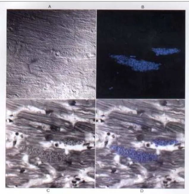
The fundamental property of confocal systems that allow optical sectioning through the sample associated with image digitization are the basis of the potential of this technique. Very informative i m a g e s r e g a r d i n g t h e s t a t e o f p a r a s i t e differentiation as well as a structural relationship with the host cell tissue in particular in the myocardium can be obtained by a combination o f i m m u n o s t a i n i n g w i t h s t a g e - s p e c i f i c
monoclonal antibodies1 and DNA probes such as
DAPI (4’,6’-diamino-2-phenylindole, dilactate) that intensely label the parasites’ nuclei and kinetoplasts. Also, most confocal systems allow simultaneous transmitted image acquisition that in combination with either phase contrast or Nomarski differential interference contrast (DIC) can also aid the unequivocal identification of Trypanosoma cruzi amastigote as well as trypomastigote forms within the myocardial fibers.
With ver y simple procedures that are routinely used in pathology laboratories, paraffin-embedded specimens can be retrospectively examined4 using standard immunofluorescence
80
Mortara RA et al
(that can be a specific monoclonal antibody), rinsed, and visualized by a fluorescence-conjugated secondary antibody in combination with DAPI staining. Slides are imaged directly in
the confocal microscope that may be coupled with image processing and documentation accessories that can directly provide output of publication-standard print-outs.
81
Revista da Sociedade Brasileira de Medicina Tropical 33:79-82, jan-fev, 2000
REFERENCES
Figure 2 - Confocal imaging of myocardial tissue from chagasic patient: A: Nomarski or differential interference contrast
1. Barros HC, Verbisck NV, Silva S, Araguth MF, Mortara RA. Distribution of epitopes of Trypanosoma cruzi amastigotes during the intracellular life cycle within mammalian cells. Journal of Euk. Microbiology 46:332-344, 1997.
82
Mortara RA et al
3. Lichtman JW. Confocal Microscopy. Science American 271:30-35, 1994.
4. Mortara RA, Silva S, Patrício FR, Higuchi ML, Lopes ER, Gabbai AA, Carnevale P, Rocha A, Ferreira MS, Souza MM et al. Imaging Trypanosoma cruzi within tissues from chagasic patients using confocal microscopy with monoclonal antibodies. Parasitology Research 85:345-393, 1999.
5. Paddock SW. Confocal images to go? Curr. Biology 4:857-868, 1994.
6. Procópio DO, Silva S, Cunningham CC, Mortara RA. Trypanosoma cruzi: Effect of protein kinase inhibitors and cytoskeletal protein organization and expression on host cell invasion by amastigotes and metacyclic trypomastigotes. Experimental Parasitology 90:1-13, 1998.