Journal of Evolution of Medical and Dental Sciences/ Volume 2/ Issue 20/ May 20, 2013 Page 3450
A COMPARATIVE STUDY OF AURAMINE STAINING USING LED
FLUORESCENT MICROSCOPY WITH ZIEHL-NEELSEN STAINING IN THE
DIAGNOSIS OF PULMONARY TUBERCULOSIS
Saroj Golia, Vivek Hittinahalli, Nirmala A.R, Sangeetha K.T, Asha S Kamath B.
1. Professor & HOD, Department of Microbiology, Dr. B.R Ambedkar Medical College. 2. Associate Professor, Department of Microbiology, Dr. B.R Ambedkar Medical College. 3. Postgraduates Student, Department of Microbiology, Dr. B.R Ambedkar Medical College. 4. Postgraduates Student,Department of Microbiology, Dr. B.R Ambedkar Medical College. 5. Postgraduates Student, Department of Microbiology, Dr. B.R Ambedkar Medical College.
CORRESPONDING AUTHOR:
Dr Saroj Golia,
No.39, Ganga Seventh cross, H M T Layout, R. T Nagar, Bangalore-560032.
E-mail: [email protected]
ABSTRACT: Background: Tuberculosis remains world’s leading cause of death from a single infectious agent. Pulmonary tuberculosis is the most common presentation of tuberculosis (TB). For developing countries with a large number of cases and financial constraints, evaluation of rapid and inexpensive diagnostic methods has a great importance. OBJECTIVE: To study the efficacy of Fluorescent Light Emitting Diode (LED) microscopy in the diagnosis of pulmonary TB in comparison to Ziehl-Neelsen (ZN) staining of sputum samples from patients suspected of pulmonary Tuberculosis of Dr.B.R.Ambedkar Medical College, Bangalore. MATERIALS AND METHODS: 634 sputum samples collected from suspected cases of pulmonary Tuberculosis were processed and subjected to ZN and Auramine-O (AO) staining for detection of TB. Positive smears were graded according to standard WHO criteria. RESULTS: Out of 634 sputum smears, 10.57% and 16.56% were positive by ZN and AO respectively. AO was found to be superior to ZN in detecting TB cases and also AO staining was able to detect more paucibacillary cases than ZN.
KEY WORDS: Ziehl-Neelsen staining, Tuberculosis, Auramine-O, LED microscopy
INTRODUCTION: Tuberculosis continues to be the world’s most important infectious cause of morbidity and mortality among adults. Nearly 9 million people develop TB disease each year, and estimated 1.6 million die from the disease [1].
Microscope remains the most appropriate method till date, but it suffers from low sensitivity for paucibacillary samples, found more frequently among immune compromised patients co- infected with HIV [2]. This seriously jeopardizes the quality of Ziehl Neelsen (ZN) microscopy, further reducing its yield [3].
Journal of Evolution of Medical and Dental Sciences/ Volume 2/ Issue 20/ May 20, 2013 Page 3451 when the patient is co-infected with HIV. Technical error is also more common in cases of ZN staining.[6]
The advantages of fluorescence staining procedure are that it is simpler and can be examined at a lower magnification than ZN (40x Vs 100x). It has been estimated that Fluorescent microscope (FM) may take upto 75% less time than ZN [4]. This advantage would be a tremendous benefit for overburdened laboratory system in many low resource settings.
Recently it has been demonstrated that low cost LEDs could be a viable alternative to Mercury vapour lamps used in FM. LEDs can produce very narrow spectrum of light and are able to excite auramine stain without the production of UV light. LEDs have an expected bulb life up to 50000h (compared with 200h for a conventional mercury bulb), produce minimal heat and contain no hazardous materials and hence the power consumption is much lower. Finally it has been reported the image quality remains good outside of a dark room [7].
In view of this, the need for quicker and more sensitive methods than those currently used for diagnosis of TB has become imperative. We, therefore, performed a study comparing the reliability of LED and ZN stained sputum smears in the diagnosis of pulmonary tuberculosis.
MATERIALS AND METHODS: This comparative study was conducted in the department of microbiology Dr. B.R.AMC, Bangalore from September 2012-to January -2013. A total of 634 sputum samples were collected from patients suspected of pulmonary TB.
2 sputum samples were collected on 2 consecutive days from each patient (One spot and one early morning sample) in clean, sterile, heat proof, wide mouth containers. The processing of samples was carried out in a bio-safety cabinet. Each sample was then subjected to ZN staining and Fluorescent Auramine –O (AO) staining.
ZN stained smears were examined using oil immersion objective (x1000) and AO stained smears using 40x objective (x400) under fluorescence microscope [8]. The results were graded in a defined manner as per the guidelines of International Union against Tuberculosis.
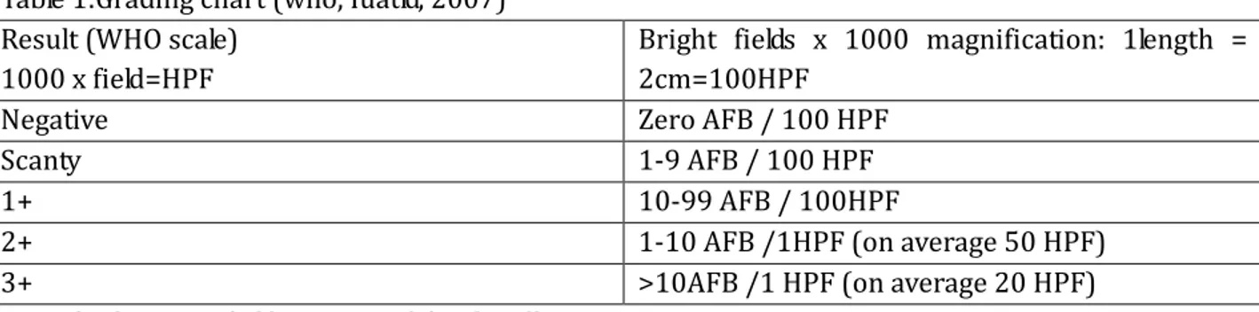
Table 1:Grading chart (who, Iuatld, 2007) Result (WHO scale)
1000 x field=HPF
Bright fields x 1000 magnification: 1length = 2cm=100HPF
Negative Zero AFB / 100 HPF
Scanty 1-9 AFB / 100 HPF
1+ 10-99 AFB / 100HPF
2+ 1-10 AFB /1HPF (on average 50 HPF)
3+ >10AFB /1 HPF (on average 20 HPF)
Journal of Evolution of Medical and Dental Sciences/ Volume 2/ Issue 20/ May 20, 2013 Page 3452 Table 2: Grading chart (who, Iuatld, 2007) for led fluorescent microscopy:
Result (WHO scale)
1000 x
field=HPF
LED fluorescent microscopy (400 x :1 length =40 fields =200HPF)
Minimum number of fields to be examined
Negative Zero AFB / 1 length 40
Scanty 1-19 AFB / 1 length 40
1+ 20-199 AFB / 1 length 40
2+ 5-50 AFB / 1 field on average 20
3+ >50 AFB / 1 field on average 8
For the present study 2+ and 3+ were classified as multibacillary and 1+ and scanty as paucibacillary [9].
RESULTS: Out of 634 sputum samples collected, 67 and 105 sputum samples were found to be positive for AFB by ZN and AO staining respectively. The ZN smear positivity rate and the AO smear positivity rate in this study was 10.57% and 16.56% respectively [Table 1 and Chart 1].
Table 3: Comparison of ZN and Auramine staining reports:
Staining method used No. of Positive smears No. of Negative smears ZN stain 67 (10.57%) 567 (89.43%)
Auramine stain 105 (16.56%) 529 (83.44%)
Table 4: Distribution of positive slides by grading and technique used:
Grading ZN Staining Auramine Staining
Scanty 1(1.49%) 18(17.14%)
1+ 2(2.99%) 8(7.62%)
2+ 28(41.8%) 35(33.33%)
3+ 36(53.73%) 44 (41.90%)
Total 67 105
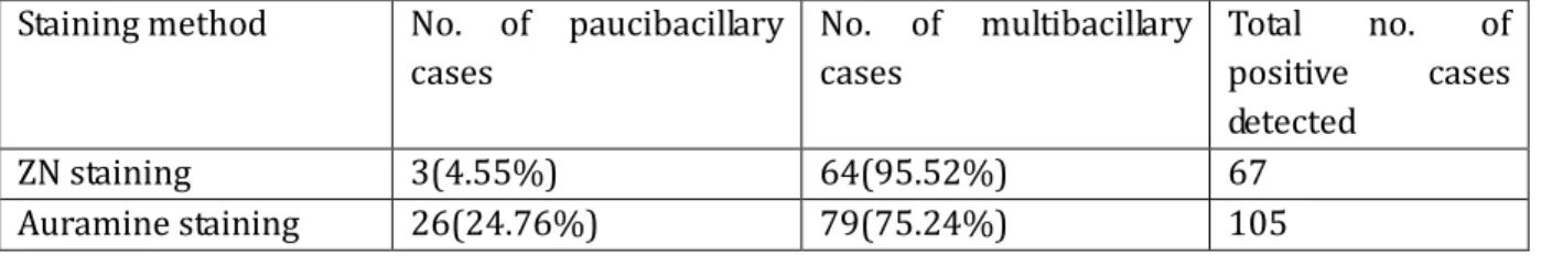
Table 5: Distribution of paucibacillary and multibacillary cases detected by ZN staining and LED fluorescent microscopy.
Staining method No. of paucibacillary cases
No. of multibacillary cases
Total no. of positive cases detected
ZN staining 3(4.55%) 64(95.52%) 67
Journal of Evolution of Medical and Dental Sciences/ Volume 2/ Issue 20/ May 20, 2013 Page 3453 Table 6: Comparison of LED fluorescent microscopy (Auramine staining) with ZN staining method:
Type of staining method used:
Fluorescent
microscopy (Auramine stain)smear positive
Fluorescent
microscopy (Auramine stain)smear negative
Total
ZN smear positive 66(10.41%) 1(0.16%) 67
ZN smear negative 39(6.15%) 528(83.28%) 567
Total 105 529
DISCUSSION: Current recommendations for the control of Tuberculosis emphasize early case detection so as to allow treatment of patients and there by limit the transmission of bacilli.
ZN stain can detect bacilli when they are in the order of 105/ml of the sputum, where as a more sensitive AO stain can detect in the order of 104/ml of sputum. In the present study out of the 634 samples examined, 67(10.57%) and 105(16.56%) TB cases were detected by ZN and AO staining methods respectively. Similar results have also been reported by studies done by Suria kumar et al, 2012[10]. Whereas higher smear positivity rates were shown by K Prasanthi et al, 2005(50% by ZN, 69% by AO) [9] and Ulukanligil et al (67.6% by ZN, 85.2% by AO)[11].
The results of this study suggest that there is no need for culture as the gold standard, given the high consistency between results obtained with the two techniques. The high consistency of the two methods resolves a pre-eminent question of quality assurance, using a system of internal quality control without recourse to culture or external proficiency testing. Nevertheless, recourse to culture could be valuable, particularly for microscopically low scanty results, both for confirmation of microscopic findings and for internal quality control.[12]
It was observed that a total no. of 39 sputum smears which were negative by ZN method were positive by AO staining method and that AO staining with LED Microscopy was more efficient over ZN stain in determining paucibacillary cases have also been proved in this study. AO staining could detect 26 paucibacillary cases, whereas ZN staining detected only 3 of them. This was in concordance with studies done by Laifangbam et al 2009[13]. Failure to detect and hence to treat paucibacillary cases can be effectively prevented by the use of fluorescent LED microscopy. The false positivity with fluorescent staining was very low (0.16%) in our study which was in accordance with study done by Khatun et al (2011).[14]
In this study AO staining was found to be 6.15% more effective than ZN staining. This shows that fluorochrome staining of sputum smears in comparison to that of ZN staining is a better method of microscopy. FM LED therefore appears to be a more sensitive technique than ZN due to its ability to detect low bacilli load especially in HIV positive subjects.
CONCLUSION: Hence our study concludes that Fluorochrome staining with LED is more efficient over ZN stain in detecting Tuberculosis bacilli in sputum, especially the paucibacillary cases and also FM LED has been found to be less time consuming as compared to ZN method (1000x) in the diagnosis of TB. FM with LED is easier to use, quicker and cheaper especially in centers where large numbers of sputum specimens are processed.
Journal of Evolution of Medical and Dental Sciences/ Volume 2/ Issue 20/ May 20, 2013 Page 3454 1. WHO,Global Tuberculosis Control Surveillance, Planning.WHO Report 2008.WHO,
Geneva,Switzerland, 1-242(2008)
2. Elliott A M, Namaambo K, Allen B W, et al. Negative sputum smear results in HIV positive patients with pulmonary tuberculosis in Lusaka, Zambia. Tubercle Lung Dis 1993; 74: 191-194.
3. Rieder H L, Van Deun A, Kam K M, Kim S J, Chonde T M, Trebucq A, Urbanczik R. Priorities for tuberculosis bacteriology services in low income countries. 2nd ed. Paris, France: International Union against Tuberculosis and Lung Disease, 2007.
4. Steingert KR, Henry M, vi vienne. Fluorescence versus conventional sputum smear microscopy for tuberculosis, a systematic review. Lancet Infect Dis 2006; 6:570-81
5. Murry SJ, Barret A, Magee JG, Freeman R. Optimization of acid fast smears for the direct detection of mycobacteria in clinical samples. J Clin Pathol 2003; 56: 613-15
6. Dinnes J, Deeks J, Kunst H, Gibson A, Gummins E, Waugh N, Drobniewski F, Lalvani A. A systematic review of rapid diagnostic tests for the detection of tuberculosis infection. Health Technol Assess 2007; 11(3):1-196
7. Van Hung N, Sy DN, Anthony RM, Cobelons FG, van Soolingen D. Fluorescence microscopy for tuberculosis diagnosis. Lancet Infect Dis.2007; 7(4): 238-239
8. Forbes BA, Sahm DF, Weissfeld AS. Mycobacteria; In Bailey and Scotts’ Diagnostic Microbiology. 12th edition. Missouri: Mosby, 2007: 491-492.
9. Prasanthi K, Kumari AR. Effi cacy of fluorochrome stain in the diagnosis of pulmonary tuberculosis co-infected with HIV. 2005. Indian J Med Microbiol 2005; 23: 179–81.
10.Dr. J. Suria Kumar. Dr. C. Chandrasekar. Dr. S. Rajasekaran. Comparison ofconventional and fluorescent staining methods in diagnosis of pulmonary tuberculosis among HIV seropositive individuals.Jr of evolution of med and dental sci.2012; Vol1, Issue4:463-466. 11.Ulukanligil M, Aslan G, Tasci S. A comparative study on the different staining methods and
number of specimens for the detection of acid-fast bacilli. Mem Inst Oswaldo Cruz. 2000;
95:855-8.
12.Ba. F, Rieder H.L A comparison of fluorescence microscopy with the Ziehl-Neelsen technique in the examination of sputum for acid-fast bacilli.1999 INT J TUBERC LUNG DIS 3(12):1101– 1105.
13.Laifangbam S, Singh HL, Singh NB, Devi KM, Singh NT. A comparative study of fluorescent microscopy with Ziehl-Neelsen staining and culture for the diagnosis of pulmonary tuberculosis. Kathmandu University Medical Journal (2009), Vol. 7, No. 3, Issue 27, 226-230. 14.Z.Khatun, M. Kamal, C K Roy, T Sultana , M.Q Rahman , MBAS Azad, ANN Ahmed.Usefulness of Light Emitting Diode (LED) fluorescent microscopy as a tool for rapid and effective method for the diagnosis of pulmonary tuberculosis.2011 Bangladesh Med Res Counc Bull
Journal of Evolution of Medical and Dental Sciences/ Volume 2/ Issue 20/ May 20, 2013 Page 3455
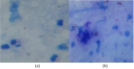
(a) (b)
Figure 1: Photomicrograph of Ziehl-Neelsen stain sputum smear showing TB Bacilli (1000x) as bright pink to red, beaded or barred forms, while the tissues cells and other organism are stained blue. : (a) AFB++ and (b) AFB +++
(a) (b)
Figure 2: Photomicrograph of Auramine-O stain sputum smear by LED microscopy (400x). The bacilli are seen as yellow luminous organism in a dark field (a) AFB++ and (b) AFB+++
Journal of Evolution of Medical and Dental Sciences/ Volume 2/ Issue 20/ May 20, 2013 Page 3456 Chart 2: