855 855 855 855 855 Mem Inst Oswaldo Cruz, Rio de Janeiro, Vol. 93(6): 855-860, Nov./Dec. 1998
Experimental Infection of
Culex (Culex) quinquefasciatus
and
Aedes (Stegomyia) aegypti
with
Wuchereria bancrofti
Cláudia ML Calheiros, Gilberto Fontes, Paul Williams*, Eliana MM Rocha/
+Departamento de Patologia, Centro de Ciências Biológicas, Universidade Federal de Alagoas, Praça Afranio Jorge s/no, 57010-020 Maceió, AL, Brasil *Departamento de Parasitologia, Instituto de Ciências Biológicas,
Universidade Federal de Minas Gerais, Belo Horizonte, MG, Brasil
A study was conducted to determine the susceptibility of local strains of Culex quinquefasciatus and
Aedes aegypti to infection with the strain of Wuchereria bancrofti that occurs in Maceió, State of Alagoas, Brazil. Cx. quinquefasciatus blood fed simultaneously on the same microfilariae carrier in-gested more blood and 2-3x more microfilariae than Ae. aegypti. Survival rates of both species of insects living for 21 days after blood feeding on microfilaraemic patients were not significantly different from the survival rates of mosquitoes that blood fed on amicrofilaraemic individuals. W. bancrofti para-sites underwent normal development in Cx. quinquefasciatus, with third stage larvae first being re-corded on the 11th day post infection, and their numbers increasing thereafter. Development of filariae in Ae. aegypti did not proceed beyond the first larval stage, and there was a progressively increasing number of non-viable larvae with the passage of time. It is concluded that Ae. aegypti is not involved in the transmission of W. bancrofti in Maceió.
Key words: Culex quinquefasciatus - Aedes aegypti - experimental infection - Wuchereria bancrofti filariasis -microfilariae
Culex quinquefasciatus is the principal vector of Bancroftian filariasis in areas where Wuchere-ria bancrofti has nocturnal periodicity, but, in other places, other species of mosquitoes may serve as insect hosts of the parasite (White 1989, WHO 1992). In Brazil, Causey et al. (1945), Rachou et al. (1955) and Rachou (1956) incriminated spe-cies of Aedes and Anopheles as hosts of secondary importance.
In Maceió, capital of the State of Alagoas, northeast Brazil, Deane et al. (1953) and Fontes et al. (1994) established that Cx. quinquefasciatus is the most important insect host of W. bancrofti. However, Ae. aegypti is frequently found in hu-man habitations in Maceió and it was considered necessary to determine if this species plays a role in the transmission of W. bancrofti in the city.
MATERIALS AND METHODS
Routine maintenance of mosquito colonies -Adult females of Cx. quinquefasciatus and Ae. aegypti were collected inside houses in urban areas of Maceió. After identification, each species was placed in a separate cage containing bowls of
dechlo-+Corresponding author. Fax: +55-82-221.2501. E-mail:
emmr@fapeal.br Received 4 March 1998 Accepted 2 July 1998
rinated water for oviposition. The mosquitoes were maintained in an insectary at 27 ± 2ºC and of 80±10% RH, and provided a 10% glucose solution. After eclosion, larvae were fed with ground food for laboratory mice. The cubes of mouse food had been previously sterilized by autoclaving. Pupae were transferred to water bowls and placed in clean insect cages for eclosion of adults. Emerged adult females of both species were pro-vided a blood meal on pigeons weekly.
The established colony of Cx. quinquefasciatus
has been maintained since March 1993. For public health safety, related to the possible transmission of Yellow Fever and Dengue, the Ae. aegypti
colony was maintained only for the duration of the study (January to August 1996).
856 856 856 856
856 Infection of Cx. quinquefasciatus and Ae. aegypti with W. bancrofti CML Calheiros et al.
Experimental infection of mosquitoes - Nul-liparous 2-5 day-old females of both species were used in the experiments. The sugar solution was removed 24 hr before the blood meal to increase blood-feeding. Females of both species were ex-posed simultaneously to either the upper surface of the hand or on the forearm of the same volun-teer between 23 hr and midnight. The time of blood feeding coincided with the peak of microfilaraemia recorded in Maceió (Rocha et al. 1991). To reduce discomfort, blood feeding times were limited to 5-10 min for Ae. aegypti and to 10-20 min for Cx. quinquefasciatus. Female mosquitoes that did not engorge were discarded.
Volume of ingested blood - The amount of in-gested blood was determined by weighing 20 unfed females of each species immediately before blood feeding, and weighing 20 engorged females im-mediately after their blood meals. The volume of blood ingested was estimated by dividing the weight differences by 1,055 (1,055 mg/ml being the approximate density of human blood).
Number of ingested microfilariae - The ex-pected number of microfilariae ingested by the mosquitoes was calculated by multiplying the microfilariaemia of each volunteer by the mean volume of ingested blood. The actual numbers of ingested microfilariae were calculated by dissect-ing out the contents of the gut of a sample of the blood fed adults. The midgut was opened and a smear was made of the contained blood. The smear was fixed in methanol, stained with Giemsa, and the number of ingested microfilariae determined.
Development of W. bancrofti in Cx. quin-quefasciatus and Ae. aegypti - Blood fed mosqui-toes were maintained in the insectary for up to 21 days and provided with a 10% glucose solution as previously described (the usual time needed for complete development of W. bancrofti in an in-sect host is less than the period of observations in the present study).
After blood feeding, 2-5 females of both spe-cies of mosquitoes were removed daily, stunned by placing them in a freezer at -10oC for 30 sec, and then dissected in a drop of buffered saline on a slide. After removal of the wings and legs, the head, thorax and abdomen were separated. Each of the body parts was transferred to separate drops of buffered saline and macerated to liberate filaria larvae. The numbers of larvae were recorded and their stage of development was noted.
Assessment of mosquito mortality - Mortality was recorded twice daily and dead mosquitoes dis-sected and examined for W. bancrofti larvae, as previously described. Survival rates were calcu-lated by dividing the numbers of mosquitoes sur-viving for 21 days after the infective blood meal
by the number of mosquitoes that took complete blood meals. Control mosquitoes of both species were allowed to blood feed on uninfected persons. The accumulative numbers of dead mosquitoes were plotted against time, and the correlation co-efficients were calculated and compared.
RESULTS
Survival rates of mosquitoes - Observations on mortality and survival are summarized in Table I. Of the 1,084 Cx. quinquefasciatus mosquitoes which blood fed on W. bancrofti microfilaraemic volunteers, 476 survived for more than 21 days, a survival rate of 0.66. For 926 female Ae. aegypti
exposed to infection, the survival rate was 0.71. Survival rates for mosquitoes that blood fed on individuals without microfilaraemia were 0.73 for
Cx. quinquefasciatus and 0.79 for Ae. aegypti.
Among species, survival rates were not signifi-cantly different in mosquitoes exposed to infec-tion (c2 = 3.21, p>0.05 for mosquitoes exposed to infection, and c2 = 0.49, p>0.05 for mosquitoes that blood fed on individuals without microfilariae). Likewise, there was no significant differences in the survival of the same species after blood feed-ing on persons with or without microfilariae (Cx. quinquefasciatus: c2 = 2.17, p>0.05; Ae. aegypti:
c2 = 2.49, p>0.05).
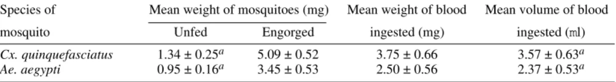
Fig. 1 shows the accumulative daily mortality of mosquitoes that blood fed on subjects with or without microfilariae. The four curves have the same correlation coefficient (r = 0.99).
Volume of ingested blood - The mean volume of blood ingested was 3.37±0.63 ml and 2.37±0.53
ml for Cx. quinquefasciatus and Ae. aegypti, re-spectively (Table II).
Number of ingested microfilariae - Sixty-one
Cx. quinquefasciatus were examined soon after they had taken an infective blood meal. Fifty-nine (97%) harboured 3-102 microfilariae in the mid-gut (mean 19.8±19.5 microfilariae/female). Simi-larly, 76 female Ae. aegypti were examined after blood feeding. Sixty-four (84%) were infected with 1-22 microfilariae (mean 5.7±5.6 microfilariae/fe-male).
Females of both species of mosquito ingested more microfilariae than the density in the periph-eral blood of the volunteers. The proportion of fe-male Cx. quinquefasciatus containing microfilariae was significantly higher than that recorded for fe-male Ae. aegypti (c2 = 4.5, p<0.05). Also, the num-ber of microfilariae ingested by female Cx. quinquefasciatus was significantly higher than the number ingested by female Ae. aegypti (p<0.05).
857 857857 857857 Mem Inst Oswaldo Cruz, Rio de Janeiro, Vol. 93(6), Nov./Dec. 1998
female Ae. aegypti (Table III). For both species of mosquito, the highest “concentration factor” was recorded in specimens that blood fed on the indi-vidual with 1,168 microfilariae/ml in the periph-eral blood.
Development of W. bancrofti in Cx. quinquefasciatus - One day after the infecting blood meal, “young” first stage larvae (=exsheathed microfilariae) were seen. Most of them (78.7%) were observed in the abdomen while 21.3% had reached the thorax. By the 4th day post infection, 94.5% of the parasites were observed in the tho-rax, but were still in the “young” L1 stage. Trans-formation into the sausage-shaped L1 larvae oc-curred between the 5th and 7th day post infection. After the 8th day of infection, larvae were observed undergoing the L1/L2 moult. The L2/L3 moult was first recorded on the 11th day post-infection and,
T
ABLE I
Summary of
Culex quinquefasciatus
and
Aedes aegypti
survival after blood feeding on
W
ucher
eria bancr
ofti
microfilaraemic volunteers
Numbers of mosquitoes
Species
of
mosquito
No. taking
No. dissected
No. killed
No. found
No. maintained
No. surviving
Survival rate
b
blood meal
immediately
daily
dead
a
in the insectary
more than
after blood meal
21 days
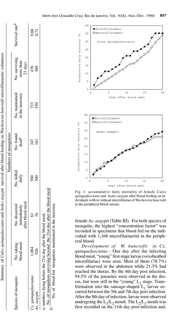
Cx. quinquefasciatus
1,084
61
300
247
723
476
0.66
Ae. aegypti
9
2
6
76
3
0
0
1
6
1
5
5
0
3
8
9
0.71
a
: numbers dying before the 21st day after the blood meal;
No. of mosquitoes surviving beyond the 21st day after the blood meal
b
:
No. of blood fed mosquitoes maintained in the insectary
0 5 10 15 20
Days after blood meal 0
5 10 15 20 25 30 35 40
Accumulative daily mortality (%)
Microfilaraemic Amicrofilaraemic
0 5 10 15 20
Days after blood meal 0
5 10 15 20 25 30 35 40
Accumulat
i
ve da
i
ly mortal
i
ty (%)
Microfilaraemic Amicrofilaraemic
Mortalidade diária de Aedes após repa sanguíneo Culex quinquefasciatus
Aedes aegypti
Fig. 1: accumulative daily mortality of female Culex
quinquefasciatus and Aedes aegypti after blood feeding on
in-dividuals with or without microfilariae of Wuchereria bancrofti
858 858 858 858
858 Infection of Cx. quinquefasciatus and Ae. aegypti with W. bancrofti CML Calheiros et al.
thereafter, the numbers of L3 (infective larvae) creased. Twenty-one days after exposure to in-fection 216 out of 476 dissected Cx. quin-quefasciatus mosquitoes yielded 860 infective lar-vae.
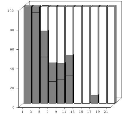
Development of W. bancrofti in Ae. aegypti -During the first four days post-infection, the ap-pearance of W. bancrofti in Ae. aegypti was simi-lar to their appearance in Cx. quinquefasciatus. After the 5th post-infection day, development of
W. bancrofti showed little progress. A small num-ber of exsheathed microfilariae developed into sau-sage shaped L1 larvae, but many of these had a shrunken, wrinkled appearance and were mostly immobile and were therefore considered dead. Development of the Maceió strain of W. bancrofti
in the Maceió strain of Ae. aegypti is shown in Fig. 2. None of the W. bancrofti larvae progressed be-yond the L1 stage, dead larvae were recorded as early as 5-7 days post-infection, and, with a single exception, only dead larvae were recorded after the 13th post-infection day.
DISCUSSION
Because the recommendations of Asahina (1964) and Consoli and Lourenço-de-Oliveira (1994) were followed, it is reasonable to assume that the colonies of Cx. quinquefasciatus and Ae. aegypti were adapted to laboratory conditions be-fore the presently reported experiments were un-dertaken.
Neither species of mosquito showed adverse effects due to the presence of W. bancrofti. This contrasts with studies of Wharton (1960) and Crans (1973) in Malasya and East Africa, respectively. However, in these earlier studies, patients had much higher microfilaraemia than did the volunteers in Maceió. It is possible that migration of high num-bers of developing filarial larvae within the mos-quito body have an effect on vector survival.
Previous authors (Manson 1884, O’Connor & Beatty 1936, Burton 1964, Crans 1973, McGreevy et al. 1982), working in different and widely sepa-rated parts of world, have recorded the disparity between the density of microfilariae in the periph-eral blood of human hosts and the numbers of mi-crofilariae ingested by arthropod hosts of the para-sites. Various factors have been suggested to ex-plain the apparent ability of mosquitoes to “con-centrate” microfilariae (Hairston & Jachowski 1968, Eberhard et al. 1988, Dickerson et al. 1989, Southgate & Bryan 1992). The simplest explana-tion is the manner in which mosquitoes acquire blood meals. The classical paper of Gordon and Lumsden (1939), confirmed by Griffiths and Gor-don (1952), shows that Ae. aegypti either obtains blood directly from a vessel or by lacerating ves-sels and, thus, forming a subcutaneous haemorrhage (pool feeding). Pool feeders take longer to engorge than do vessel feeders. When a mosquito pool feeds, there is more time for mi-crofilariae to seep from the lacerated vessel into TABLE II
Estimated weight and volume of blood ingested by females Culex quinquefasciatus and Aedes aegypti
Species of Mean weight of mosquitoes (mg) Mean weight of blood Mean volume of blood
mosquito Unfed Engorged ingested (mg) ingested (ml)
Cx. quinquefasciatus 1.34 ± 0.25a 5.09 ± 0.52 3.75 ± 0.66 3.57 ± 0.63a
Ae. aegypti 0.95 ± 0.16a 3.45 ± 0.53 2.50 ± 0.56 2.37 ± 0.53a
a: confidence limits 95%; significant differences (p<0.05).
TABLE III
Differences between the expected and observed numbers of microfilariae ingested by females of
Culex quinquefasciatus and Aedes aegypti
Microfilaraemia Mean number of microfilariae/mosquito Difference
of volunteers Observed Expected (No. observed/No. expected)
(mf/ml) Culex Aedes Culex Aedes Culex Aedes
906 8.8 3.5 3.4 2.0 2.6 1.8
1,168 24.4 5.5 3.2 2.0 7.6 2.8
1,215 13.8 6.8 4.3 3.4 3.2 2.0
859 859859 859859 Mem Inst Oswaldo Cruz, Rio de Janeiro, Vol. 93(6), Nov./Dec. 1998
the subcutaneous haemorrhage. However, it is rec-ognized that further studies are necessary to un-derstand the “concentration effect” of microfilariae in mosquitoes (Lowrie et al. 1989).
Complete development of W. bancrofti in Cx. quinquefasciatus was expected because females of this species have been found naturally infected in Maceió (Fontes et al. 1994).
Experimental infection of Ae. aegypti with W. bancrofti has given variable results. In some cases (Ponnudurai et al. 1971, McGreevy et al. 1982), the parasites have developed to the L3 stage, whereas other researchers (Hunt & Gunders 1990, Anosike & Onwuliri 1992) obtained negative re-sults. The present results are most similar to those of MacDonald and Ramachandran (1965) and Anosike and Onwuliri (1992), with larval devel-opment only progressing to the sausage-shaped phase.
The available literature provides evidence that susceptibility of Ae. aegypti to infectionwith fi-larial parasites is controlled by a sex-linked reces-sive gene (MacDonald & Ramachandran 1965,
Ponnudurai et al. 1971, McGreevy et al. 1974). Although it has not yet been possible to make ge-netic studies on Ae. aegypti in Maceió, it is rea-sonable to suspect that the Maceió strain of Ae. aegypti is genetically incapable of supporting the development of W. bancrofti. For this reason, it is concluded that Ae. aegypti is not involved in the transmission of Bancroftian filariasis in Maceió.
REFERENCES
Anosike JC, Onwuliri CO 1992. Experimental
Wuchere-ria bancrofti infection of Culex quinquefasciatus and
Aedes aegypti.Angew Parasitol 33: 139-142.
Asahina S 1964. Food material and feeding procedures for mosquito larvae. Bull WHO 31: 465-466. Burton GJ 1964. The intake of microfilariae of
Wuchere-ria bancrofti by Culex pipiens fatigans in British
Guiana. Ann Trop Med Parasitol58: 333-338. Causey OR, Deane MP, Costa O, Deane LM 1945.
Stud-ies on the incidence and transmission of filaria
Wuchereria bancrofti, in Belém, Brazil. Am J Hyg
41: 143-149.
Chularerk P, Desowitz RS 1970. A simplified membrane filtration technique for the diagnosis of microfilare-1 3 5 7 9 11 13 15 17 19 21
Days after the infecting blood meal 0
20 40 60 80 100
% of L1 larvae in the thorax
young L1 larvae (=unsheathed microfilariae)
sausage form L1 larvae
non-viable L1 larvae (young and sausage)
860 860 860 860
860 Infection of Cx. quinquefasciatus and Ae. aegypti with W. bancrofti CML Calheiros et al.
mia. J Parasitol56: 623-624.
Consoli RAGB, Lourenço-de-Oliveira R 1994. Principais Mosquitos de Importância Sanitária no
Brasil, Editora Fiocruz, Rio de Janeiro, 228 pp.
Crans WJ 1973. Experimental infection of Anopheles
gambiae and Culex pipiens fatigans with
Wuchere-ria bancrofti in Coastal Africa. J Med Entomol 10:
189-193.
Deane LM, Rosa D, Rachou RG, Martins JS, Costa A, Gomes HM, Carvalho ME 1953. A filariose bancroftiana em Maceió, Alagoas: resultado de um inquérito realizado em 1952. Rev Bras Malariol D Trop5: 17-22.
Dickerson JW, Eberhard ML, Lammie PJ, Roberts JM 1989. Further evidence of a skewed distribution of microfilariae in capillary blood. Trop Med Parasitol 40: 472-473.
Eberhard ML, Roberts JM, Lammie PJ, Lowrie Jr RC 1988. Comparative densities of Wuchereria bancrofti microfilariae in paired samples of capillary and venous blood. Trop Med Parasitol39: 295-298. Fontes G, Brito AC, Calheiros CML, Antunes CMF,
Rocha EMM 1994. Situação atual da filariose bancroftiana na cidade de Maceió, Estado de Alagoas, Brasil. Cad Saúde Públ 10 (Supl. 2): 293-300. Gordon RM, Lumsden WHR 1939. A study of the
behaviour of the mouth-parts of mosquitoes when taking up blood from living tissue; together with some observations on the ingestion of microfilariae.
Ann Trop Med Parasitol33: 259-278.
Griffiths RB, Gordon RM 1952. An apparatus which enables the process of feeding by mosquitoes to be observed in the tissues of a living rodent, together with an account of the ejection of saliva and its sig-nificance in malaria. Ann Trop Med Parasitol 46: 311-319.
Hairston NG, Jachowski LA 1968. Analysis of the
Wuchereria bancrofti population in the people of
American Samoa. Bull WHO38: 29-59.
Hunt RH, Gunders AE 1990. The susceptibility of five laboratory colonies of mosquitoes to the human nematode Wuchereria bancrofti (Cobbold). Ann Trop
Med Parasitol 84: 381-386.
Lowrie RC, Eberhard ML, Lammie PJ, Raccurt CP, Katz SP, Duverseau YT 1989. Uptake and development
of Wuchereria bancrofti in Culex quinquefasciatus
that fed on Haitian carriers with different microfi-laria densities. Am J Trop Med Hyg 41: 429-435. MacDonald WW, Ramachandran CP 1965. The
influ-ence of gene fm of Aedes aegypti to seven strains of
Brugia, Wuchereria and Dirofilaria.Ann Trop Med
Parasitol59: 64-73.
Manson P 1884. The metamorphosis of Filaria sanguinis
hominis in the mosquito. Trans Linn Soc Lond 2:
366-390.
McGreevy PB, Theis JH, Lavoipierre MMJ, Clark J 1974. Studies on filariasis. III. Dirofilaria immitis: emergence of infective larvae from the mouthparts
of Aedes aegypti. J Helminthol 48: 221-228.
McGreevy PB, Kolstrup N, Tao J, McGreevy MM, Marshal TF 1982. Ingestion and development of
Wuchereria bancrofti in Culex quinquefasciatus,
Anopheles gambiae and Aedes aegypti after feeding
on humans with varying densities of microfilariae in Tanzania. Trans R Soc Trop Med Hyg76: 288-296.
O’Connor FW, Beatty HA 1936. The early migrations
of Wuchereria bancrofti in Culex fatigans. Trans R
Soc Trop Med Hyg30: 125-127.
Ponnudurai T, Denham DA, Nelson GS 1971. The use of a membrane feeding technique for infecting mos-quitoes with filarial worms transported between labo-ratories. J Helminthol 45: 415-418.
Rachou RG 1956. Transmissores da filariose bancroftiana no Brasil. Rev Bras Malariol D Trop8: 267-268.
Rachou RG, Lima MM, Neto JAF, Martins CM 1955. Inquérito epidemiológico de filariose bancroftiana em uma localidade de Santa Catarina, como fase preliminar de uma prova profilática. Constatação de transmissão extradomiciliária por um novo vetor,
Aedes scapularis. Rev Bras Malariol D Trop7:
51-70.
Rocha EMM, Fontes G, Vergetti G, Santos ACB, Fire-man FAT, Dreyer G 1991. Periodicidade de microfilárias de Wuchereria bancrofti em filarióticos autóctones de Maceió-Alagoas. Rev Inst Med Trop
S Paulo33 (Supl. 8): 35.
Southgate BA, Bryan JH 1992. Factors affecting trans-mission of Wuchereria bancrofti by anopheline mos-quitoes. 4. Facilitation, limitation and proportional-ity and their epidemiological significance. Trans R
Soc Trop Med Hyg86: 523-530.
Wharton RH 1960. Studies on filariasis in Malaya: field and laboratory investigations of the vectors of a ru-ral strain of Wuchereria bancrofti.Ann Trop Med
Parasitol54: 78-91.
White GB 1989. Lymphatic filariasis, p. 23-34. In Geografical Distribution of Arthropod-borne
Dis-eases and their Principal Vectors, World Health
Or-ganization, Vector Biology and Control Division, WHO/VBC/89.967.
WHO - World Health Organization 1987. Control of Lymphatic Filariasis: A Manual for Health
Person-nel, WHO, Geneva, 89 pp.
WHO - World Health Organization 1992. Lymphatic
Filariasis: the Disease and its Control, Fifth report