Mitodepressive and clastogenic effects of aqueous extracts of the lichens
Myelochroa lindmanii
and
Canoparmelia texana
(Lecanorales, Parmeliaceae)
on meristematic cells in plant bioassays
José Marcello Salabert de Campos
1, Lisete Chamma Davide
1, Geraldo Luiz Gonçalves Soares
2and Lyderson Facio Viccini
31
Departamento de Biologia, Universidade Federal de Lavras, Lavras, MG, Brazil.
2
Departamento de Botânica, Universidade Federal do Rio Grande do Sul, Porto Alegre, RS, Brazil.
3
Departamento de Biologia, Universidade Federal University de Juiz de Fora, Juiz de Fora, MG, Brazil.
Abstract
The cytotoxicity effect of aqueous extracts of the lichens speciesMyelochroa lindmanii and Canoparmelia texana (Lecanorales, Parmeliaceae) were evaluated using meristematic cells of lettuce (Lactuca sativa) and maize (Zea mays). The seasonal effect was also evaluated. Extracts of M. lindmanii and C. texana inhibited root growth and/or percentage germination, possibly due to alterations in the cell cycle. TheM. lindmanii extract showed anti-mitotic ef-fects and blocked the cell cycle in metaphase so that c-mitosis and cells with chromosome duplication were pro-duced. TheC. texana extract appeared to hinder cell division, increasing the number of interphase cells. In addition, both extracts caused an increase in percentage of cell death. Clastogenic effects were also observed, such as sticky chromosomes, bridges, fragments and later segregation. Both lichen species are thus potential sources of biologi-cally active substances with possible applications in biology, medicine and agronomy.
Key words: Canoparmelia texana, cytotoxicity, mitotic index,Myelochroa lindmanii, plant bioassays. Received: April 12, 2007; Accepted: September 25, 2007.
Lichens are symbiotic organisms and together with some marine organisms and frog venom, are some of the most important sources of biologically active compounds (Barnes, 2000) with antitumour, antiviral, antimycobac-terial, antiinflamatory, antiherbivoral, immunostimulating, cytotoxic and mito-depressive effects (Perryet al., 1999; Vijayakumaret al., 2000; Cochiettoet al., 2002). However, few studies have been done with lichens and screening bioassays are essential tools for identifying the effects of li-chens. Using plant cytogenetic tests, the effects can be observed at chromosome level (clastogenesis) through al-terations in chromosome structure and number (aneu-ploidy, polyploidy) (Grant, 1994). A wide range of plant screening procedures are available, notable among them lettuce (Lactuca sativaL.), used in root growth and germi-nation tests (Chonet al., 2005), and maize (Zea maysL.), commonly used as a cytotoxicity evaluation model (Pavel and Creanga, 2005).
In recent unpublished work we observed that aqueous extracts of the lichens Myelochroa lindmanii(Lynge) Elix & Hale andCanoparmelia texana (Tuckermann) Elix & Hale (Lecanorales, Parmeliaceae) inhibited the root growth ofL. sativain an allelopathic test, an observation which led to the work described in the present paper.
Considering that little knowledge about these species is available, we designed the present study to evaluate the effects of theM. lindmaniiandC. texanaextracts on germi-nation, root growth and chromosome alterations in lettuce and maize.
We collectedM. lindmaniiandC. texanatwice in the rainy season (January, summer) and twice in the dry season (August, winter) in Juiz de Fora city, Minas Gerais state, Brazil (-21.77° S, -43.35° W). The climatological details of these seasons were obtained from the Laboratory of Clima-tology and Environmental Analysis at the Federal Univer-sity of Juiz de Fora (UFJF) and are shown in Table 1. Voucher specimens were identified according to Ribeiro CH (MSc dissertation, UNESP, Botucatu, SP, 1998) and deposited at the Leopoldo Krieger herbarium (UFJF) asM. lindmanii (Voucher: 31656) and C. texana (Voucher: 31658).
Send correspondence to José Marcello Salabert de Campos. Uni-versidade Federal de Lavras, Departamento de Biologia, Campus Universitário, 37200-000 Lavras, MG, Brazil. E-mail: jmscampos@ yahoo.com.br.
An aqueous extract was prepared from each lichen by infusing macerated lichen tissue in distilled water at a li-chen-to-water ratio of 1:10 (w/v) during 24 h at 25 °C. The extracts were filtered through a Whatman number 1 filter paper and stored at 4 °C.
To evaluate the effects of the extract on percentage germination, root growth and cytogenetic alterations we used Grand Rapids variety lettuce seeds and inbred maize line L-922 seeds (UFV).
For germination and root growth analysis we used a completely random design with 4 replicates. For each repli-cate, we placed 30 lettuce seeds and 30 maize seeds in sepa-rate petri dishes, added 5 mL of extract to each dish and incubated them at 25 °C for 72 h, after which we assessed the percentage germination and root growth in millimeter. Control experiments replaced the extract with the same vol-ume of distilled water.
For cytogenetic analysis seeds were treated with dis-tilled water as described above until root emergence and then 5 mL of lichen extract was added and the seeds incu-bated at 25 °C for 36 h. After incubation, eight root tips (5 to 10 mm long) were collected from each dish, washed in distilled water and fixed in fresh cold 3:1 methanol:acetic acid (v/v). Slides were made by the air drying technique with enzymatic maceration (Carvalho and Saraiva, 1993) using 1:10 (v/v) Pectinex (Novo Nordisk):sodium citrate buffer, incubated at 34 °C and stained with 5% Giemsa. Mitotic and metaphase indices and the percentage of cyto-genetic abnormalities (bridges, fragments, sticky chromo-somes, later segregation, chromosome duplication, c-meta-phase and cell death) were assessed by scoring a minimum of 500 cells from each slide, with almost 12000 cells being analyzed per treatment.
The results for each season (dry or rainy) were ob-tained by calculating the average of the two collections. Statistical analysis was accomplished using the Tukey test (p < 0.05).
TheM. lindmaniiextract reduced root growth in both
L. sativa and Z. maysbut did not affect percentage germina-tion, whereas theC. texanaextracts reduced root growth in
L. sativaonly and percentage germination in bothL. sativa
andZ. mays, mainly when the lichens were collected during the rainy season (Table 2).
In general, inhibition of root growth and percentage germination were more evident for extracts made from
ma-terial collected during rainy season. We used cytogenetic analysis to investigate the mechanisms involved in reduc-tion of root growth and found that M. lindmanii extract caused an increase in mitotic index in bothZ. maysandL. sativa, due to an increase of the metaphase number, whereasC. texanaextract produced a reduction in both mi-totic index and metaphase index, principally for the extracts produced from material collected in the rainy season as was observed for theM. lindmaniiextract (Table 2).
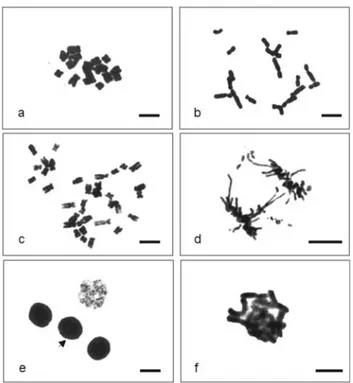
The extracts of both lichens induced cytologic and chromosome alterations. In L. sativa meristematic cells treated withM. lindmanii extract prepared from material collected in the rainy season there was a increase of 2.4, 10.7 and 4.1 times respectively for bridges, cell death and c-metaphase (Table 2). Similar, but smaller, effects being seen with material collected in the dry season (Table 2). The effect ofM. lindmaniiextract was similar inZ. mays
meristematic cells, with significant effects on c-meta-phases, anaphase/telophase bridges, chromosome frag-ments and cell death. Some chromosome/cell abnormalities being show in Figure 1.
We also observed a large increase in the percentage of
Z. maysanaphase/telophase bridges. The increase being 7 times forM. lindmaniiextract prepared from material col-lected in the rainy season and 2.8 times for extract made from material collected in the dry season. In addition, some
Z. mayscells with duplicated chromosome numbers were observed after treatment withM. lindmanii extract made from material collected in the both seasons (Figure 1).
TheC. texana extract produced less intense clasto-genic effects than theM. lindmaniiextract, with only sticky chromosomes and cell death being observed inL. sativa
andZ. maysmeristematic cells (Table 2, Figure 1). Unlike the effect of M. lindmanii extracts, sticky chromosomes constitute an important consequence of theC. texana ex-tract. In general, we verified that collections from the rainy season caused larger effects.
Information about the effects of lichen extracts on plant cells is still scarce considering the large number of li-chen species. In this paper we have reported cytotoxic ef-fects of lichen extracts onL. sativaandZ. maysat both the macroscopic level (percentage germination and root growth) and the microscopic level (cytogenetic analysis), with, in general, the extracts producing effects in all the tests. Macroscopically, we observed that the extracts tested
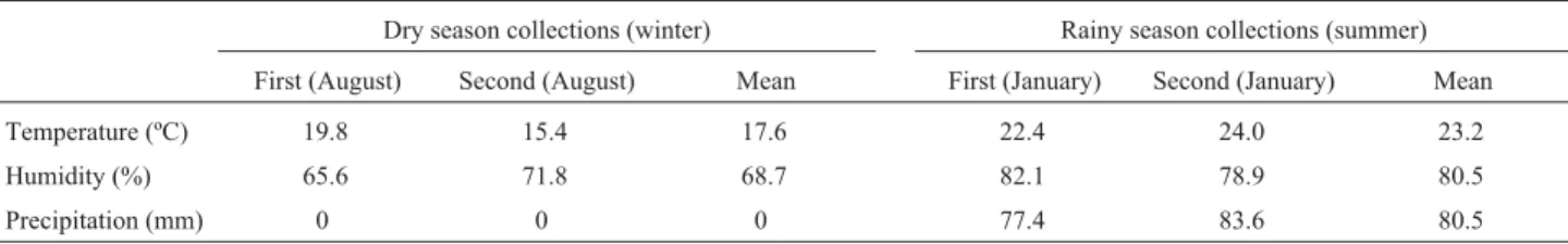
Table 1- The climatological data during the collection of the lichensMyelochroa lindmaniiandCanoparmelia texanain the city of Juiz de Fora (Minas Gerais. Data from the Laboratory of Climatology and Environmental Analysis, Federal University of Juiz de Fora, Brazil).
Dry season collections (winter) Rainy season collections (summer)
First (August) Second (August) Mean First (January) Second (January) Mean
Temperature (ºC) 19.8 15.4 17.6 22.4 24.0 23.2
Humidity (%) 65.6 71.8 68.7 82.1 78.9 80.5
interfered with the percentage germination and/or root growth of bothL. sativaandZ. mays. The effect of the ex-tracts appeared to be directly related to interference in the cell division process, including blocking cell division and the induction of c-metaphases. The increase in mitotic in-dex caused byM. lindmaniiextract could have been due to the interruption of the cell cycle and the accumulation of metaphase stage cells. TheM. lindmaniiextract seemed to act as an antimitotic, some of which affect the dynamics of microtubules which carry out important cell functions such as chromosome migration, cell structure and cell wall for-mation during both growth and the mitotic cycle (Jordan and Wilson, 1999). Treatment withM. lindmaniiextract ap-peared to affect the normal microtubule function, an hy-pothesis reinforced by the observed increase in the number of c-metaphases, increased late segregation of chromo-somes and the presence of some cells with chromosome du-plication. The observation of some cells with micronuclei and multipolar anaphases (data not shown) reinforces the hypothesis that this extract can act on microtubules. Verhoevenet al.(1990) demonstrated that micronuclei can
start to appear in condensed metaphase chromosomes spread in c-metaphase cells. Ramuluet al.(1995) reported that when microtubules are not formed, micro-nucleation occurs as a consequence of modified mitosis in which metaphase chromosomes form the micronuclei directly without the division of centromeres and sister chromatid separation.
TheM. lindmaniiextract produced no effect on ger-mination but it is important to note that in our study germi-nation was considered to have occurred when the radicle emerged, and at this initial stage of germination mitosis might not have been affected. Previous studies of several species have shown that specific inhibitors of mitosis did not hinder the initial stage of germination and suggests that mitosis is not always essential for germination (Labouriau and Spillmann, 1989), indicating that germination could be controlled by metabolic mechanisms that precede cell divi-sion. Even so, in our experiments,M. lindmanii extracts produced a marked inhibition in root growth in subsequent stages of germination. TheC. texanaextracts appear to act through a different mechanism, because our experiments
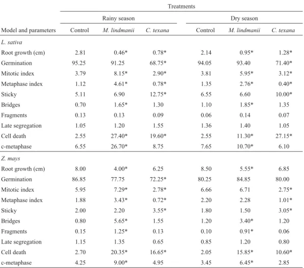
Table 2- Root growth (cm), germination (%) and cytogenetic alterations (%) inLactuca sativaandZea maystreated with aqueous extracts ofMyelochroa lindmaniiandCanoparmelia texanacollected in the city of Juiz de Fora (Minas Gerais state, Brazil) during the rainy season (January, summer) and dry season (August, winter).
Treatments
Rainy season Dry season
Model and parameters Control M. lindmanii C. texana Control M. lindmanii C. texana
L. sativa
Root growth (cm) 2.81 0.46* 0.78* 2.14 0.95* 1.28*
Germination 95.25 91.25 68.75* 94.05 93.40 71.40*
Mitotic index 3.79 8.15* 2.90* 3.81 5.95* 3.12*
Metaphase index 1.12 4.61* 0.78* 1.35 2.76* 0.40*
Sticky 5.11 6.90 12.75* 6.55 6.60 10.00*
Bridges 0.70 1.65* 1.30 1.10 1.85* 1.35
Fragments 0.13 0.13 0.09 0.06 0.14 0.07
Late segregation 1.05 1.20 1.55 1.36 1.40 1.05
Cell death 2.55 27.40* 19.60* 2.55 11.30* 27.15*
c-metaphase 6.55 26.70* 8.75 7.65 10.70* 6.10
Z. mays
Root growth (cm) 8.00 4.00* 6.25 8.50 5.55* 6.85
Germination 86.85 77.75 72.25* 80.25 84.85 80.00
Mitotic index 5.95 7.29* 2.78* 6.66 6.71 2.75*
Metaphase index 1.88 3.43* 0.72* 2.20 2.28 1.01*
Sticky 2.00 2.20 3.55* 1.80 1.50 3.05*
Bridges 0.80 5.65* 1.55 1.20 3.40* 1.20
Fragments 0.15 1.25* 0.13 0.10 0.91* 0.06
Late segregation 1.15 1.35 0.65 0.85 1.20 0.80
Cell death 2.70 20.35* 16.65* 2.05 15.85* 10.60*
c-metaphase 4.25 9.00* 4.95 3.45 6.45* 2.85
showed that these extracts provoked a decrease in mitotic index due to an increased number of interphase or dead cells. However, theC. texanaextracts did not seem to affect microtubule dynamics. Vyvyan (2002) has suggested that the accumulation of interphase cells may be due to inhibi-tion of DNA synthesis.
The clastogenic effects caused by the extracts from both lichen species included anaphase/telophase bridges, chromosome fragments and sticky chromosome. Babichet al. (1997) reported that metaphases with sticky chromo-somes lose their normal appearance and appear to have a sticky “surface” which causes chromosome agglomeration, possibly due to effects on chromatin and chromosome or-ganization. Singh (2003) states that the presence of chro-mosome fragments is an indication of chrochro-mosome breaks, and can be a consequence of anaphase/telophase bridges. Our observations suggests that the lichen extracts not only interfered with the cell cycle but also affected chromatin or-ganization and caused chromosome breaks.
Reigosaet al.(1999) state that, in plants, the amount and quality of allelopathic substances vary due to internal and external factors. Several studies have evaluated the sea-sonal effect of natural substances which can be influenced by different environmental factors, including temperature, light intensity, rainfall and the length of previous dry peri-ods. Some studies have shown the influence of ecological
factors on the concentration of secondary metabolites, with Nash and Olafsen (1995) having reported that climate change can influence the ecophysiological response of li-chens.
TheM. lindmaniiandC. texanaextracts used in our study were produced from material collected during both the dry and the rainy season so that we could investigate the effect of environmental changes. Our results indicate that, in general, the effects ofM. lindmaniiandC. texana ex-tracts were intensified when using material collected dur-ing the rainy season.
Information on the biochemistry of these M. lindmaniiandC. texanais still scarce, but the lichen para-depside atranorin is known to be a chemical taxonomic marker for these species (Ribeiro CH, MSc dissertation, UNESP, Botucatu, SP, 1998). Maranteet al. (2003) sug-gested that the paradepsides atranorin and chloroatranorin, could act as “parent metabolites” for the enzymatic and/or chemical transformation of a series of compounds that are released into the environment during rainy weather, when conditions favor their hydrolysis and the lichen needs to combat the germination and growth of plants. Maranteet al. (2003) also describe the chemical composition and phytotoxicity and antioxidative activity of allelochemicals from the lichenLethariella canariensis, also a member of the Parmeliaceae, and reported that a phytotoxic mixture of compounds from this lichen is leached into the environ-ment by rainwater where they accumulate by resisting mi-crobiological degradation and act as allelochemicals after penetrating the cell walls of seeds. Furthermore, Maranteet al.(2003) present results which suggest that atranorin and chloroatranorin undergo hydrolysis to produce monocyclic fragments that are present in varying amounts in the thallus ofL. canariensis and should be considered as true stress metabolites whose concentrations fluctuate with the clima-tic conditions. The paradepsides accumulate in the dry sea-son, whereas in the rainy season environmental humidity increases and enhances the production of the monocyclic fragments (principally methyl-β-orsellinate, atranol and chloroatranol), with rainwater leaching the phenolic metab-olites into the soil where they accumulate. The presence of atranorin inM. lindmaniiandC. texanasuggests a relation-ship between these substances and the effects observed in our study. The seasonal effects observed by us regarding the increased activity of the extracts prepared from material collected during the rainy season reinforcing this hypothe-sis. These are the challenges for further investigations in our laboratory, where work is underway to elucidate the re-lationship between the seasonal chemical composition of
M. lindmaniiandC. texanaand the genetic and cellular ef-fects described in the present paper.
Acknowledgments
The authors thank the Brazilian Federal agency Coor-denação de Aperfeiçoamento de Pessoal de Nível Superior (CAPES) for financial support and Juliana Lanna for assis-tance in the identification and collection of lichens.
References
Babich H, Segall MA and Fox KD (1997) TheAllium test– A sim-ple, eukaryote genotoxicity assay. Am Biol Teach 59:580-583.
Barnes J (2000) Pharmacognosy in the 21st century. Pharm J 264:701-703.
Carvalho CR and Saraiva LS (1993) A new heterochromatin banding patterns revealed by modified HKG banding tech-nique in maize chromosomes. Heredity 70:515-519. Chon SU, Jang HG, Kim DK, Kim YM, Boo HO and Kim YJ
(2005) Allelopathic potential in lettuce (Lactuca sativaL.) plants. Sci Hortic 106:309-317.
Cocchietto M, Skert N, Nimis PL and Sava G (2002) A review on usnic acid, an interesting natural compound. Naturwissen-schaften 89:137-146.
Grant WF (1994) The present status of higher plant bioassay for detection of environmental mutagens. Mutat Res 310:175-185.
Jordan MA and.Wilson L (1999) The use and action of drugs in analyzing mitosis. Method Cell Biol 61:267-295.
Labouriau LG and Spillmann FV (1989) Germination of seeds in solutions of antimitotics. An Acad Bras Cienc 61:355-371. Marante FJT, Castellano AG, Rosas FE, Aguiar JQ, Barreira JB
(2003) Identification and quantification of allelochemicals
from the lichen Lethariella canariensis: Phytotoxicity and antioxidative activity. J. Chem Ecol 29:2049-2071. Nash TH and Olafsen AG (1995) Climate change and the
ecophy-siological response of arctic lichens. Lichenologist 27:559-565.
Pavel A and Creanga DE (2005) Chromosomal aberrations in plants under magnetic fluid influence. J Magn Magn Mater 289:469-472.
Perry NB, Benn MH, Brennan NJ, Burgess EJ, Ellis G, Galloway DJ, Lorimer SD and Tangney D (1999) Antimicrobial, Anti-viral and Cytotoxic activity of New Zealand lichens. Lichen-ologist 31:627-636.
Ramulu KS, Dijkhuis P, Rutgers E, Blass J, Verbeek WHJ, Herhoeven HA and Colijn-Hooymans CM (1995) Micro-protoplast fusion technique: a new tool for gene transfer be-tween sexually-incongruent plant species. Euphytica 85:255-268.
Reigosa MJ, Sánches-Moreiras A and Gonzáles L (1999) Eco-physiological approach in allelopathy. Crit Rev Plant Sci 18:575-608.
Singh RJ (2003) Plant cytogenetics. CRC Press, Boca Raton, 463 pp.
Verhoeven HA, Ramulu KS and Dijkhuis PA (1990) Comparison of the effects of various spindle toxins on metaphase arrest and formation of micronuclei in cell-suspension cultures of
Nicotiana plumbaginifolia.Planta 182:408-411.
Vijayakumar CS, Viswanathan S, Kannappa RM, Parvathavar-thini S, Kundu AB and Sukumar E (2000) Antiinflammatory activity of (+)-usnic acid. Fitoterapia 71:564-566.
Vyvyan JR (2002) Allelochemicals as leads for new herbicides and agrochemicals. Tetrahedron 58:1631-1646.