Quinolone resistance and ornithine decarboxylation activity in lactose-negative
Escherichia coli
Franciane Gomig
1, Carolina Weigert Galvão
1, Denis Leandro de Freitas
1,
Larissa Labas
1, Rafael Mazer Etto
2, Luiz Antonio Esmerino
3,
Marcelo Andrade de Lima
4, Marcia Helena Appel
1, Silvio Marques Zanata
5,
Maria Berenice Reynaud Steffens
6, Helena Bonciani Nader
4, Rafael Bertoni da Silveira
11
Departamento de Biologia Estrutural, Molecular e Genética, Universidade Estadual de Ponta Grossa, Ponta Grossa, PR, Brazil. 2
Departamento de Química, Universidade Estadual de Ponta Grossa, Ponta Grossa, PR, Brazil. 3
Departamento de Análises Clínicas, Universidade Estadual de Ponta Grossa, Ponta Grossa, PR, Brazil. 4
Departamento de Bioquímica, Universidade Federal de São Paulo, São Paulo, SP, Brazil. 5
Departamento de Patologia Básica, Universidade Federal do Paraná, Curitiba, PR, Brazil. 6
Departamento de Bioquímica, Universidade Federal do Paraná, Curitiba, PR, Brazil.
Submitted: December 6, 2013; Approved: October 30, 2014.
Abstract
Quinolones and fluoroquinolones are widely used to treat uropathogenicEscherichia coliinfections. Bacterial resistance to these antimicrobials primarily involves mutations ingyrAandparCgenes. To date, no studies have examined the potential relationship between biochemical characteristics and quinolone resistance in uropathogenicE. colistrains. The present work analyzed the quinolone sensi-tivity and biochemical activities of fifty-eight lactose-negative uropathogenicE. colistrains. A high percentage of the isolates (48.3%) was found to be resistant to at least one of the tested quinolones, and DNA sequencing revealed quinolone resistant determining regiongyrAandparCmutations in the multi-resistant isolates. Statistical analyses suggested that the lack of ornithine decarboxylase (ODC) activity is correlated with quinolone resistance. Despite the low number of isolates examined, this is the first study correlating these characteristics in lactose-negativeE. coliisolates.
Key words:ODC,gyrA,parC, uropathogenic.
Introduction
Urinary tract infections (UTIs) are the second cause of antimicrobial prescriptions in South Brazil and are one of the major causes of office visits and hospitalization in the United States; these infections primarily affect women, pregnants and elderly people (Foxman, 2002; Tavares et al., 2008).Escherichia coliis the main agent of UTIs, espe-cially in community-acquired UTIs, and quinolones and fluoroquinolones have been used extensively to treat these infections (Ronald, 2003; Van Bambekeet al., 2005). Since these antimicrobials agents were introduced, resistant strains have emerged and spread around the world (Schito et al., 2009). Many studies have sought to understand the
mechanisms of resistance, to develop more efficient antibi-otics, and more recently, to relate resistance to biochemical or genetic characteristics (Lemoset al., 2011; Rodriguez-Martínezet al., 2011). The main mechanism of quinolone resistance involves mutations in the quinolone resistance determining region (QRDR) ofgyrAandparCgenes. The most common mutations are within the Ser83 and Asp87 codons in gyrA and within the Gly78, Ser80 and Glu84 codons inparC. ThegyrAandparCgenes encode subunits of DNA gyrase and topoisomerase IV, two enzymes in-volved in DNA supercoiling and DNA decatenation, re-spectively (Hooper, 2000). InE. coli, DNA gyrase is more susceptible to inhibition by quinolones than topoisomerase
ISSN 1678-4405 www.sbmicrobiologia.org.br
DOI: http://dx.doi.org/10.1590/S1517-838246320131291
Send correspondence to R.B. Silveira. Departamento de Biologia Estrutural, Molecular e Genética, Universidade Estadual de Ponta Grossa, 84030-900 Ponta Grossa, PR, Brazil. E-mail: rafaelbertoni@yahoo.com.br.
IV. In gram-negative bacteria, a single mutation ingyrA can reduce the susceptibility of DNA gyrase, furthermore additional mutations ingyrAor ingyrBandparCcan in-crease resistance to the antibiotic (Jacoby, 2005; Minarini et al., 2012).
The frequency of lactose-negative (lac-)E. coli phe-notype has shown to be very low, ranging from 5 to 10% (Winnet al., 2006; Oliveiraet al., 2006). To characterize this group, the present work selected and evaluated uro-pathogenic E. coli isolates from UTIs regarding their quinolone resistance and biochemical activity profiles.
Materials and Methods
Bacterial sample
Fifty-eightlac-E. coliisolates from the urine of UTI patients from the Ponta Grossa, Brazil region were ana-lyzed. These isolates were selected from inpatients and outpatients from 2008 to 2010. The urinary bacterial con-centration used to diagnose a urine infection was > 105 col-ony-forming units per milliliter. The bacteria were stored until their use at -20 °C in BHI medium (Himedia, Mumbai, India) containing 15% glycerol. Thelac- phenotype was confirmed in MacConkey agar (BD, Sparks, MD, EUA).E. coliATCC 25922 was used as a control in the antibiotic susceptibility and biochemical tests.
Antibiotic susceptibility tests
The susceptibility of theE. coliisolates to the quino-lones nalidixic acid (Nal), ciprofloxacin (Cip), norfloxacin (Nor) and ofloxacin (Ofx), and the beta-lactams cefotaxime (Ctx), ceftazidime (Ctz), aztreonam (Atm) and amoxicillin clavulanate (Amc) was determined using the disk diffusion method following the recommendations of the Clinical and Laboratory Standards Institute (2010). Disks (Laborclin, Pinhais, PR, Brazil) were stored at -20 °C until their use.
Biochemical characterization
The E. coli biotype was determined using the Enterobacteriaceae identification kit (Newprov, Pinhais, PR, Brazil) according to the manufacturer’s instructions. This kit, approved by the Agência Nacional de Vigilância Sanitária (ANVISA), provides the following ten differen-tial biochemical tests: L-tryptophan deamination; sulfidric acid, indole and gas production; glucose and rhamnose
fer-mentation; lysine and citrate utilization; ornithine decarboxylation and motility.
Detection ofgyrAandparCmutations
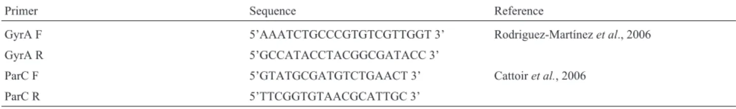
Multiplex PCR was used to amplify the gyrA and parCregions. SingleE. colicolonies grown on MacConkey agar (BD, Sparks, MD, USA) were suspended in 15mL of sterile deionized water and disrupted after 15 min of incu-bation at 95 °C. Then, the following reagents were added to a final volume of 30mL: Taq DNA polymerase Invitrogen buffer (1), magnesium chloride (2.5 mM), deoxyribo-nucleotide triphosphates (dNTPs) (Invitrogen, Carlsbad, California, USA) (0.2 mM), primers (IDT, Coralville, Iowa, USA) (0.2mM of GyrA primer and 0.4mM of ParC primer) and Taq DNA polymerase (0.75 U). The primer se-quences are listed in Table 1.
The genes were amplified using the following the thermal cycling profile: 2 min at 95 °C and 35 cycles of 30 s at 95 °C, 60 s at 55.4 °C and 60 s at 72 °C. The PCR prod-ucts were separated on a 1 x TAE, 2% agarose gel and quantified using UVP Labwork Software (UVP Inc.).
The amplification products (20mL) were treated with 10 U of exonuclease I (Biolabs, Ipswich, New England) and 1.0 U of alkaline phosphatase (USB, Cleveland, Ohio, USA) at 37 °C for 90 min. Then, the enzymes were inacti-vated at 80 °C for 30 min (Werleet al., 1994).
The treated PCR products (5mL) were sequenced us-ing 0.5mL of primer (10mM), 1mL of Big Dye Terminator mix (Applied Biosystems, Carlsbad, California, USA), 3mL of Big Dye Buffer (1X) and ultrapure H2O to a final volume of 10 mL using the following program: 2 min at 96 °C and 35 cycles of 45 s at 96 °C, 30 s at 55.4 °C and 4 min at 60 °C. The sequencing PCR products (10mL) were precipitated using 2 mL of ammonium acetate (7.5 M), 60mL of absolute ethanol and 10mL of ultrapure water fol-lowed by 45 min of centrifugation. The supernatant was discharged and the precipitate was washed with 70% etha-nol, dried and dissolved in deionized formamide. Sequen-cing was performed using a 24-capillary 3500xL System (Applied Biosystems, Carlsbad, California, USA). Reads were trimmed for the removal of low quality bases using the Phred program (Ewinget al., 1998). To detect nucleo-tide mutations, the DNA sequences were aligned using Clustal W (Thompsonet al., 1994) against the wild-type
Table 1- Primer sequences
Primer Sequence Reference
GyrA F 5’AAATCTGCCCGTGTCGTTGGT 3’ Rodriguez-Martínezet al., 2006
GyrA R 5’GCCATACCTACGGCGATACC 3’
ParC F 5’GTATGCGATGTCTGAACT 3’ Cattoiret al., 2006
ParC R 5’TTCGGTGTAACGCATTGC 3’
gyrAorparCgene nucleotide sequences fromE. coliK12 substr. MG1655 (accession numbers 946614 and 947499, respectively).
Statistical analysis
Statistical analyses to correlate antibiotic resistance and biochemical characteristics were performed using Yates’ chi-squared test because it is recommended for small sample numbers (Graphpad Prism 6.0). P value < 0.01 were considered statistically significant.
Results and Discussion
Forty-eight percent of thelac-E. coliisolates (28/58) were able to grow in the presence of at least one quinolone. Among the resistant isolates, seventy-nine percent (22/28) were resistant to all quinolones tested, and 21% (6/28) were resistant only to nalidixic acid. In contrast, only one isolate displayed intermediate resistance to the beta-lactams cefo-taxime and ceftazidime. Our data showed high quinolone resistance rates among the isolates. Other studies that ana-lyzed uropathogenicE. coliisolates in the same area re-vealed a norfloxacin resistance rate of 11.6% in Ponta Grossa (Bailet al., 2006) and 13.8% in Curitiba (Itoet al., 2008). In Fortaleza in the northeast of Brazil, a lower resis-tance rate of 7.5% was described for norfloxacin (Araújoet al., 2011). In Europe, a multicentric study showed a cipro-floxacin resistance rate from 1.4% to 12.9% (Schitoet al., 2009), whereas a higher rate (31%) was registered in the hospitalized population in Ribeirão Preto, Brazil (Santoet al., 2006). Because our isolates were primarily of commu-nity origin, the elevated resistance was unexpected. The available literature analyzed all uropathogenicE. coli lates without determining the lactose phenotype of the iso-lates; our work suggests that the quinolone resistance fre-quency is increased inlac-E. colistrains.
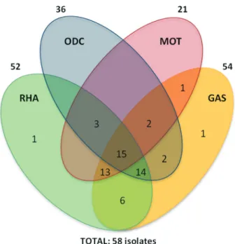
Biochemical tests identified nine E. coli biotypes. They presented a common behavior in the majority of the tests except Ornithine Decarboxylase (ODC), Motility (Mot), Gas production (Gas) and Rhamnose fermentation (Rha) (Figure 1). The most common biotypes (72%), 981, 991 and 971, were able to ferment glucose and rhamnose, to produce gas and indole and to metabolize lysine and citrate. On the other hand, they presented a distinguished profile in the ornithine decarboxylase and motility tests. Most of the ornithine decarboxylase positive (ODC+) E. coli isolates (26/36) showed to be sensitive to quinolone while most of the ODC-E. coliisolates (18/22) showed to be resistant to it. Statistical analyses revealed a relationship between ODC and quinolone resistance (p < 0.01); no relationship was identified between motility and the same resistance parame-ter (p > 0.05). Several studies relating biological and genetic characteristics with antibiotic resistance have been per-formed over the last years (Bashiret al., 2011). Ferjaniet al. (2011) showed a direct relationship among virulence deter-minants, phylogenetic groups and susceptibility to
fluoro-quinolones. A Brazilian study relatedE. colicarbohydrate fermentation to virulence factors. Dulcitol-positive and raffinose-negative isolates were found to be more virulent than other isolates (Lemoset al., 2011). Vilaet al.(2002) re-lated quinolone resistance and virulence factors, suggesting that quinolone-resistant strains are less virulent. The present study is the first to relate biochemical characteristics to quinolone resistance in lactose-negativeE. coliisolates.
Because the primary mechanism of quinolone resis-tance involves mutations in the QRDR of thegyrA and parCgenes (Jacoby, 2005), these regions of the isolates’ DNA were sequenced. One quinolone-sensitivelac-isolate was also included as a negative control. Of the twenty-eight quinolone resistant isolates analyzed, six could not be mo-lecularly analyzed due to the low quality of their sequences. Their nucleotide substitutions are shown in Table 2.
QRDR regions or other mechanisms that were not explored here, such as changes in permeability or efflux pump activ-ity (Friedmanet al., 2001).
The seventeen sequencedE. coliisolates that showed resistance to all of the tested quinolones presented GyrA substitution at codons 83 (Ser®Leu) and 87 (Asp®Asn) and ParC substitution at codon 80 (Ser ® Ile) and 84 (Glu®Val or Glu®Lys). Other studies have detected these QRDR GyrA and ParC amino acid mutations (Chen et al., 2001; Mavroidiet al., 2012). These changes were shown to reduce the affinity of the drug for their targets, thus resulting in bacterial growth even in the presence of quinolone (Bernardet al., 2001).
Silva and Mendonça (2012) suggested that the GyrA codon 83 mutation generates supercoiling DNA alterations that could modify the expression of virulence factors. In ad-dition, Weberet al.(2013) demonstrated that alterations in supercoiling affect fundamental cellular processes, includ-ing transcription. Based on these observations, it is possible that the GyrA codon 83 mutation that was detected in all of the analyzed multi-resistant isolates could be preventing the transcription of thelacoperon genes and thespeCgene, thus generating lac- and ODC- phenotypes, respectively. Because we cannot rule out the possibility that these genes are absent from the genome of the isolates, further studies are necessary.
A recent work showed that sublethal concentrations of fluoroquinolones were able to produce oxidative stress. To prevent DNA damage caused by reactive oxygen spe-cies (ROS) in the bacterial cell, ODC is upregulated, and the polyamine concentration increases (Umezawa et al., 1997; Tkachenkoet al., 2011). Therefore, it might be inter-esting to measure ROS production in the studied isolates in future experiments.
In conclusion, the studied uropathogenic lactose-negativeE. coliisolates showed a high quinolone resistance rate and indicated that there is a relationship between the absence of ornithine decarboxylase activity and quinolone resistance. The present work could serve as the basis for more comprehensive studies including a greater number of isolates from different localities to confirm our results.
Acknowledgments
We thank CNPq, CAPES, INCT-FBN/CNPq-MCT and Fundação Araucária for providing financial support for this study. We would like to thank the researcher groups from UEPG, UNIFESP and UFPR for excellent technical support.
References
Bail L, Ito CA, Esmerino LA (2006) Infecção do trato urinário: comparação entre o perfil de susceptibilidade e a terapia empírica com antimicrobianos. Revista Brasileira de Aná-lises Clínicas 38:51-56.
Bashir S, Sarwar Y, Ali Aet al.(2011) Multiple drug resistance patterns in various phylogenetic groups of uropathogenicE. coli isolated from Faisalabad region of Pakistan. Braz J Microbiol 42:1278-83.
Bernard FM, Maxwell A (2001) Interaction between DNA gyrase and quinolones: effects of alanine mutations at GyrA sub-unit residues Ser83 and Asp87. Antimicrob Agents Chemoter 45:1994-2000.
Cattoir V, Lesprit P, Lascols Cet al.(2006) In vivo selection dur-ing ofloxacin therapy of Escherichia coli with combined topoisomerase mutations that confer high resistance to oflo-xacin but susceptibility to nalidixic acid. J Antimicrob Chemother 58:1054-1057.
Chen JY, Siu LK, Chen YHet al.(2001) Molecular epidemiology and mutations at gyrA and parC genes of ciprofloxacin-resistantEscherichia coliisolates from a Taiwan medical center. Microb Drug Resist 7:47-53.
Clinical and Laboratory Standards Institute (2010). Performance standards for antimicrobial susceptibility testing; Twentieth informational supplement Document M100-S20. Wayne, Pa: Clinical and Laboratory Standards Institute.
Everett MJ, Jin YF, Ricci Vet al.(1996) Contributions of individ-ual mechanisms to fluoroquinolone resistance in 36
Esche-richia coli strains isolated from humans and animals.
Antimicrob Agents Chemoter 40:2380-2386.
Ewing B, Hillier L, Wendl Met al.(1998) Base-calling of auto-mated sequencer traces using phred. I. Accuracy assess-ment. Genome Res 8:175-185.
Ferjani S, Saidani M, Ennigrou Set al.(2011) Virulence determi-nants, phylogenetic groups and fluoroquinolone resistance
inEscherichia coliisolated from cystitis and pyelonephritis.
Pathologie Biologie 60:270-274.
Foxman B (2002) Epidemiology of Urinary Tract Infections: Inci-dence, Morbidity, and Economic Costs. Am J Med 113:5S-13S.
Friedman SM, Lu T, Drlica K (2001) Mutation in the DNA gyrase A gene ofEscherichia colithat expands the quinolone resis-tance-determining region. Antimicrob Agents Chemoter 45:2378-2380.
Hooper DC (2000) Mechanisms of Action and Resistance of Older and Newer Fluoroquinolones. Clin Infect Dis 31:24-28.
Ito CA, Gales AC, Tognim MCet al.(2008) Quinolone-resistant
Escherichia coli. Braz J Infect Dis 12:5-9.
Jacoby GA (2005) Mechanisms of Resistance to Quinolones. Clin Infect Dis 41:S120-S126.
Komp-Lindgren P, Karlsson A, Hughes D (2003) Mutation Rate and Evolution of Fluoroquinolone Resistance inEscherichia coli Isolates from Patients with Urinary Tract Infections. Antimicrob Agents Chemother 47:3222-3232.
Lemos TD, Tavares LGF, Arais LRet al.(2011) Avaliação da biotipagem na distinção de perfis de virulência de amostras
deEscherichia coliuropatogênica isoladas no hospital
uni-versitário Antonio Pedro, Niterói, RJ XXVI Congresso Brasileiro de Microbiologia, Foz do Iguaçu, PR.
Mavroidi A, Miriagou V, Liakopoulos Aet al. (2012) Cipro-floxacin-resistantEscherichia coliin central Greece:
mech-anisms of resistance and molecular identification. BMC Infect Dis 12:371-377.
Minarini LAR, Darini ALC (2012) Mutations in the quinolone re-sistance-determining regions of gyrA and parC in Entero-bacteriaceae isolates from Brazil. Braz J Microbiol 43:1309-1314.
Oliveira BG, Albini CA, Botão GMDet al.(2006) A identificação direta pelos meios cromogênicos é confiável a ponto de dispensar as provas bioquímicas? Newslab 75:130-142. Rodríguez-Martínez JM, Velasco C, Pascual Aet al.(2006)
Cor-relation of quinolone resistance levels and differences in basal and quinolone-induced expression from three qnrA-containing plasmids. Clin Microbiol Infect 12:440-445. Rodriguez-Martínez JM, Cano ME, Velasco C et al. (2011)
Plasmid mediated quinolone resistance: an update. J Infect Chemother 17:149-182.
Ronald A (2003) The etiology of urinary tract infection: tradi-tional and emerging pathogens. Disease a Month 49:71-82. Santo E, Salvador MM, Marin JM (2006) Antimicrobial
resis-tance among urinary tractEscherichia coliisolates from in-patients and outin-patients in a tertiary care center in Sao Paulo, Brazil. Int J Infect Dis 11:558-559.
Schito GC, Naber KG, Botto Het al.(2009) The ARESC study: an international survey on the antimicrobial resistance of pathogens involved in uncomplicated urinary tract infec-tions. Int J Antimicrob Agents 34:407-413.
Silva GJ, Mendonça N (2012) Association between antimicrobial resistance and virulence in Escherichia coli. Virulence 3:18-28.
Tavares NUL, Bertoldi AD, Mucillo-Baisch AL (2008) Pres-crição de antimicrobianos em unidades de saúde da família no Sul do Brasil. Caderneta de Saúde Pública 24:1791-1800. Thompson JD, Higgins DG, Gibson TJ (1994) CLUSTAL W: im-proving the sensitivity of progressive multiple sequence alignment through sequence weighting, position specific gap penalties and weight matrix choice. Nucleic Acids Res. 22:4673-80.
Tkachenko AG, Akhova AV, Shumkov MSet al.(2011) Polya-mines reduce oxidative stress inEscherichia colicells ex-posed to bactericidal antibiotics. Res Microbiol 163:83-91. Umezawa N, Arakane K, Ryu Aet al.(1997) Participation of
re-active oxygen species in phototoxicity induced by quinolone antibacterial agents. Arch Biochem Biophys 342:275-281. Van Bambeke F, Michot JM, Van Eldere Jet al.(2005)
Quino-lones in 2005: an update. Clin Microbiol Infect 11:256-280. Vila J, Simon K, Ruiz Jet al.(2002) Are Quinolone-Resistant
UropathogenicEscherichia coliLess Virulent? J Infect Dis 186:1039-1042.
Webber MA, Ricci V, Whitehead Ret al.(2013) Clinically rele-vant mutant DNA gyrase alters supercoiling, changes the transcriptome, and confers multidrug resistance. MBio 4:1-10.
Werle E, Schneider C, Renner Met al.(1994). Convenient sin-gle-step, one tube purification of PCR products for direct se-quencing. Nucleic Acids Res. 22:4354-4355.
Winn W, Allen S, Janda Wet al.(2006) Koneman’s Color Atlas and Textbook of Diagnostic Microbiology. Lippincott Wil-liams & Wilkins, Philadelphia.Associate Editor: Nilton Erbet Lincopan Huenuman