Autonomous growth of BALB/MK keratinocytes transfected
with a retroviral vector carrying the human epidermal growth factor gene
Jomuna V. Choudhuri
1, Monica B. Mathor
2, Flávia H. Silva
3,4and Sang W. Han
1,3 1Departamento de Biofísica, Universidade Federal de São Paulo, São Paulo, SP, Brazil.
2
MQA-IPEN-CNEN, São Paulo, SP, Brazil.
3
Centro Interdisciplinar de Terapia Gênica, Universidade Federal de São Paulo, São Paulo, SP, Brazil.
4Departamento de Genética, Universidade Federal do Rio Grande do Sul, Porto Alegre, RS, Brazil.
Abstract
Epidermal growth factor (EGF), which promotes epidermal regeneration and wound closure, is important for the pro-liferation and differentiation of epidermal and epithelial tissues in animals. Exogenous EGF is a promising therapeu-tic agent for wound healing, but its general use is restricted by the limited availability of this protein. In this work, we show that the transfection of mouse BALB/MK keratinocytes, which are totally dependent on EGF for growth and mi-gration, with mature cDNA for human EGF via a retroviral vector abolished the cells requirement for exogenous EGF. The transformed cells had normal morphology and a growth rate that varied according to the source of the retroviral vector used. Keratinocyte transfection with EGF cDNA provides a time- and cost-efficient means of culturing keratinocytes and yields cells that may be useful for skin grafting.
Key words:BALB/MK keratinocytes, epidermal growth factor, retroviral vector.
Received: February 7, 2008; Accepted: April 24, 2008.
Introduction
Epidermal growth factor (EGF) was first isolated from murine salivary glands (Cohen, 1962). The complete human EGF (hEGF) sequence (1,207 amino acids) includes an active EGF sequence, eight EGF-like units and a hydro-phobic sequence at the carboxy terminal end characteristic of an integral membrane protein. Active, mature EGF con-tains only 53 amino acids, six of which are cysteines (Car-penter and Cohen, 1990).
EGF stimulates the proliferation and differentiation of epidermal and epithelial tissues in animals, and is there-fore involved in wound closure and epidermal regeneration (Carpenter and Cohen, 1979; Moulin, 1995; Gibbset al., 2000). In addition, cells cultured with medium containing EGF share many of the features observed during wound re-generation (Gibbset al., 2000). EGF is synthesized by sev-eral cells involved in the regulation of wound healing, including platelets and activated macrophages. Human keratinocytes express four members of the EGF family, namely, TGF-α(transforming growth factor-α), HB-EGF (heparin-binding EGF), amphiregulin and epiregulin (Hashimoto, 2000); the expression of EGF itself in
kerati-nocytes has not yet been demonstrated. EGF receptors are expressed in cells directly involved in wound healing, in-cluding skin keratinocytes, fibroblasts, vascular endothelial cells and gastrointestinal epithelial cells (Schultz et al., 1987, 1991).
Local and systemic applications of a number of growth factors have been used to treat chronic wounds (Brownet al., 1986). Although EGF is an important factor in wound healing, this mediator also has important interac-tions with other growth factors such as PDGF (platelet-derived growth factor), IGF (insulin-like growth factor) and TGF-β(transforming growth factor-β). Additionally, EGF receptors are important for the autocrine growth of normal epidermis (Yateset al., 1991). In wounded skin, the transient but dynamic elevation of EGF receptors during wound healing contributes to the migratory potential of keratinocytes (Hudson and McCawley.,1998), thereby en-hancing re-epithelialization of the wound.
Topically applied growth factors can accelerate heal-ing by inducheal-ing the migration and proliferation of target cells, and exogenous EGF is a promising therapeutic agent in this process (Brownet al., 1986; Steed, 1998). Ideally, a continuous supply of EGF is required for optimal healing during the early stages of repair, but this is not always feasi-ble. An attractive alternative to the constant application of exogenous growth factor isex vivogene transfer by retro-viral vectors that would allow the continuous delivery of
Send correspondence to Sang Won Han. Centro Interdisciplinar de Terapia Gênica, Universidade Federal de São Paulo, Rua Mirassol 207, 04044-010 São Paulo, SP, Brazil. E-mail: sang@biofis. epm.br.
EGF to the wound. In this approach, a small piece of tissue isolated from a patient is expanded into a large number of cells in culture (Greenet al., 1979), transfected with retro-viral vectors and transplanted to the site of the lesion. Using this approach, several authors have successfully introduced a number of genes encoding proteins such as human growth hormone (Morganet al., 1987), clotting factor IX (Gerrard
et al., 1993) and apolipoprotein E (Fenjveset al., 1994). The insulin-like growth factor-1 gene (IGF-1), which en-codes another important non-autocrine growth factor for keratinocytes, has been successfully transferred to kera-tinocyes by retroviral vectors; the resulting cells were no longer dependent on exogenous IGF, but still required supplementation with exogenous EGF (Eminget al., 1996). In this report, we describe the successful transduction with a retroviral vector and expression of mature human EGF (hEGF) cDNA in mouse BALB/MK keratinocytes (Weissman and Aaronson, 1983) that are normally totally dependent on EGF for growth and migration.
Materials and Methods
Retroviral vectors
A vector containing hEGF cDNA was obtained from the American Tissue Culture Collection (ATCC, catalog no. 20658). The plasmid was digested withBamHI to re-lease a 1.7 kb insert that was subcloned into pBluescript SK (Stratagene) pretreated with the same enzyme. From this product, a cassette containing the hEGF sequence with the signal peptide sequence was isolated withEcoRI andSalI, and subcloned into the retroviral vector pLXSN (Miller and Rosman, 1989) previously digested with the same enzy-mes. This vector was denominated as LESN.
The method for virus production has been described in detail elsewhere (Milleret al., 1993). Briefly, the vectors LESN and LXSN (control) in plasmid form were used to transfect PE501 ecotropic packaging cells, and the super-natants were used to infect PA317 amphotropic packaging cells. After selection with G418, resistant colonies were isolated and expanded to generate clonal vector-producing cell lines followed by determination of the viral titer.
DNA sequencing
DNA was sequenced by the dideoxy method (Sanger
et al., 1977) using a T7 Sequenase kit (USB Amersham Life Science) according to the manufacturers instructions. Three primers based on the vector pBluescript SK (Stra-tagene) (Reverse, KS, M13-20) were used for this sequenc-ing.
Cell culture and virus production
The amphotropic retrovirus-producing cell clone PA317/LESN was cultured in Dulbeccos modified Eagle medium (DMEM) with high glucose (4.5 g/mL), supple-mented with 2 mM glutamine, 200 U of penicillin/mL,
200 mg of streptomycin/mL and 10% fetal bovine serum (FBS; Gibco) at 37 °C in a humidified atmosphere with 5% CO2.
The murine keratinocyte cell line, BALB/MK (kindly provided by Dr. Stuart A. Aaronson), was cultured in Ea-gles minimum essential medium (EMEM; Biofluids Inc, Rockvill, MD) with low Ca2+ (0.05 mM) supplemented with 10% FBS and 5 ng of EGF/mL (Sigma) at 37 °C in a humidified atmosphere with 5% CO2.
Cell migration assay
BALB/MK cells (5 x 10⁴per well) were seeded on 24-well plates and PA317/LESN or PA317/LXSN (con-trol) cells (3.5 x 10⁵ per well) were similarly seeded on 12-well plates. After 24 h, the culture medium was replaced with fresh medium. The following day, viruses were col-lected from PA317/LESN or PA317/LXSN cells and cen-trifuged at 10,000 xgfor 1 min in an Eppendorf centrifuge. In the plate containing BALB/MK cells, a yellow pipet tip was used to scrape a line through the cell monolayer in each well and the wells were then washed with PBS (phos-phate-buffered saline) to remove the detached cells. This was followed by the addition of 0.5 mL of virus solution (1 x 10⁵cfu) containing 8µg of Polybrene/mL to each well of BALB/MK cells. Cell migration was observed with an inverted microscope (Nikon). After 48 h, the cells were washed with PBS, fixed with methanol:acetone (1:1, v/v) for 4 min and stained with 0.5% Coomassie brilliant blue G in 35% ethanol and 10% glacial acetic acid for 2 min. The cells were then washed with distilled water and photo-graphed.
Results and Discussion
Vectors and PA317 packaging cell clones
In this work, BALB/MK cells were transfected with the retroviral vector LESN. Initially, the DNA of a vector containing hEGF cDNA was sequenced and found to con-tain a 1.3 kb cassette with sequences for theS. cerevisiae
signal peptide and mature hEGF betweenEcoRI andSalI restriction sites (Figure 1). The amino acid sequence for hEGF contained three changes in the N-terminus com-pared to the original sequence reported by Bell et al.
(1986). However, these changes do not affect the proteins biological activity (ATCC product information), and this was confirmed by the cell migration assay described be-low.
in-tegrated into the cellular genomic DNA (not shown), as is characteristic of retroviral vectors (assayed by Southern blotting).
Effects of EGF on BALB/MK cells modified with LESN
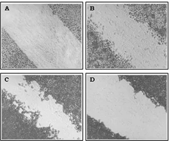
Initially, we determined effects of exogenous EGF on the growth of BALB/MK cells by using the cell migration assay. Since EGF is one of the most important factors for stimulating cell growth, we examined the ability of a com-bination of EGF (10 ng/mL) and 10% FBS to promote cell growth compared to the response triggered by EGF and FBS separately. The concentration of EGF used was chosen based on a dose-response curve for this growth factor in BALB/MK cells in which concentrations > 10 ng/mL did not significantly increase the cellular response beyond that seen with 10 ng/mL.
As expected, BALB/MK cells cultured only with EMEM showed no observable migration (Figure 2A). In contrast, cells incubated with 10 ng of EGF/mL or 10% FBS showed intermediate migration when compared to those incubated with both (Figures 2B-D). The effect of
EGF and FBS on BALB/MK cell growth differed: cells in-cubated with EGF were more dispersed (indicating moto-genic activity) whereas those incubated with FBS formed a dense layer (indicating greater mitogenicity and less moto-genicity), as previously reported for primary cultures of keratinocytes (Rheinwald and Green, 1977; Barrandon and Green, 1987).
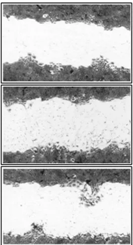
Since cellular migration was greater in medium con-taining FBS and EGF, we used medium concon-taining 10% FBS to assess the migration of BALB/MK cells modified with LESN. Of the PA317/LESN clones shown in Table 1, clones 2, 4 and 6 had more pronounced foci of cell migra-tion (Figure 3). However, the cellular migramigra-tion stimulated by the PA317/LESN clones was not totally independent of EGF since during the assay exogenous EGF was added to the medium to facilitate retroviral infection (see Materials and Methods).
To demonstrate the self-stimulated growth of trans-formed BALB/MK cells, the cells were selected with G418 and seeded onto new plates in the absence of exogenous EGF (Figure 4). Cells transformed with clone 6 showed greater growth than those transformed with viruses from PA317/LXSN (negative control) or PA317/LESN clone 9 (low cellular migration) (Figure 4). BALB/MK cells trans-formed with clone 6 reached confluence 6-7 days after infection, which was similar to the time required for non-transformed BALB/MK cells to reach confluence in the presence of exogenous EGF. In contrast, cells infected with the negative control virus (LXSN) or LESN clone 9 did not reach confluence even after two weeks in culture; indeed, most of the cells died before two weeks if EGF was not added to the culture (not shown).
Figure 1- DNA sequence of hEGF subcloned into the retroviral vector. The open reading frame of hEGF is underlined and the signal peptide is in italics. Codons that differ from the original hEGF sequence (Bellet al., 1986) are in bold.
Figure 2- Effect of EGF and/or FBS on BALB/MK cell migration. BALB/MK cells were grown as described in Methods. Except for the day of virus infection, the medium was replaced by new one without EGF and FBS (A), with 10 ng EGF/mL (B), with 10 ng EGF/mL and 10% FBS (C) or with 10% FBS (D). After incubation for 48 h, the cells were fixed and stained with Coomassie blue. Magnification: 100X.
Table 1- Viral titer of PA317/LESN clones.
PA317/LESN Titer (cfu/mL)
Clone 2 4.0 x 10⁵
Clone 3 1.0 x 10⁵
Clone 4 < 105
Clone 5 < 105
Clone 6 9.0 x 10⁵
Clone 7 3.0 x 10⁵
Clone 9 3.8 x 10⁶
For the cell migration assay, BALB/MK cells were transformed with the same volume of virus from PA317/LESN clones. Since the viral titer differed among the clones, the number of infected BALB/MK cells was also different. Consequently, cells incubated with a higher viral concentration supposedly had a greater level of EGF production and greater migration. However, PA317/LESN clone 9, which had a viral titer > 1 x 10⁶cfu/mL, grew less than clone 6, which had a lower titer (Figure 4). The most likely explanation for this discrepancy is that the viral DNA incorporated into the cells suffered mutations or rearrange-ments that resulted in a low level of EGF gene expression. This hypothesis was supported by the finding that the viral genome in PA317/LESN clone 9 was smaller than that of the other clones, as shown by Southern blotting after diges-tion withSal I, which cleaves in both LTR regions (not shown). In addition, northern blotting revealed a strong band of EGF RNA only in clone 6 (not shown). The latter finding supports the idea that a rearrangement of DNA in clone 9 resulted in low expression of EGF. Clone 4 of PA317/LESN had a viral titer < 1 x 10⁵cfu/mL but caused
greater migration than clones 3 and 9, both of which had higher titers (Table 1 and Figure 3), probably also because of rearrangements in its genomic DNA (not shown). To-gether, these results indicate the need to carefully analyze genetically modified cells in order to identify the DNA re-arrangement involved.
In conclusion, keratinocytes transformed by intro-ducing the hEGF gene can be cultured without exogenous EGF. These modified cells should be useful for short-term skin grafting because they do not require a continuous sup-ply of exogenous EGF. Since most keratinocytes have a limited life span, EGF productionin vivowill cease when the transformed cells die, thereby minimizing the danger of keratinocytes permanently expressing this growth factor. Nevertheless, since keratinocytes that have been geneti-cally modified by retroviral vectors are subject to inser-tional mutagenesis that can activate oncogenes, these modified cells require rigorous genetic analysis before their long-term usein vivois approved.
Acknowledgments
This work was funded by FAPESP (94/5778-7 and 97/03709-6).
References
Barrandon Y and Green H (1987) Cell migration is essential for sustained growth of keratinocyte colonies: The roles of transforming growth factor-alpha and epidermal growth fac-tor. Cell 50:1131-1137.
Figure 3- Cell migration stimulated by EGF produced by BALB/MK cells transfected with LESN. The cells were grown as described in Methods. After transfection, the medium was replaced with fresh solution containing only 10% FBS. After incubation for 48 h, the cells were fixed and stained with Coomassie Blue. Panels A, B and C represent clones 2, 4 and 6 respectively. Magnification: 100X.
Bell GI, Fong NM, Stempien MM, Wormsted MA, Caput D, Ku LL, Urdea MS, Rall LB and Sanchez-Pescador R (1986) Hu-man epidermal growth factor precursor: cDNA sequence, expressionin vivoand gene organization. Nucleic Acids Res 14:8428-8446.
Brown GL, Curtsinger 3rd L, Brightwell JR, Ackerman DM, Tobin GR, Polk Jr HC, George-Nascimento C, Valenzuela P and Schultz GS (1986) Enhancement of epidermal regenera-tion by biosynthetic epidermal growth factor. J Exp Med 165:1319-1324.
Carpenter G and Cohen S (1979) Epidermal growth factor. Annu Rev Biochem 48:193-216.
Carpenter G and Cohen S (1990) Epidermal growth factor. J Biol Chem 265:7709-7712.
Cohen S (1962) Isolation of a mouse submaxillary gland protein accelerating incisor eruption and eyelid opening in the new-born animal. J Biol Chem 237:1555-1562.
Eming SA, Snow RG, Yarmush ML and Morgan JR (1996) Tar-geted expression of insulin-like growth factor to human keratinocytes: Modification of the autocrine control of kera-tinocyte proliferation. J Invest Dermatol 107:113-120. Fenjves ES, Smith J, Zaradic S and Taichman LB (1994) Systemic
delivery of secreted protein by grafts of epidermal kera-tinocytes: Prospects for keratinocyte gene therapy. Hum Gene Ther 5:1241-1248.
Gerrard AJ, Hudson DL, Brownlee GG and Watt FM (1993) To-wards gene therapy for haemophilia B using primary human keratinocytes. Nat Genet 3:180-183.
Gibbs S, Silva Pinto AN, Murli S, Huber M, Hohl D and Ponec M (2000) Epidermal growth factor and keratinocyte growth factor differentially regulate epidermal migration, growth and differentiation. Wound Repair Regen 8:192-203. Green H, Kehinde O and Thomas J (1979) Growth of cultured
hu-man epidermal cells into multiple epithelia suitable for grafting. Proc Natl Acad Sci USA 76:5665-5668.
Hashimoto K (2000) Regulation of keratinocytes function by growth factors. J Dermatol Sci 24:S46-S50.
Hudson LG and McCawley LJ (1998) Contributions of the epider-mal growth factor receptor to keratinocyte motility. Microsc Res Tech 43:444-455.
Miller AD and Rosman GJ (1989) Improved retroviral vectors for gene transfer and expression. Biotechniques 7:980-986. Miller AD, Miller DG, Garcia JV and Lynch CM (1993) Use of
retroviral vectors for gene transfer and expression. Methods Enzymol 217:581-599.
Morgan JR, Barrandon Y, Green H and Mulligan RC (1987) Ex-pression of an exogenous growth hormone gene by trans-plantable human epidermal cells. Science 237:1476-1479. Moulin V (1995) Growth factors in skin wound healing. Eur J Cell
Biol 68:1-7.
Rheinwald JG and Green H (1977) Epidermal growth factor and the multiplication of cultured human epidermal keratino-cytes. Nature 265:421-424.
Sanger F, Nicklen S and Coulson AR (1977) DNA sequencing with chain-termination inhibitors. Proc Natl Acad Sci USA 74:5463-5467.
Schultz G, Rotatori DS and Clark W (1991) EGF and TGF-alpha in wound healing and repair. J Cell Biochem 45:346-352. Schultz GS, White M, Mitchell R, Brown G, Lynch J, Twardzik
DR and Todaro GJ (1987) Epithelial wound healing en-hanced by transforming growth factor-alpha and vaccinia growth factor. Science 235:350-352.
Steed DL (1998) Modifying the wound healing response with ex-ogenous growth factors. Clin Plast Sur 25:397-405. Weissman BE and Aaronson SA (1983) Balb and Kirsten murine
sarcoma viruses alter growth and differentiation of EGF-dependent Balb/c mouse epidermal keratinocyte lines. Cell 32:599-606.
Yates RA, Nanney LB, Gates RE and King Jr LE (1991) Epider-mal growth factor and related growth factors. Int J Dermatol 30:687-694.
Associate Editor: Carlos F.M. Menck