Frédéric Bustos Gaspar
Dissertation presented to obtain the Ph.D degree in Biology
Instituto de Tecnologia Química e Biológica | Universidade Nova de Lisboa
Oeiras,
June, 2012
Insert here an image
with rounded corners
Non-clinical isolates bring new findings
on enterococcal virulence
Supervisors:
Maria de Fátima Gonçalves Ribeiro dos Santos Silva Lopes
Investigadora Auxiliar | ITQB, Portugal.
Maria Teresa Ferreira de Oliveira Barreto Goulão Crespo
Investigadora Sénior | IBET, Portugal.
Examining committee:
Maria Constança Matias Ferreira Pomba
Professora Associada | Departamento de Clínica, Faculdade de
Medicina Veterinária, Universidade Técnica de Lisboa, Portugal.
Lélia Mariana Marcão Chambel
Professora Auxiliar | Faculdade de Ciências da Universidade de
Lisboa, Portugal.
Pascale Serror
Chargé de Recherche 1ère classe | Micalis, INRA, Jouy-en-Josas, France.
Ana Pimenta da Gama da Silveira Viana Semedo
Investigadora Auxiliar | Departamento de Química e Bioquímica,
"Thinking is no more than a tiny aspect of the totality of
consciousness, the totality of who you are."
Acknowledgments
Every single person with whom my path has crossed during my PhD
has influenced me. I have learned so much and everyone was an essential
piece of the puzzle that this journey ended up being.
Without the support of Dr Fátima Lopes and Dr Teresa Crespo this
journey would not have even been able to start. I am forever grateful to
have been given the ability to fulfil several of my visions and dreams. I was
encouraged to think on my own. I was allowed to develop my ideas. I grew
up.
Pascale Serror, Michael Gilmore, Johannes Huebner: I am
extremely thankful for all the opportunities that each of you offered me in
your labs, where I was so kindly welcomed, and where I learned so much. It
was an amazing experience.
ITQB, IBET & IGC @Oeiras.pt / FCUL @Lisbon.pt / INRA
@Jouy-en-Josas.fr / Harvard Medical School @Boston.us / University of Freiburg
@Freiburg.de / The support that I received from the members of my labs,
the division, the research institutes, the universities, and from everyone
with whom I have had the opportunity to interact and work was absolutely
xii
The completion of this work has equally relied on the hands of many
other people for whom I feel great esteem, appreciation, gratitude, and also
love. @ Lisbon, Cascais, Almada, Oeiras .pt / @ Copenhagen .dk /
@ London .uk / @ Madrid, Barcelona .es / @ New York, Boston, San
Diego, Santa Barbara, Los Angeles, San Francisco .us / @ Jouy-en-Josas,
Paris .fr / @ Amsterdam .nl / @ Ljubljana .si / @ Freiburg .de / Where I
worked, where I was, where we met, where you have been, where you
are…
I also want to acknowledge everyone that I feel so close to and that
I consider belonging to my families: the acquired and the natural one.
Especially my maman. To whom I owe my Life… And to whom I owe my
Abstract
Enterococci are Gram-positive lactic acid bacteria, widespread in
the environment, present in water, soil, plants and animals, including
humans. They typically colonize the skin and mucous membranes, namely
the gastrointestinal tract. However, enterococci, and most notably
Enterococcus faecalis and Enterococcus faecium, have become
problematic causative agents of several nosocomial infections, including
urinary tract infections, bacteraemia, surgical sight infections, and
endocarditis. Besides being opportunistic pathogens, the resilient bacteria
of the genus Enterococcus are key factors contributing to the ripening,
flavour, and the organoleptic properties of fermented food products.
The ubiquitous nature of enterococci derives from a number of
features, which can be intrinsic to the genus or specific to some species or
even strains. These traits allow probing the environment in order to adapt,
enabling a survival and fitness advantage. They are encoded in numerous
genes that can be easily transferable due to the high genomic promiscuity
of enterococci. These genes have been ascribed a role in virulence as they
xvi
adhesion, colonization, invasion, evasion of the immune system and spread
through the hostʼs tissues. Enterococcal virulence factors can be either
secreted (cytolysin, proteases, hyaluronidase, superoxide), surface
associated (enterococcal surface protein, aggregation substance,
extracellular polymeric substances, pilin gene clusters, enterococcal
microbial surface component recognizing adhesive matrix molecules), or
intracellular. At the time this thesis work began, researchers were starting
to realize that virulence factors in enterococcal clinical isolates were also
present in isolates from other environments, in particular, where
enterococci play beneficial roles, namely food. Since dissemination of
virulence factors among food isolates was no longer crucial, other issues
started to become relevant in the still debated enterococcal virulence.
A thorough search in the literature clearly shows recurrent
incongruent results between genotype and phenotype of the virulence
factor cytolysin. Moreover, the phenotypic assays are not performed under
the same conditions and only a few cyl genes are screened for in the
majority of the experiments. Therefore, in the first part of this work, we
developed a new genotypic and phenotypic screening for cytolysin. Based
on our methodology, a much higher correlation was obtained compared to
any previously described screening. Complete agreement between
genotypic and phenotypic assays was seen for all 55 strains tested, which
belong to four enterococcal species, namely E. faecalis, E. faecium,
Enterococcus durans and Enterococcus hirae. The proposed PCR
screening for the complete cyl locus gives a measure of the gene reservoir
of the strain, while the phenotypic assay is still the only test that allows
Abstract
In the second part of this work, the goal was to evaluate the roles of
fsrB and gelE, two genes that encode for well-characterized enterococcal
virulence factors, in the potential virulence of E. faecalis food strains.
Virulence of unrelated Enterococcus isolates, including dairy strains
carrying fsr and gelE operons, was compared in the Galleria mellonella
insect model. E. faecalis dairy strains were able to kill larvae and were as
virulent as one of the most widely used strains for virulence studies. In
contrast, E. durans and E. faecium strains were avirulent or poorly virulent
in G. mellonella. To evaluate the role of fsrB and gelE in the virulence of E.
faecalis dairy strains, both genes were deleted independently in two strains.
Although both mutations significantly attenuated virulence in G. mellonella,
the fsrB mutant strains were more strongly attenuated. Our work
demonstrates that the presence of functional fsrB, and to a lesser extent
gelE, significantly contributes to the virulence of E. faecalis food isolates,
and that the presence of these genes in dairy enterococci should be
considered with caution. The simple G. mellonella animal model may
provide insights for risk assessment of food isolates.
Next, we further characterized the non-starter E. faecalis cheese
isolate QA29b, which harbours virulence genes and proved to be virulent in
a G. mellonella virulence model. We looked at traits relevant to the host
pathogen interaction, in particular adhesion, colonization and infection.
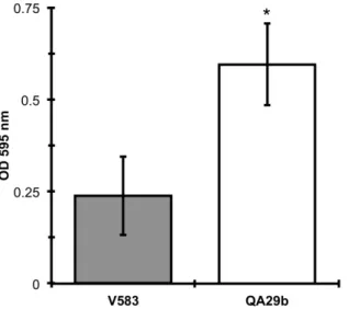
QA29b demonstrated high ability to form biofilms, to adhere to epithelial
cells and was readily eliminated by J774.A1 macrophage cells. This work
illustrates for the first time that cps genes, that are associated with
virulence, may be differentially transcribed between isolates, and therefore
xviii
traits important for interaction, colonization and infection in the host
performed on a good food isolate representative of E. faecalis. Overall,
QA29b characterization shows that, despite its virulence potential in an
insect model, this food strain is readily eliminated by mammalian
macrophages, indicating that fine-tuned approaches combining cellular and
mammalian models are needed to address and elucidate the multifactorial
aspect of virulence potential associated with food isolates.
In the last part of this work, we were able to associate the
extracellular production of autoinducer-2 (AI-2) in E. faecalis VE14089, a
plasmid-cured derivative of E. faecalis V583, to the presence and
expression of luxS gene. This gene is also present in other clinical and
commensal isolates as well as in food isolate QA29b. AI-2-mediated
quorum sensing has been extensively studied in relation to the regulation of
microbial behaviour and has been recognized as an interspecies
communication molecule, which may influence community structure and
function. When compared with the wild type, the luxS mutant had no
apparent phenotype regarding growth, biofilm formation, adhesion to
Caco-2 cells, resistance to oxidative stress and survival inside macrophages.
However, microarray comparison of gene expression revealed that the luxS
mutation caused pleiotropic effects in gene expression, affecting genes
involved in DNA, fatty acid and intermediary metabolites metabolism, which
could not be complemented by extracellular AI-2 addition. This study shows
that, in E. faecalis, differential gene expression related to the luxS mutation
cannot be ascribed to quorum sensing and that LuxS has, at least, a role in
Abstract
In conclusion, the main findings presented in this thesis reveal that
there is, until now, no virulence trait exclusive to strains isolated from
clinical settings, and that the sole presence of these traits does not allow
the identification of a strains' habitat. We showed that the presence of
virulence associated genes, and therefore an associated virulence
potential, does not necessarily translate to a virulent and pathogenic
behaviour in E. faecalis food isolates. Also, we showed that to determine
the outcome of the persistence and resilience of enterococci, not only the
presence of a specific gene is important but also the whole genome plays a
pivotal role. We revealed for the first time that the LuxS enzyme from the
genus Enterococcus is responsible for the production of the interspecies
communication molecule, AI-2. Taken together, the data reported in this
thesis showed the potential for virulence of enterococcal dairy strains in
specific virulence models, while being avirulent in other models. This
highlights the problem that their pathogenic potential in humans cannot be
entirely excluded, and therefore, reiterating the need to keep a close
Resumo
Os enterococos são bactérias lácticas, disseminadas no meio
ambiente, presentes na água, solo, plantas e animais, incluindo o ser
humano. Para além da colonização da pele e de mucosas, tal como o
tracto gastrintestinal, os enterococos, especialmente as espécies
Enterococcus faecalis e Enterococcus faecium, têm-se tornado agentes
responsáveis por diversas infecções nosocomiais nomeadamente do tracto
urinário, bacterémias, infecções cirúrgicas, e endocardites. Para além de
serem agentes patogénicos oportunistas, as resilientes bactérias do
género Enterococcus são agentes essenciais que contribuem para a
maturação, sabor e outras propriedades organolépticas de produtos
alimentares fermentados.
A natureza ubíqua dos enterococos pode ser atribuída a diversas
características, quer intrínsecas do género quer específicas de certas
espécies, que conferem uma vantagem nas suas capacidades de
adaptação e sobrevivência. Facilmente transferíveis devido à elevada
promiscuidade genómica dos enterococos, estes factores estão
xxiv
ambiente de modo a adaptar o seu comportamento quer às condições
ambientais quer à densidade celular. Estes genes desempenham um papel
na virulência pois são relevantes nas diferentes etapas do processo de
infecção, nomeadamente na adesão, colonização, invasão, evasão do
sistema imunitário e disseminação nos tecidos do hospedeiro. Envolvidos
neste processos estão vários factores de virulência, incluindo aqueles
estudados nesta tese, nomeadamente citolisina, Fsr, gelatinase, cápsula.
No início do trabalho que levou a esta tese, os investigadores começavam
a aperceber-se que os factores de virulência presentes em enterococos
isolados de ambientes clínicos também podiam ser encontrados em
enterococos isolados de outros ambientes, tais como de produtos
alimentares onde os enterococos desempenham um papel benéfico. Uma
vez que a pesquisa da disseminação de factores de virulência deixara de
ser crucial, outros aspectos começaram a tornar-se relevantes na ainda
debatida virulência em enterococos, nomeadamente, a incongruência entre
genótipo e fenótipo relativamente ao factor de virulência citolisina (Capítulo
2), o potencial de virulência de estirpes alimentares de E. faecalis
associado à presença dos genes fsrB e gelE (Capítulo 3), o papel de
alguns factores de virulência na relação com o hospedeiro de estirpes de
E. faecalis alimentares (Capítulo 4) e a contribuição para a virulência da
proteína LuxS, responsável pela produção extracelular do autoindutor-2
(AI-2) possivelmente envolvido certamente na comunicação entre E.
faecalis e outras bactérias dentro e fora do hospedeiro humano (Capítulo
5).
Na primeira parte deste trabalho, e relativamente ao factor de
Resumo
só os ensaios fenotípicos não eram realizados em condições idênticas
como também, na maioria dos trabalhos publicados, poucos dos oito genes
do operão cyl eram pesquisados, levando frequentemente a resultados
incongruentes entre o genótipo e o fenótipo. Entre as 55 estirpes testadas
nesta tese, pertencentes a quatro espécies diferentes de enterococos,
nomeadamente E. faecalis, E. faecium, Enterococcus durans e
Enterococcus hirae, e usando uma nova metodologia de pesquisa
genotípica e fenotípica, foi possível obter uma concordância perfeita entre
os ensaios genotípicos e fenotípicos.
Na segunda parte deste trabalho, o objectivo foi avaliar o papel dos
genes fsrB e gelE, dois genes que codificam para factores de virulência
bem caracterizados em enterococos clínicos, no potencial de virulência de
estirpes alimentares de E. faecalis. A virulência de isolados não
relacionados do género Enterococcus, incluindo estirpes lácteas
portadoras dos operões fsr e gelE, foi então comparada no insecto modelo
Galleria mellonella. As estirpes lácteas de E. faecalis foram capazes de
matar as larvas sendo tão virulentas quanto uma das estirpes mais
utilizadas em estudos de virulência. Por oposição, as estirpes das espécies
E. durans e E. faecium mostraram-se avirulentas ou pouco virulentas em
G. mellonella. De modo a avaliar o papel dos genes fsrB e gelE na
virulência de estirpes alimentares de E. faecalis, os dois genes foram
independentemente deletados em duas estirpes. Embora ambas as
mutações tenham significativamente atenuado a virulência em G.
mellonella, esse efeito foi mais pronunciado nas estirpes mutantes no gene
fsrB. Este trabalho demonstrou que a presença de um gene fsrB funcional,
e de uma forma menos acentuada de um gene gelE, contribui
xxvi
que a presença desses genes em enterococos lácteos deve ser
considerada com precaução. G. mellonella, sendo um modelo simples,
pode apresentar vantagens na avaliação do risco associado a isolados
alimentares.
O isolado de queijo E. faecalis QA29b, portador de genes de
virulência e previamente identificado como virulento no modelo de
virulência G. mellonella, foi estudado no que respeita a fenótipos
relevantes para a interacção com o hospedeiro que podem ter impacto na
capacidade de provocar infecções e colonizar o hospedeiro humano. Este
trabalho constituiu o primeiro estudo deste tipo de características num E.
faecalis alimentar, que pertence ao Complexo Clonal 72, sendo, por isso,
um bom representante dos E. faecalis alimentares. QA29b demonstrou,
por um lado, uma elevada capacidade de formar biofilmes e aderir a
células epiteliais, e por outro foi prontamente eliminado por células de
macrófago J774.A1. Este trabalho evidenciou, pela primeira vez, que os
genes cps, associados à virulência, podem ser transcritos de forma
diferencial entre diferentes isolados, podendo não ser fenotipicamente
expressos. Apesar de demonstrar ter um potencial de virulência num
modelo de insecto, este isolado alimentar mostrou ser prontamente
eliminado por macrófagos de mamíferos. Este trabalho mostra que
optimizar métodos que combinem modelos celulares e animais é
necessário de modo a abordar e elucidar o aspecto multifactorial do
potencial de virulência associado a isolados alimentares.
Na última parte deste trabalho a produção extracelular de AI-2 em
E. faecalis VE14089, um derivado de E. faecalis V583 curado dos seus
Resumo
gene encontra-se também presente em outros isolados clínicos e
comensais bem como no isolado alimentar QA29b. A percepção de
quórum mediada por AI-2 tem sido extensamente estudada na regulação
do comportamento microbiano e foi reconhecida enquanto molécula de
comunicação entre espécies, podendo influenciar a estrutura e função da
comunidade. Quando comparada com a estirpe selvagem, o mutante luxS
não demonstrou ter nenhum fenótipo quanto ao crescimento, formação de
biofilme, adesão a células Caco-2, resistência ao stress oxidativo ou
sobrevivência no interior de macrófagos. No entanto, a análise por
microarray da expressão génica revelou que a mutação luxS teve efeitos
pleiotrópicos, afectando genes envolvidos no metabolismo de DNA, ácidos
gordos e intermediários metabólicos, não sendo complementados pela
adição extracelular de AI-2. Este estudo mostrou que, em E. faecalis, a
expressão diferencial génica relacionada com a mutação luxS não pode
ser atribuída à percepção de quórum e que o LuxS tem, pelo menos, um
papel no metabolismo.
Concluindo, o trabalho apresentado nesta tese evidenciou, pela
primeira vez, que uma estirpe láctea de E. faecalis tem tanto potencial de
virulência quanto uma estirpe clinica da mesma espécie. No entanto, ficou
também claro com este trabalho que a presença de genes associados
com virulência em E. faecalis alimentares não se traduz necessariamente
num comportamento virulento e patogénico e que a persistência e
resiliência de enterococos depende tanto do papel desempenhado pela
presença de genes específicos quanto pelo restante genoma. Tendo em
conta que os enterococos alimentares, bem como os comensais e clínicos,
xxviii
comunicação inter-espécies poderá desempenhar um papel de relevo na
capacidade para colonizar ou infectar o hospedeiro, estudou-se o papel do
LuxS em vários fenótipos relevantes no processo de colonização e
infecção por E. faecalis. Mostrou-se pela primeira vez que o enzima LuxS,
presente no género Enterococcus, também em estirpes lácteas, é
responsável pela produção da molécula de comunicação entre espécies,
AI-2. A totalidade dos dados relatados nesta tese revelam que o potencial
patogénico de Enterococcus em humanos não pode ser inteiramente
excluído, reiterando a necessidade de manter uma estreita vigilância da
List of Publications
Gaspar, F.B., Crespo, M.T.B., Lopes, M.F.S., 2009. Proposal for a
reliable enterococcal cytolysin production assay avoiding apparent
incongruence between phenotype and genotype. J. Med. Microbiol. 58,
1122–1124.
Gaspar, F., Teixeira, N., Rigottier-Gois, L., Marujo, P.,
Nielsen-LeRoux, C., Crespo, M.T.B., Lopes, M. de F.S., Serror, P., 2009. Virulence
of Enterococcus faecalis dairy strains in an insect model: the role of fsrB
Abbreviations
°C degree Celsius
2×YTGlu 2×YT supplemented with 0.5 % glucose
A adenine (in a nucleotide sequence)
A alanine (in an amino acid sequence)
Ace adhesin of collagen of E. faecalis
Acm adhesin of collagen of E. faecium
AI autoinducer
AMC activated methyl cycle
AS aggregation substance
ATP adenosine triphosphate
Bap biofilm-associated protein
BCAA branched-chain amino acid
BCKDH branched-chain alpha-keto acid dehydrogenase
BHI brain heart infusion
BLAST Basic Local Alignment Search Tool
bop biofilm on plastic surfaces
bp base pair
BT bacterial translocation
C cysteine (in an amino acid sequence)
C cytosine (in a nucleotide sequence)
CC clonal complex
CcpA catabolite control protein A
CCR carbon catabolite repression
cDNA complementary deoxyribonucleic acid
CFU colony-forming unit
CGH comparative genome hybridization
xxxvi
CPS capsular polysaccharide
cre catabolite responsive element
Cyl cytolysin
CYS L-cysteine
CYSTA L-cystathionine
D Aspartic acid
Dam DNA adenine methylase
Dcm DNA cytosine methylase
DNA deoxyribonucleic acid
DPD (S)-4,5-dihydroxypentan-2,3-dione
DS diffusion sensing
E glutamic acid
ebp endocarditis and biofilm-associated pili
EcbA E. faecium collagen binding protein A
ECF extracytoplasmic function
eDNA extracellular DNA
EDTA ethylenediaminetetraacetic acid
EfaA E. faecalis antigen A
Ehk enterococcal histidine kinase
EI enzyme I
EII enzyme II
Epa enterococcal polysaccharide antigen
EPS exopolysaccharide
Err enterococcal response regulator
Ers enterococcal regulator of survival
ES efficiency sensing
Esp enterococcal surface protein
F phenylalanine
FASII fatty acid synthase II
FDR false discovery rate
FRET fluorescence resonance energy transfer
Fsr E. faecalis regulator
G glycine (in an amino acid sequence)
G guanine (in a nucleotide sequence)
GBAP gelatinase biosynthesis-activating pheromone
GEI genomic island
GelE gelatinase
GI gastrointestinal
Gsp general stress protein
H histidine
HCY L-homocysteine
Abbreviations
HJ Holliday junction
HK histidine kinase
HPr histidine protein
HR homologous recombination
HSE L-homoserine
Hyl Hyaluronidase
HypR hydrogen peroxide regulator
I isoleucine
ICE integrative and conjugative element
int intergenic
IS insertion sequence
JCVI J. Craig Venter Institute
K lysine
L leucine
LAB lactic acid bacteria
LB Luria–Bertani
LPS lipopolysaccharide
LTA lipoteichoic acid
M methionine
M17AGlu M17 agar supplemented with 0.5 % (w/v) glucose
M17BGlu M17 broth supplemented with 0.5 % (w/v) glucose
M17Glu M17 supplemented with 0.5 % (w/v) glucose
MET L-methionine
MGE mobile genetic element
mL millilitre
MLST multi-locus sequence typing
MnSOD manganese superoxide dismutase
MOI multiplicity of infection
MRSA methicillin-resistant Staphylococcus aureus
MSCRAMM Microbial Surface Component Recognizing Adhesive Matrix
Molecule
N asparagine
NAD nicotinamide adenine dinucleotide
NCBI National Center for Biotechnology Information
ng nanogram
nm nanometre
OD optical density
ORF open reading frame
P proline
PAI pathogenicity island
PBS phosphate-buffered saline
xxxviii
PEP phosphoenolpyruvate
PerA pathogenicity island-encoded regulator A
PerR peroxide regulator
PFGE pulsed field gel electrophoresis
PGC pilin gene cluster
pH hydrogen potential
PTS phosphoenolpyruvate transport system
Q glutamine
QS quorum sensing
R arginine
RBS ribosome-binding site
RIVET recombinase-based in vivo expression technology
RNA ribonucleic acid
RNase ribonuclease
rpm rotations per minute
RR response regulator
rRNA ribosomal RNA
RT-PCR reverse transcription PCR
s second
S serine
S/I similarity/identity
SAH S-adenosyl-L-homocysteine
SAM S-adenosyl-L-methionine
Scm second collagen adhesin of E. faecium
SI survival index
spp. species
SprE serine protease
SRH S-ribosyl-L-homocysteine
ST sequence type
T threonine (in a amino acid sequence)
T thymine (in a nucleotide sequence)
TCA tricarboxylic acid
TCS two-component system
Tpx thiol peroxidase
UTI urinary tract infection
UV ultraviolet radiation
V valine
v/v volume/volume
VBNC viable but nonculturable
VRE vancomycin-resistant Enterococcus
W tryptophan
Abbreviations
WT wild type
Y tyrosine
μg microgram
μL microliter
Table of Contents
Acknowledgments ix
!
Abstract xiii
!
Resumo xxi
!
List of Publications xxix
!
Abbreviations xxxiii
!
Table of Contents xli
!
List of Figures and Tables xlvii
!
List of Figures xlix
!
List of Tables li
!
Chapter 1 — General Introduction 1
!
WHO 5
!
WHERE / Enterococci can be found in diverse habitats 8
!
WHY & HOW / Characteristics and mechanisms that allow enterococci
to colonize and survive diverse habitats 15
!
WHEN & WHAT / Is there a straightforward enterococcal associated
xliv
Chapter 2 — Proposal for a reliable enterococcal cytolysin production
assay avoiding apparent incongruence between phenotype and genotype
47
Acknowledgements 55
!
Chapter 3 — Virulence of Enterococcus faecalis dairy strains in an insect
model: the role of fsrB and gelE 57
!
Abstract 61
!
Introduction 62
!
Materials and Methods 64
!
Results 69
!
Discussion 73
!
Acknowledgments 77
!
Chapter 4 — Incongruence between the cps type 2 genotype and
host-relate phenotypes of an Enterococcus faecalis food isolate 79
!
Abstract 83
!
Introduction 84
!
Material and Methods 86
!
Results 91
!
Discussion 98
!
Acknowledgments 100
!
Chapter 5 — Role of LuxS in Enterococcus faecalis 103
!
Abstract 107
!
Introduction 108
!
Materials and Methods 111
!
Results and Discussion 118
!
Conclusion 161!
Table of Contents
Chapter 6 — General Discussion 165
!
Problem with enterococci 169
!
Clinical enterococci and infection 170
!
Presence of virulence factors in commensal and food strains 170
!
Expression of virulence traits in food isolates 171
!
Colonization as a stage of infection 172
!
Putative virulence factors or complete genome responsible for
pathogenic and virulent behaviour 173
!
Virulence models for an effective virulence assessment 175
!
Many described virulence associated features are essential for the
hostʼs health 177
!
Health, disease and the host 180
!
Finishing notes 182
!
List of Figures and Tables
List of Figures
Figure 1.1. WHERE enterococci are found: habitat occurrence and
features, and flow of presence perpetuation. 11
!
Figure 1.2. HOW enterococci are able to interact with their habitat: intrinsic
and acquired characteristics and mechanisms. 16
!
Figure 2.1. (A) Organization of the cyl locus and location of the primers Cf
and Cr used for the PCR. (B) The E. faecalis cytolysin senses and destroys
target cells. 51
!
Figure 3.1. Killing of G. mellonella larvae by various enterococci isolates.70
!
Figure 3.2. Role of E. faecalis gelatinase in killing of G. mellonella larvae.
72
!
Figure 3.3. Effects of gelE and fsrB inactivation on killing of G. mellonella.
72
!
Figure 4.1. CGH analysis of QA29b strain. 93
!
l
Figure 4.3. QA29b and VE14089 adhesion to Caco-2 cells. 95
!
Figure 4.4. Time course of intracellular survival of QA29b and VE14089
strains within murine J774A.1 macrophages. 96
!
Figure 5.1. Sequence alignments of putative LuxS protein from E. faecalis
V583 (ENTFA) and selected organisms for which there is evidence of the
existence of LuxS at the protein level. 118
!
Figure 5.2. E. faecalis V583 ef1182 (luxS) promoter and terminator
sequences. 120
!
Figure 5.3. (A) Mechanism for ligand-induced fluorescence resonance
energy transfer (FRET) changes in the cyan fluorescent protein (CFP),
LuxP, yellow fluorescent protein (YFP) fusion. (B) Calibration curve
performed with AI-2 samples of known concentration. 123
!
Figure 5.4. Growth curves and AI-2 production from VE14089 (A) and
VE14089ΔluxS (B). 124
!
Figure 5.5. Phenotypic characterization of luxS mutant: VE14089 in grey
bars with full line outline and VE14089ΔluxS in white bars with a dashed
line outline. (A) Biofilm formation on polystyrene microtiter plates. (B)
VE14089 and VE14089ΔluxS adhesion to Caco-2 cells. (C) Percentage (±
standard deviation) survival of growing cells of E. faecalis VE14089 and
VE14089ΔluxS at 2, 4 and 6 h of a challenge with 7 mM H2O2. (D) Time
course of intracellular survival of VE14089 and VE14089ΔluxS strains
within murine J774A.1 macrophages. 127
!
Figure 5.6. Growth of E. faecalis strains VE14089 and VE14089ΔluxS in
2×YT medium, after growth conditions optimization. 128
!
Figure 5.7. Distribution by number of differentially expressed genes,
List of Figures and Tables
shown as negative), and by JCVI (http://cmr.jcvi.org) cellular mainrole found
to be affected by the luxS mutation, which was independent of DPD
addition, when compared to the parental strain VE14089. 141
!
Figure 5.8. AMC in V583. 144
!
List of Tables
Table 1.1. Compilation of enterococcal species names, groups, isolation
materials, and date. 10
!
Table 2.1. Results from the genetic and phenotypic screening of the
cytolysin system. 54
!
Table 3.1. Strains used in this study 65
!
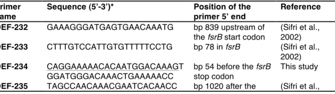
Table 3.2. Primers used in this study 66
!
Table 4.1. E. faecalis strains used in this study. 87
!
Table 4.2. Macrophage Survival Assay: MOI, uptake percentage at 0 h of
infection, and standard deviation values. 97
!
Table 5.1. Strains used in this study 111
!
Table 5.2. Primers used in this study 113
!
Table 5.3. Macrophage Survival Assay: MOI, uptake percentage at 0 h
post-killing, and standard deviation values. 126
!
Table 5.4. List of differentially regulated genes in at least one of the
pairwise comparisons: VE14089 and VE14089ΔluxS, and VE14089 and
lii
Table 5.5. Differential expression of genes associated with the interaction
with transcriptional regulators, with the consensus motif localized in the
intergenic region (int) or in the coding region (cod) in the VE14089ΔluxS
mutant when compared to VE14089, independently of extracellular added
DPD. 158
!
Table 5.6. Differential expression of potential virulence factors (according
to Manson et al. (Manson and Gilmore, 2006b)) in the VE14089ΔluxS
mutant when compared to VE14089 with no extracellular addition of DPD.
Chapter 1
Chapter 1
WHO 5
!
WHERE / Enterococci can be found in diverse habitats 8
!
Animals, plants and environment / Habitat diversity 8
!
Perpetuation of enterococci in nature and man-made environments 12
!
WHY & HOW / Characteristics and mechanisms that allow enterococci to
colonize and survive diverse habitats 15
!
Intrinsic characteristics of the genus 17
!
Bacteriocins 18
!
Putative virulence traits 19
!
Secreted factors 20
!
Cytolysin (Cyl) 20
!
Proteases: Gelatinase (GelE) and Serine Protease (SprE) 21
!
Hyaluronidase (Hyl) 23
!
Superoxide 23
!
Surface-associated factors 24
!
Enterococcal Surface Protein (Esp) 24
!
4
Extracellular polymeric substances 25
!
Pilin Gene Clusters (PGCs) 28
!
Enterococcal Microbial Surface Component Recognizing Adhesive
Matrix Molecules (MSCRAMMs) 29
!
Intracellular factors 30
!
Emergence of new genetic material 33
!
Sensing and probing the environment 34
!
Two-component systems (TCS) 34
!
Quorum sensing (QS) 35
!
WHEN & WHAT / Is there a straightforward enterococcal associated human
health risk? 38
!
In human disease 39
!
Chapter 1
WHO
Description of the genus Enterococcus (ex Thiercelin and Jouhaud
1903)
Enterococcus (En.te.ro.cocʻcus. Gr. n. enteron intestine; Gr. n. coccus a
grain, berry; M.L. masc. n. Enterococcus intestinal coccus) cells are ovoid,
occur singly, in pairs, or in short chains, and are frequently elongated in the
direction of the chain. Gram positive. Endospores are not formed. May be
motile. Facultatively anaerobic. Optimum growth temperature, ca. 35 °C.
Strains grow at 10 and 45 °C. Most strains survive heating at 60 °C for
30 min. Grow in 6.5 % NaCl and at pH 9.6. Hydrolyse
pyrrolidonyl-β-naphthylamide. Chemoorganotrophs. Metabolism fermentative. The
predominant end product of glucose fermentation is L-lactic acid. Oxygen or
other hydrogen acceptors may alter the end products of carbohydrate
metabolism. Hydrogen peroxide may or may not accumulate in the
presence of oxygen. Do not contain haem compounds. Benzidine negative
6
pseudocatalase. Some strains synthesize cytochromes or catalase or both
when they are provided with haemin. The minimal nutritional requirements
are generally complex. React with group D antisera; some strains also react
with group Q antisera.
Some strains possess respiratory quinones (menaquinones or
demethylmenaquinones). Long-chain fatty acids are predominantly of the
straight-chain saturated or monounsaturated types; some strains produce
cyclopropane ring acids.
Peptidoglycan type: Lys-D-Asp or Lys-Ala2-3.
The G+C content of the DNA ranges from 37 to 45 mol %.
Type species: Enterococcus faecalis.
Nucleic acid hybridization studies, in particular DNA-rRNA hybridization
studies, demonstrate that members of the genus Enterococcus are closely
related to each other but not to members of the genus Streptococcus.
Enterococci can easily be differentiated from streptococci by their ability to
grow in 6.5 % NaCl and at pH 9.6. Moreover, in contrast to most
streptococci (exceptions are Streptococcus lactis, Streptococcus cremoris,
and Streptococcus uberis), they can grow at 10°C.
(Schleifer et al., 1984)
With the previous description Schleifer and Kilpper-Bälz (Schleifer et
al., 1984) revived in 1984 the genus Enterococcus. However, Thiercelin
was the first to use, in 1899, the term “enterocoque” to indicate the
intestinal origin of a Gram-positive diplococcus, and 4 years later, together
with Jouhaud, the genus Enterococcus was proposed as new (Domig et al.,
2003; Khan et al., 2010). Nonetheless, Enterococcus as a new genus did
Chapter 1
“enterocoque” as a group of streptococci so characteristic of the human
intestine that the term "Streptococcus faecalis" could be applied to it
(Andrewes and Horder, 1906). Based on a serological typing system for
haemolytic streptococci developed by Lancefield (Lancefield, 1933),
Lancefield, this time with colleague Hare, continued, in 1935, that
association between the two genus, relating cultures of the intestinal
enterococci to group D haemolytic streptococci (Lancefield and Hare,
1935). Sherman was the first to clearly mention, in 1937, that the term
"enterococcus" had a somewhat variable and hazy meaning (Sherman,
1937), existing in the literature a synonymous usage of enterococci, faecal
streptococci and group D streptococci. At the same time he also stated
that, despite being hard at that time to justify a generic segregation of the
enterococci from the other streptococci making them an independent
genus, enterococci represent one of the clearly defined primary divisions of
the streptococci, probably the most clearly marked subdivision of the whole
genus, the other three being the pyogenic, viridans and lactic groups
(Sherman, 1937).
The genus Enterococcus was revived and separated from
Streptococcus sensu lato based on genomic studies, which relied on
DNA-DNA and DNA-DNA-rRNA hybridization studies (Schleifer et al., 1984). This
separation was further confirmed by 16S ribosomal RNA (rRNA) sequence
analysis, which showed that, within the previously described
Streptococcus-Enterococcus group, organisms fall into three clusters
defined by Enterococcus, the lactic acid streptococci and streptococci of
the pyogenic and oral groups (Ludwig et al., 1985). Since the revival of the
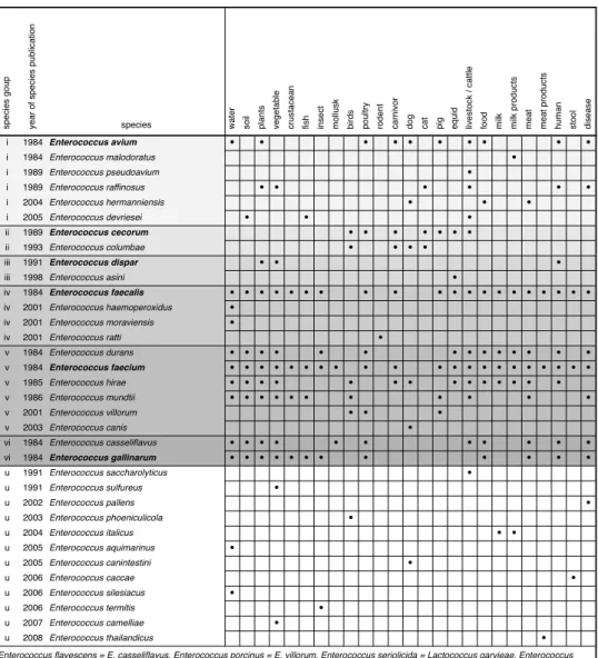
genus a total of 40 enterococcal species have been described (Table 1.1),
8
previously described enterococcal species or belonging to other genus
((Euzéby, 1997), related website last visited: 2012.03.20)).
Even if theyʼre not in presence of optimal growth conditions, the
enterococcal phenotypic characteristics allow them to grow in diverse and
broad range of conditions, conferring them the ability to colonize a wide
variety of environments.
WHERE can they be found?
WHERE / Enterococci can be found in diverse habitats
Bacteria of the genus Enterococcus are ubiquitous in nature and
their role is often unclear. They can occupy the most varied niches if their
viability or basic growth needs are met. Even if some species specificity
can be seen in certain habitats (Table 1.1), they are naturally found in
animals, plants, and in the environment (Figure 1.1).
Animals, plants and environment / Habitat diversity
In opposition to parasitism, where one organism benefits at the
expense of its host, and mutualism, where both organisms benefit,
commensalism is not as easy to define as the previous ones. By definition,
commensals benefit from their relationship with their host without causing
harm neither benefiting it.
Enterococci have been identified as human commensals since their
presence in the gastrointestinal (GI) tract was reported, still in the 19th century. In healthy humans they are mainly associated with the GI tract but,
although they are less commonly found at other body sites, enterococci can
also be recovered from the skin, the vagina, and the oral cavity (Fisher and
Chapter 1
healthy animals: from warm-blooded animals, such as other mammals and
birds (including production animals, such as livestock and fowl), to other
vertebrates, like fish (Bulushi et al., 2010; Albesharat et al., 2011),
amphibians and reptiles (Cox and Gilmore, 2007), and even invertebrates,
like molluscs (Valenzuela et al., 2010) and insects (Devriese and Baele,
2006; Cox and Gilmore, 2007).
Certain host-specific variations in the occurrence of different
enterococcal species in different animal hosts are known to exist. Other
species variations have also been described regarding the hostʼs age,
compartments colonized and the species distribution, as well as the effect
due to feeding on species colonization (Devriese and Baele, 2006).
Plants represent a harsh environment with physicochemical
conditions that fluctuate widely and rapidly over short periods of time, such
as temperature and osmotic conditions within the same day. The aerial
plant surfaces are mainly aerobic, overall poor in nutrients, and exposed to
UV radiation. Even in these considered non-host conditions, so different
from the usual stable temperature, shielded from UV rays, anaerobic and
nutrient rich environment of the GI tract of animals where they are
commensal, enterococci can find a secondary habitat (Albesharat et al.,
2011). Enterococci have been found on the most diverse plant materials,
like fruit, root and bulbous vegetables, as well as salads and cereals
(Schwaiger et al., 2011).
Enterococci are also widely present in natural environments such as
soil, sediments, sand and water (Whitman et al., 2003; Devriese and Baele,
2006; Brownell et al., 2007; Albesharat et al., 2011; Wright et al., 2011).
Several enterococcal species have been isolated from these niches, such
as E. faecium, E. faecalis, E. casseliflavus, E. hirae, E. durans and E.
10
Table 1.1. Compilation of enterococcal species names, groups, isolation materials,
and date. As described by Williams et al. (Williams et al., 1991), De Graef et al. (De
Graef et al., 2003) and Koort et al. (Koort et al., 2004) enterococci can be classified
in 6 different groups that are indicated by roman numbering (u: for unknown), with
the type species of each group indicated in bold type. The species in need of
renaming are at the bottom of the table ((Euzéby, 1997), related website last
visited: 2012.03.20). sp e ci e s g o u p ye a r o f sp e ci e s p u b lica ti o n
species water so
il plants ve g e ta b le cru st a ce a n fi
sh inse
ct mo llu sk b ird s p o u lt ry ro d e n t ca rn ivo r dog ca t
pig equid live
st o ck / ca tt le food mi lk mi lk p ro d u ct s me a t me a t p ro d u ct s h u ma n st o o l d ise a se
i 1984 Enterococcus avium ! ! ! ! ! ! ! ! ! !
i 1984 Enterococcus malodoratus !
i 1989 Enterococcus pseudoavium !
i 1989 Enterococcus raffinosus ! ! ! ! ! !
i 2004 Enterococcus hermanniensis ! ! !
i 2005 Enterococcus devriesei ! ! !
ii 1989 Enterococcus cecorum ! ! ! ! ! ! !
ii 1993 Enterococcus columbae ! ! ! !
iii 1991 Enterococcus dispar ! ! !
iii 1998 Enterococcus asini !
iv 1984 Enterococcus faecalis ! ! ! ! ! ! ! ! ! ! ! ! ! ! ! ! ! ! ! !
iv 2001 Enterococcus haemoperoxidus !
iv 2001 Enterococcus moraviensis !
iv 2001 Enterococcus ratti !
v 1984 Enterococcus durans ! ! ! ! ! ! ! ! ! ! ! ! ! !
v 1984 Enterococcus faecium ! ! ! ! ! ! ! ! ! ! ! ! ! ! ! ! ! ! ! ! !
v 1985 Enterococcus hirae ! ! ! ! ! ! ! ! ! ! ! ! ! !
v 1986 Enterococcus mundtii ! ! ! ! ! ! ! ! ! ! !
v 2001 Enterococcus villorum ! ! !
v 2003 Enterococcus canis !
vi 1984 Enterococcus casseliflavus ! ! ! ! ! ! ! ! ! ! !
vi 1984 Enterococcus gallinarum ! ! ! ! ! ! ! ! ! ! ! !
u 1991 Enterococcus saccharolyticus !
u 1991 Enterococcus sulfureus !
u 2002 Enterococcus pallens !
u 2003 Enterococcus phoeniculicola !
u 2004 Enterococcus italicus ! !
u 2005 Enterococcus aquimarinus !
u 2005 Enterococcus canintestini !
u 2006 Enterococcus caccae !
u 2006 Enterococcus silesiacus !
u 2006 Enterococcus termitis !
u 2007 Enterococcus camelliae !
u 2008 Enterococcus thailandicus !
Chapter 1
Figure 1.1. WHERE enterococci are found: habitat occurrence and features, and
flow of presence perpetuation.
In addition to natural environments, in man-made environments
enterococci can also resort to their survival mechanisms. Enterococci are
capable of surviving on contaminated environmental surfaces for prolonged
time periods (Devriese and Baele, 2006; Boyce, 2007). They have been
shown to survive for one week to two months on countertops, for greater
than seven days on fabric chairs, for seven days to four months on dry
polyvinyl chloride surfaces, and for a few days to more than three months
12
Perpetuation of enterococci in nature and man-made
environments
Even if they are mainly known as warm-blooded animal
commensals, we have seen that enterococci have a great diversity in their
ecology and can be found retaining viability in numerous other
environments. From the GI tract of animal where they can be found in
impressive concentrations, < 107 CFU/g stool (Fisher and Phillips, 2009), they are excreted and the faeces end up in the environment. These organic
wastes of human or production animal origin can be used as manure to
fertilize soil for farming. This is a potential source of bacterial
cross-contamination of vegetables, along with sewage sludge or irrigation with
wastewater (Kühn et al., 2003). Anyhow, they end up being found on
plants, in soil, and in the water. Sludge and sewer water may eventually
reach lakes, rivers, or the sea, ending up on sandy terrains. The
interconnection between all these environments perpetuates the presence
of enterococci in nature, from water to soil, from plants to animals (Figure
1.1).
In order to respond to hostile environment and preserve their
viability, enterococci can activate several survival strategies including
starvation and the viable but nonculturable (VBNC) state. The VBNC state
is defined as a survival mechanism activated by bacteria in response to
multiple environmental stresses and allowing microorganisms to conserve
their viability despite the loss of their own culturability (Lleò et al., 2005).
VBNC aquatic enterococci, present in drinking or swimming water, ingested
by humans, maintaining the adhesive properties, are capable of binding
intestinal cells, resuscitating, and then colonizing the GI tract (Signoretto
Chapter 1
The members of the genus Enterococcus can also be found in
many food products of vegetable, meat and dairy origin (Ogier and Serror,
2008).
The presence of enterococci in the GI tract of animals may lead to
contamination of meat at the time of slaughtering. They have been
consistently isolated from beef, poultry, and pig carcasses, as well as from
fresh raw meat from those animals. Besides raw meats, enterococci have
been associated with processed meats, like several types of fermented
sausages, where heating of processed meats during production may confer
enterococci a selective advantage.
Enterococci are also recognized to be an essential component of
the natural microbiota of many dairy products, predominating in some of
them over lactobacilli and lactococci. Dairy products, like traditional
European cheeses made from raw ewes' or goats' milk (Khan et al., 2010),
contain many different species of enterococci, where E. faecalis and E.
faecium are the species most commonly isolated with E. durans being also
frequently isolated (Fortina et al., 2008; Ogier and Serror, 2008).
Their recovery and persistence in a variety of cheeses, also
produced from pasteurized milk, is justified by their ability to survive under
adverse conditions, such as temperatures and salinity (Fortina et al., 2008).
Previous studies have shown that in addition to cheese technology,
environmental contamination of the milk is also a relevant factor in
microbial development in cheeses, particularly traditional raw-milk cheeses
(Ogier and Serror, 2008). Contaminating microorganisms may enter the
milk either directly from the faecal matter of animals or even humans or
indirectly from contaminated water sources, from the surface of the
animals, from milking equipment, and from bulk milk holding tanks
14
Enterococci have been identified as having a role in ripening, flavour
development, and bacteriocin production in cheeses. During milk curdling
and cheese ripening, complex interactions occur in the microbial community
of artisanal products (Carraro et al., 2011). Probably by proteolysis,
lipolysis, and citrate breakdown (Ogier and Serror, 2008; Khan et al., 2010),
the presence of enterococci throughout ripening positively affects taste,
colour, and the sensory profile of the full-ripened cheese (Devriese and
Baele, 2006; Ogier and Serror, 2008). In addition to their technological
properties and potential contribution to the organoleptic properties of
fermented food products, many strains of enterococci may also act as
protective agents against various pathogens, such as Listeria
monocytogenes, Staphylococcus aureus, Clostridium botulinum,
Clostridium perfringens, and Vibrio cholera (Devriese and Baele, 2006;
Ogier and Serror, 2008; Khan et al., 2010).
As well as being considered as normal parts of the food microbiota
and an important component of artisanal cultures, enterococci have also
been involved in food intoxication and spoilage (Foulquié Moreno et al.,
2006; Pérez-Pulido et al., 2006), have the ability to produce biogenic
amines in cheese, fermented sausages (Ogier and Serror, 2008) and cured
meat products (Foulquié Moreno et al., 2006), and have also been
associated as indicators for poor hygiene in the cheese production process
(Klein, 2003; Foulquié Moreno et al., 2006). However, in the mammary
gland of the healthy animal, milk maintains a microbial load. Fresh milk
drawn from a healthy animal normally contains a low microbial load (less
than 103 CFU/mL) (Fotou et al., 2011). Even if, until recently, the species diversity and relative abundance of bacteria naturally present in milk were
largely unknown, it is now clear that the presence of a microbial load in milk
Chapter 1
application of proper sanitary conditions in the milking practice and
fermentation processes assures the avoidance of contamination while
preserving the natural flora of the milk and hence providing its special
characteristics (Fotou et al., 2011).
Even if they have previously been only associated with faecal
contamination, enterococci have a great diversity in ecology where they are
now considered naturalized. The species distribution seems to be niche
dependent, even if we cannot exclude biased isolation and identification
results coming from bacteria in the VBNC state, low initial CFU, suboptimal
growth conditions or even difficulty in identification of more recent species,
which could result in the enrichment of some species. Nevertheless,
enterococci are organism whose ubiquitous presence in nature perpetuates
its own presence feeding back the cycle.
WHY & HOW / Characteristics and mechanisms that
allow enterococci to colonize and survive diverse
habitats
Enterococci are able to inhabit the most diverse habitats. This
ubiquitous nature of enterococci is possibly due to a number of features,
which can either be intrinsic to the genus or specific to some species or
even strains, and that enable a survival and fitness advantage. These
characteristics are encoded in numerous genes, which have been
associated with virulence and the ability to cause infection, can easily be
transferable due to high genomic promiscuity of the genus, and allow to
probe the environment in order to adapt the behaviour both to the
16
Figure 1.2. HOW enterococci are able to interact with their habitat: intrinsic and
acquired characteristics and mechanisms. Hyl: hyaluronidase; O2
—
: superoxide
anion; Cyl: cytolysin; CylLs: small cytolysin subunit; CylLL: large cytolysin subunit;
QS: quorum sensing; AI-2: autoinducer-2; GBAP: gelatinase
biosynthesis-activating pheromone; Fsr: E. faecalis regulator; GelE: gelatinase; SprE: serine
protease; Esp: Enterococcal Surface Protein; AS: Aggregation Substance; EPS:
exopolysaccharide; PGCs: Pilin Gene Clusters; MSCRAMMs: Enterococcal
Microbial Surface Component Recognizing Adhesive Matrix Molecules; TCSs:
Two-component systems; ECF: extracytoplasmic function.
The Enterococcus genus has been unevenly studied: some strains
have not been identified to the species level, there is understandably far
less information regarding the newer species, and only two species, E.
Chapter 1
been biased towards fitness characteristics and mechanisms of E. faecalis
and E. faecium since, besides their commensal coexistence with humans,
they have been associated with pathogenic behaviours and regarded as a
health concern for humans, which will be further discussed in the part
WHEN & WHAT of this introduction.
Intrinsic characteristics of the genus
Enterococci are able to resist hostile condition since strains can
grow both in aerobic and anaerobic conditions, between 10 and 45 °C,
survive heating at 60 °C for 3 min, and grow in 6.5 % NaCl, at pH 9.6 and
tolerate the presence of 40 % (w/v) bile salts (Fisher and Phillips, 2009).
They are intrinsically resistant to or tolerant to many antibiotics and are
readily able to acquire more resistances (Giridhara Upadhyaya et al.,
2010). There are nonetheless several exceptions to these general genus
characteristics. Some species, specially the newly identified ones, lack one
or more of these features (Benachour et al., 2005), which can make them
difficult to identify. However, even lacking some of these extreme survival
features, enterococci remain hardy bacteria able to survive in extreme
conditions.
In-depth studies revealed more details about E. faecalis ability to
survive under adverse conditions. Like S. aureus, E. faecalis is one of the
few bacteria that are completely lysozyme resistant (Le Jeune et al., 2010).
Exponentially growing cells can resist stresses such as heat, high
osmolarity, and the presence of ethanol, detergents, hydrogen peroxide,
sodium hypochlorite, and heavy metals, with a cation homeostasis which is
thought to contribute to its resistance to pH, salt, metals and desiccation
(Benachour et al., 2005; Fisher and Phillips, 2009). Moreover, adaptation
18
increase in resistance to the corresponding, usually lethal, stress
(Benachour et al., 2005). When E. faecalis is grown at non-stress
temperatures, subsequently cultured cells do not have the resilience to
warm and cold environments that would occur if the first generation were
grown at stressful temperatures (Fisher and Phillips, 2009). Starvation
promoted by exhaustion of the carbon and energy source glucose or
incubation in an oligotrophic microcosm strongly enhances the resistance
of E. faecalis to environmental stresses and can be correlated with the
increased synthesis of many proteins (Benachour et al., 2005).
Bacteriocins
Diverse enterococcal species, such as E. faecalis, E. faecium, and
E. mundtii, have already been associated with the ability to produce an
impressive array of bacteriocins (Franz et al., 2007; Fisher and Phillips,
2009). Bacteriocins are substances elaborated by specific strains of
bacteria that are lethal against other strains of the same or related species.
They are ribosomally synthesized, small, cationic, amphiphilic (rather
hydrophobic), extracellularly released antimicrobial peptides (Foulquié
Moreno et al., 2006; Fisher and Phillips, 2009). Their production is favoured
during lower growth rates, such as in stressful growth conditions (Fisher
and Phillips, 2009), and have the cytoplasmic membrane as their primary
target (Foulquié Moreno et al., 2006).
Termed enterocin, cytolysin, enterolysin or mundticin, these
enterococcal bacteriocins are preferentially active against Gram-positive
bacteria. The lytic effect together with the wide range of targets makes
these enterococcal bacteriocins valuable for the fermented food industry,
inhibiting Gram-positive food-spoilage and foodborne pathogenic bacteria
Chapter 1
peptides provides a competitive advantage to enterococci when competing
for an ecological niche and, in combination with the previously mentioned
intrinsic characteristics of the genus, may explain why these bacteria are so
robust in nature and why they occur in such a wide variety of ecological
niches.
Putative virulence traits
As indicated by case fatality rates and/or the ability to invade the
tissues of the host, the degree by which a microorganism is able to cause
disease, in man, animals, or plants, beyond that intrinsic to the species
background (Michaux et al., 2011), is determined by its virulence factors.
Even if they are not required for the viability per se of bacteria, these
virulence factors have already been described and recognized as being
involved in the different steps by which an organism is able to cause
disease: adhesion, colonization, invasion, immune response inhibition
(immune evasion and/or immunosuppression), obtaining nutrition from the
host, and toxin production.
Coded by genes in chromosomal DNA, bacteriophage DNA or
plasmids, several virulence factors have already been identified in the
genus Enterococcus, mainly regarding E. faecalis species, but also in E.
faecium. The association of these factors with virulence has mainly been
made through the use of virulence models. However, none of the
enterococcal virulence models adequately mimic the particular
physiopathological conditions that would clearly distinguish between
pathogenic and non-pathogenic strains (Ogier and Serror, 2008), and the
virulence attributed to a virulence factor, in its particular virulence model,
cannot be necessarily generalized to other virulence models, such as
20
Virulence factors are not exclusive to enterococcal strains isolated
from diseased hosts. They are also present in commensal isolates and
strains found in other ecological niches, without presenting any pathogenic
effect clearly associated to them (Franz et al., 2001; 2003; Lepage et al.,
2006; Solheim et al., 2009; Giridhara Upadhyaya et al., 2010). It is still not
clear whether the presence of these factors in E. faecalis isolates from
clinical and commensal isolates contributes to the virulence in humans
(Giridhara Upadhyaya et al., 2010), or even if some of them can be
beneficial in the commensal relationship with the human host. Enterococcal
virulence is subtle and complex, involving both pathogen and host factors,
and all of these factors rarely occur in a single strain (Michaux et al., 2011).
Even if virulence factors have been associated with a pathogenic
potential, they are mainly fitness factors for bacteria. The previously
described enterococcal virulence factors in the literature can be either
secreted, surface associated, or intracellular.
Secreted factors
Several secreted factors have been associated with enterococcal
virulence in virulence models. Some of these secreted factors have been
associated with toxin production, giving a fitness advantage when
competing with other organisms for an ecological niche as well as giving an
arsenal for attack or defence when in presence of other organisms, but also
with obtaining nutrition from the environment, the colonization process, and
immune evasion.
Cytolysin (Cyl)
The Cyl (hemolysin/ bacteriocin) system involves production of an
extracellular product, the Cyl, that consists of two small lantibiotic-like