Involvement of Cerebral Nervous System Areas
and Cytokines on Antihyperalgesic and
Anti-Inf lammatory Activities of
Kielmeyera rugosa
Choisy (Calophyllaceae) in Rodents
M.S. Melo,1R.G. Brito,1P.L. Santos,1P.C.L. Nogueira,3V.R.S. Moraes,3M.C.P. Matos,3 J.N.S. Ferro,4E.O. Barreto,4W. de Lucca Junior,2M.A. Botelho5and L.J. Quintans Junior1* 1Department of Physiology, Federal University of Sergipe, São Cristóvão, Sergipe, Brazil
2Department of Morphology, Federal University of Sergipe, São Cristóvão, Sergipe, Brazil 3
Department of Chemistry, Federal University of Sergipe, São Cristóvão, Sergipe, Brazil 4Center for Multidisciplinary Research, Federal University of Alagoas, Maceió, Alagoas, Brazil 5The Northeast Biotechnology Network, University of Potiguar, Natal, Rio Grande do Norte, Brazil
Kielmeyera rugosa is a medicinal plant known in Northeastern Brazil as ‘pau-santo’, and it is used in the treatment of several tropical diseases such as malaria, schistosomiasis, and leishmaniasis. We evaluated antihyperalgesic and anti-inflammatory activities of methanol stem extract ofK.rugosa(MEKR) in mice. The mechanical hyperalgesia induced by carrageenan and tumor necrosis factor-alpha (TNF-α), prostaglandin E2, and dopamine were assessed. We also investigated the anti-inflammatory effect of MEKR on carrageenan-induced pleurisy and paw edema. Ninety minutes after the treatment, the animals were submitted to an imunofluorescence for Fos protein. MEKR (100, 200, and 400 mg/kg; p.o.) inhibited the development of mechanical hypernociception and edema. MEKR significantly decreased TNF-αand interleukin 1βlevels in pleural lavage and suppressed the recruitment of leukocytes. MEKR (1, 10, and 100 mg/mL) did not produce cytotoxicity, determined using the methyl-thiazolyl-tetrazolium assayin vitro. The locomotor activity was not affected. MEKR activated significantly the bulb olfactory, piriform cortex, and periaqueductal gray of the central nervous system. Our results provide first time evidence to propose that MEKR attenuates mechanical hyperalgesia and inflammation, in part, through an activation of central nervous system areas, mainly the periaqueductal gray and piriform cortex areas. Copyright © 2014 John Wiley & Sons, Ltd.
Keywords: Kielmeyera rugosa;Calophyllaceae; hyperalgesia; pain; inflammation; Fos.
INTRODUCTION
Pain is the most common reason why individuals seek medical attention, yet the pain sensation is highly necessary to protect the organism from potentially tissue-damaging stimuli. Moreover, pain is one of the classic signs of the in-flammatory process, whose treatment represents a major problem due to the use of available medications and their side effects (McCurdy and Scully, 2005; da Silvaet al., 2012). Inflammatory hyperalgesia, commonly associated with hypernociception in animals, is an increased response to a stimulus which is normally painful (Verri et al., 2006; Cunhaet al., 2008a). For this, compounds derived from natural products have been utilized since the beginning of time for the treatment of inflammatory pain, as a chal-lenge to reduce side effects of pain medications currently used (McCurdy and Scully, 2005; Guimarãeset al., 2013). Natural products have been important in the develop-ment of modern analgesics (Guimarães et al., 2014). Henceforth, the discovery of new analgesics may also be derived from recent work carried out with plant
extracts and compounds (Balunas and Kinghorn, 2005; Quintans et al., 2013). The genus Kielmeyera, family Calophyllaceae, is present in the vast majority of the 47 species occurring exclusively in Brazil (Pinheiro
et al., 2003; Sela et al., 2010). Some species, such as
Kielmeyera coriacea, are popularly known in Brazil as
‘pau-santo’, used by the native population of Brazil in the treatment of several tropical diseases such as malaria, schistosomiasis, leishmaniasis, and fungal or bacterial infections (Audiet al., 2002).
Considering that this genus is explored in chemical studies of natural products due to its potential use in phytochemical and pharmacological products (Pinheiro
et al., 2003), the speciesKielmeyera rugosa Choisy has been the focus of biological evaluation. Xanthones, besides 4-alkyl and 4-phenylcoumarins, are important chemotaxonomic markers within the genusKielmeyera, in a specific study on the composition parts ofK.rugosa
(Nogueira et al., 2009). Recently, it was demonstrated that the stem extract of K.rugosa possesses significant antitumoral activity (Ribeiro et al., 2012). It has also been observed in this genus that microinjections of xanthone from dichloromethane fraction ofK.coriacea
stems in the intra-median raphe nucleus reduce immo-bility time in the forced swimming test model; it also has an antidepressant effect on rats submitted to the test (Sela et al., 2010). In recent research, extracts from * Correspondence to: L.J. Quintans Junior, Laboratório de Farmacologia
Pré-Clínica, Universidade Federal de Sergipe-UFS, Av. Marechal Rondom, s/n, São Cristóvão, Sergipe, Brazil.
E-mail: lucindojr@gmail.com
Phytother. Res.(2014)
Published online in Wiley Online Library (wileyonlinelibrary.com)DOI: 10.1002/ptr.5205
stems ofK.rugosashowed positive result in a cytotoxic screening and antitumor activity on sarcoma 180 (Oliveiraet al., 2013).
In this context, the aim of this study was to investigate the effect of methanol stem extract of K.rugosa
(MEKR) on inflammatory hyperalgesia induced by carrageenan (CG), tumor necrosis factor-alpha (TNF-α), prostaglandin E2 (PGE2), and dopamine (DA). In addition, the current study was designed to clarify the characteristics of pain-associated neuronal activities by immunofluorescence localization of the c-Fos protein on central nervous system areas.
MATERIALS AND METHODS
Plant material and preparation of extract.Stems of K.
rugosa were collected in May 2010 from a ‘restinga’
(the vegetation mosaic found on Brazilian coastal sandy plains) near the Pomonga River [coordinates: 10°47′
23.7″S 36°58′31.4″W] in the Municipality of Santo Amaro das Brotas, Sergipe State, Brazil. The species was identified by Dr. Volker Bittrich and Dr. Maria C. E. Amaral, plant taxonomists of the Institute of Biology at the State University of Campinas (UNICAMP). A voucher was registered under the code 206.
The methanolic extract of K.rugosa was obtained according to what has been described(Nogueira et al., 2009). The stems (397.4 g) ofK.rugosa were extracted at room temperature with methanol (1,5 L, twice). The solvent was removed under reduced pressure to give the correspondent crude extract (20.9 g).
LC-grade methanol (Tedia, Fairfield, OH, USA) and formic acid (JT Baker, Philipsburg, PA, USA) were used for high-performance liquid chromatography (HPLC) analysis. Deionized water was purified by a Milli-Q system (Millipore, São Paulo, SP, Brazil). All the solvents were filtered through nylon 0.45μm membranes (MFS) and degassed by ultrasonic bath before use.
High-performance liquid chromatography– photodiode-array detection analysis. High-performance liquid chro-matography analysis was performed using a Shimadzu-model (Kyoto, Japan) prominence liquid chromatograph equipped with a vacuum degasser (DGU-20A3), autosampler (SIL-10A), two high-pressure pumps (LC-6A), and a SPDM20Avp photodiode array detector (DAD) system coupled with a CBM 20A interface. Data collection was carried out using LC Solution software. Analysis was performed in an analytical Phenomenex KinetexTMC18 column (250 × 4.6 mm i.d., 5μm of particle diameter, Torrance, CA, USA) with a C18 guard column (4 × 3 mm, 4μm, Phenomenex, Torrance, CA, USA) under the following conditions: flow rate at 0.8 mL/min, injection volume of 20μL and a mobile phase consisting of 0.1% aqueous formic acid (v/v, A) and methanol (B). The gradient elution for MEKR sample was as follows: 10–30% (B) in 12 min, 30–100% (B) in 70 min, remaining at 100% (B) for 10 min. Photodiode array detector was set at 254 nm for acquiring chromatogram and ultraviolet spectra were recorded between 200 and 500 nm. Identification was based on comparisons of ultraviolet (UV) absorption.
A sample of MEKR was dissolved in methanol at a concentration of 2.25 mg/mL and then was submitted to filtration in a cellulose membrane (pore diameter of 0.45μm) before HPLC injection.
Drugs and reagents.λ-Carrageenan, TNF-α, PGE2, DA, Tween 80, fluoromount G, glycine and bovine serum albumin were purchased from Sigma (USA). Indometha-cin and dipyrone were obtained from União Química (São Paulo, Brazil). Diazepam (DZP) was purchased from Cristália (Brazil). Rabbit anti-Fos and donkey anti-rabbit Alexa Fluor 594 was obtained from Santa Cruz Biotechnology (USA). The extract was freshly prepared with 0.9% saline and Tween 80 0.02% (vehicle) for pharmacological experiments. The other substances were solubilized with distilled water or saline.
Animals.Adult (3-month-old) male albinoSwissmice (28–32 g) were randomly housed in appropriate cages at 21 ± 2°C on a 12 h light/dark cycle (lights on from 06:00 a.m. to 6:00 p.m.), with free access to food (Purina®, Brazil) and water. All experiments were car-ried out between 09:00 a.m. and 14:00 p.m. in a quiet room. All hyperalgesic tests were carried out by the same visual blinded observer. Experimental protocols were approved by the Animal Care and Use Committee at the Federal University of Sergipe (CEPA/UFS 102/11) and handling procedures were in accordance with the Inter-national Association for the Study of Pain guidelines for the use of animals in pain research (Zimmermann, 1983).
Hyperalgesia induced by carrageenan, tumor necrosis factor-alpha, prostaglandin E2, and dopamine. Mouse paw hyperalgesia was performed as previously described (Cunhaet al., 2004; Vivancoset al., 2004). The mice were divided into five groups (n= 6, per group), which were treated with vehicle (saline + Tween 80 0.02%, v/v, p.o.), MEKR (100, 200, or 400 mg/kg, p.o.), indomethacin (10 mg/kg, i.p.) or dipyrone (60 mg/kg, i.p.). Thirty mi-nutes after treatment, 20μL of CG (300μg/paw), PGE2 (100 ng/paw), DA (30μg/paw), or TNF-α (100 pg/paw) were injected subcutaneously into the subplantar region of the hind paw. The degree of hyperalgesia was evaluated at 0.5, 1, 2, and 3 h after the injection of hyperalgesic agents.
Measurement of mechanical hyperalgesia. Mechanical hyperalgesia was tested in mice as reported by Cunha
taken with minimal intervals of 3 min. The animals were tested before and after treatments. The results are presented as the∆withdrawal threshold (g), calculated by the difference between the values obtained after the treatments and before the treatment.
Carrageenan-induced pleurisy.Pleurisy was induced by intrathoracic (i.t.) injection of CG (300μg; 0.1 mL) diluted in sterile saline. Control animals received the same volume of vehicle. The animals were pretreated with MEKR (100, 200, and 400 mg/kg; p.o.) or vehicle (saline + Tween 0.02% v/v; p.o.) 30 min before injection of the inflammatory agent. Four hours after stimulation, the animals were sacrificed in a CO2chamber; the pleural cavities were opened and washed with 1 mL of PBS(1×) containing EDTA (10 mM). Total counts of leukocyte collected in the pleural lavage were performed on a Neubauer chamber under an optical microscope. The samples were diluted (40×) in Türk solution. The differen-tial leukocyte analysis was performed under a light micro-scope with immersion oil objective in cytocentrifuged smears colored with May–Grunwald–Giemsa, where 100 cells per slide were counted. The amount of TNF-αand IL-1βproduced in the pleural cavity were assessed 4 h after injection of CG. The recovered pleural lavage was centrifuged at 770 × g for 10 min. TNF-αand IL-1βwere quantified on supernatant free of cells by enzyme immu-noassay (ELISA) following the manufacturer’s protocol (BD-Bioscience Pharmingen).
Measurement of paw edema. The effect of MEKR on edema formation caused by the intraplantar injection of CG was analyzed according to the method previously reported (Levy, 1969). A separate group of mice was di-vided into five groups (n= 6, per group) that were treated with vehicle (saline + Tween 80 0.02%; p.o.), MEKR (100, 200, and 400 mg/kg; p.o.), or indomethacin (10 mg/kg; i.p.). Right paw volume was measured by the dislocation of the water column of a plethysmometer (Insight®, Brazil) before (time zero) and at 1, 2, 3, 4, 5, and 6 h after subplantar injection of 50μL of CG(1%). Paw edema was expressed (in milliliter) as the difference between the volume of the paw after and before CG injection. The area under the curve (AUC[0–240 min]; in milliliter per minute) was also calculated using the trapezoidal rule.
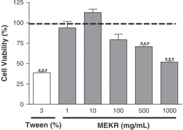
Methyl-thiazolyl-tetrazolium cell viability assay. The cytotoxic effect of MEKR on macrophages was deter-mined using the methyl-thiazolyl-tetrazolium (MTT) assay method according to Mosmann (1983) (Mosmann, 1983). Murine peritoneal macrophages (2.5 × 10⁵ cells) were treated with MEKR at concentrations ranging from 1 mg/mL to 1000 mg/mL and were later cultured in RPMI-1640 supplemented with 10% FBS for 24 h. Thereafter, the medium was replaced with fresh RPMI containing 5 mg/mL of MTT. After additional 4 h of incubation at 37°C, the supernatant was discharged, and dimethyl sulfoxide solution (150μL/well) was added to each culture plate. After 15 min of incubation at room temperature, absorbance of solubilized MTT formazan product was spectrophotometrically measured at 540 nm.
Five individual wells were assayed per treatment and percentage of viability was determined in relation to con-trols [(absorbance of treated cells/absorbance of untreated cells) × 100].
Evaluation of the motor activity.In order to investigate the motor activity of the animals treated with MEKR and the consequent impairment of the mechanical hyperalgesic assessment, the motor activity of the ani-mals was evaluated in rota-rod apparatus, according to Dunham and Miya (1957) (Dunham and Miya, 1957) with some modifications. Initially, the mice able to re-main on the Rota-rod apparatus (AVS®, Brazil) longer than 180 s (7 rpm) were selected 24 h before the test. Thirty minutes after the administration of either MEKR (100, 200, and 400 mg/kg, p.o.), vehicle (saline + Tween 80 0.02%; p.o.), or DZP (1.5 mg/kg, i.p.), each animal was tested on the Rota-rod apparatus and the time (s) remained on the bar for up to 180 s was recorded at 0.5, 1, and 2 after the administration.
Immunofluorescence.To evaluate the action of the test drug on the central nervous system, 90 min after the in-jection of MEKR (100, 200, or 400 mg/kg; p.o.) or vehi-cle (Saline + Tween 80 0.02%, p.o.), the animals (n= 6, per group) were perfused, and the brains were collected and cryoprotected for immunofluorescence processing to Fos protein.
Frozen serial transverse sections (20μm) of all brain were collected on gelatinized glass slides. The tissue sec-tions were stored at 80°C until use. The sections were washed with phosphate buffer (0.01 M) saline isotonic (PBS) 5× for 5 min and incubated with 0.1 M glycine in PBS for 10 min. Non-specific protein binding was blocked by incubation of the sections for 30 min in a solution containing 2% bovine serum albumin. After that, the sections were incubated overnight with rabbit anti-Fos as primary antibodies (1:2000). Afterwards, the sections were incubated for two hours with donkey anti-rabbit Alexa Fluor 594 as secondary antibodies (1:2000). The cover slip was mounted with Fluoromount G. As an immunofluorescence control for non-specific labeling, sections were incubated without primary anti-body. After each stage, slides were washed with PBS 5× for 5 min.
A striking attribute of Fos is that it is rapidly expressed in central neurons after noxious stimuli. As that is a very used way to visualize the pathways involved in the integration of noxious input. For this reason, we evaluated the action of the test drug on the central nervous system, 90 min after the injection of MEKR (100, 200, or 400 mg/kg; p.o.) or vehicle (Saline + Tween 80 0.02%; p.o.), after injection CG (300μg/paw), 60 min after the treatment.
(written by the authors) that uses the same level of label intensity to select and count the Fos positive cells.
Statistical analysis. Data were evaluated using GraphPad Prism Software Inc. (SanDiego, California, USA) version 5.0, through the analysis of variance followed by Tukey’s test. The results are presented as mean ± SEM. In all cases, the differences were consid-ered significant ifp<0.05.
RESULTS
High-performance liquid chromatography– photodiode-array detection analysis
High-performance liquid chromatography-DAD analy-sis of the methanol extract from stems of K.rugosa
(MEKR) (Fig. 1) revealed the presence of peaks with phenolic compounds-like UV spectra. On the basis of elution order in C18 column and their UV absorption spectra, peaks 1–3 (Rt= 11.0 min,Rt= 21.0 min, andRt= 40.8 min, respectively) were similar to hydroxybenzoic acids such as vanillic acid, protocatechuic acid, and syringic acid, respectively (Sunet al., 2007). Peaks 4–8 (Rt= 52.8 min,Rt= 68.1 min,Rt= 76.2 min,Rt= 79.1 min,
and Rt= 81.0 min, respectively) presented UV spectra similar to the one observed for alkyl and phenylcoumarins (Garazdet al., 2003; Scioet al., 2003). Peak 4 (Rt= 52.8 min) was identified by matching the re-tention time with that of an authentic sample of the disprorinol A, a 4-propylcoumarin previously isolated fromK.rugosa(Nogueiraet al., 2009).
Effect of methanol stem extract ofKielmeyera rugosa on carrageenan-induced mechanical hyperalgesia and mouse paw edema
Injection of CG in the subplantar region of the mouse paw induced a marked mechanical hyperalgesia characterized by an increased sensitivity as the intensity of stimulus was decreased, which remained throughout the 3 h. MEKR demonstrated an antihyperalgesic effect in this model, as mice treated with MEKR (100, 200, or 400 mg/kg; p.o.) 0.5 h before CG administration exhibited a significant reduction in mechanical hyperalgesia induced by CG at all evaluated times, when compared with animals of the control group that received only vehicle (Fig. 2A). These doses produced an effect similar to indomethacin (10 mg/kg). The group of animals that received saline in the subplantar region, instead of CG, did not present any alteration on the threshold of sensitivity to mechanical stimuli (data not shown). Mouse paw edema induced by CG administration was also evaluated. As shown in Fig. 3, CG injection in-creased the mouse paw volume from 1 to 6 h after injec-tion, and the treatment of mice with MEKR significantly decreased the edema. The doses of 100, 200, and 400 mg/kg of MEKR were able to maintain the reduc-tion of the edema during the 6-h evaluareduc-tion period, as did indomethacin. Animals that received only the vehi-cle of CG (sterile saline) did not present significant al-teration in paw volume (data not shown).
7
5
4 6
1 2
3
8
Figure 1. High-performance liquid chromatography-diode array detector chromatogram at 254 nm of methanolic extract from
Kielmeyera rugosastems.
0 4 8 12
Vehicle MEKR (100 mg/kg) MEKR (200 mg/kg) MEKR (400 mg/kg) IND (10 mg/kg)
Time (min)
Intensity of hypernociception (∆
of withdrawal threshold, g) 0
4 8 12
Vehicle MEKR (100 mg/kg) MEKR (200 mg/kg) MEKR (400 mg/kg) IND (10 mg/kg)
Time (min)
Intensity of hypernociception (∆
of withdrawal threshold, g)
0 4 8 12
Vehicle MEKR (100 mg/kg) MEKR (200 mg/kg) MEKR (400 mg/kg) DIPY (60 mg/kg)
Time (min)
Intensity of hypernociception (∆
of withdrawal threshold, g) 0
4 8 12
Vehicle MEKR (100 mg/kg) MEKR (200 mg/kg) MEKR (400 mg/kg) DIPY (60 mg/kg)
0 30 60 120 180 0 30 60 120 180
0 30 60 120 180 0 30 60 120 180
Time (min)
Intensity of hypernociception (∆
of withdrawal threshold, g)
A B
C D
Figure 2. Effect of acute administration of vehicle, methanolic extract of stem ofKielmeyera rugosa(MEKR; 100, 200, or 400 mg/kg, p.o.), indomethacin (IND, 10 mg/kg, i.p.), or dipyrone (DIPY, 60 mg/kg, i.p.) on mechanical hyperalgesia induced by carrageenan (A), tumor necro-sis factor-alpha (B), prostaglandin E2(C), or dopamine (D). Each point represents the mean ± SEM of the paw withdrawal threshold (in grams)
Effect of methanol stem extract ofKielmeyera rugosaon tumor necrosis factor-alpha, dopamine, or prostaglandin E2-induced mechanical hyperalgesia
The inhibitory effect of MEKR on the mechanical hyperalgesia induced by TNF-α is shown in Fig. 2B. MEKR (100, 200, and 400 mg/kg, p.o.) was able to reduce mechanical hyperalgesia induced by TNF-α, when compared with animals of the vehicle group, similarly to indomethacin.
The MEKR antihyperalgesic effects on PGE2 -in-duced and DA-in-in-duced hyperalgesia are shown in Fig. 2C and D, respectively. Acute treatment with MEKR (100, 200, and 400 mg/kg) can reduce the mechanical hyperalgesia induced by PGE2 and DA when compared with vehicle group animals.
Effect of methanol stem extract ofKielmeyera rugosa on carrageenan-induced mouse pleurisy
All doses of MEKR were able to significantly suppress the recruitment of leukocytes to the mouse pleural cavity, as shown in Fig. 4A. Pretreatment with MEKR also significantly attenuated the number of neutrophils (Fig. 4B). MEKR (100, 200, and 400 mg/kg) also signifi-cantly reduced the TNF-α(Fig. 4C) and IL-1β(Fig. 4D) levels in the pleural exudates collected at 4 h after CG injection.
Lack of cytotoxicity effect of methanol stem extract of Kielmeyera rugosa
Increasing concentrations of MEKR (1, 10, 100, 500, and 1000 mg/mL) were unable to cause alteration of mu-rine peritoneal macrophages viability of RPMI control 1–100 mg/mL), indicating that MEKR treatment did not significantly affect mitochondrial reduction of MTT to formazan resulting in an undetectable cytotoxic effect, in these concentrations (Figure 5).
Figure 3. Anti-inflammatory effect of methanol stem extract of
Kielmeyera rugosatreatment on carrageenan-induced paw inflam-mation. Methanolic extract of stem of K.rugosa (MEKR; 100, 200, or 400 mg/kg, p.o.), saline (control group, p.o.), or indometh-acin (IND, 10 mg/kg, i.p.), was administered 6 h before carra-geenan. Paw edema measured at 1, 2, 3, 4, 5, and 6 h after the carrageenan injection. Data are expressed as mean ± SEM; *p<0.05, **p<0.01, ***p<0.001 compared with control group (analysis of variance followed by Tukey’s test).
A B
C D
Lack of effect of methanol stem extract ofKielmeyera rugosaon motor activity
Fig. 6 shows the motor activity of mice treated with different doses of MEKR. In this test, MEKR, in all doses, was unable to cause a significant decrease of ambulation (number of crossings) at 0.5, 1, and 2 h after administration, unlike DZP.
Effect of methanol stem extract ofKielmeyera rugosa on immunofluorescence
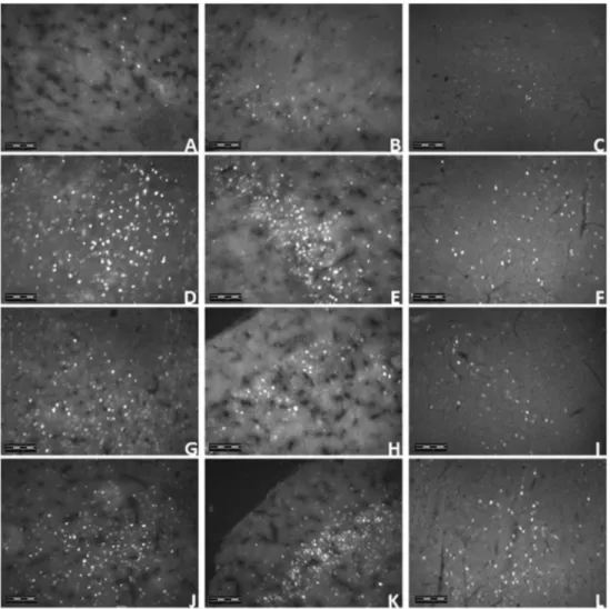
In the olfactory bulb (p<0.01), piriform cortex (100, 400 mg/kg, p<0.01; 200 mg/kg, p<0.05) and in the periaqueductal gray (p<0.01) of the animals brains, the average number of neurons showing Fos protein was significantly increased by an oral injection of MEKR when compared with control (Figs 7 and 8).
DISCUSSION
Because the chemical profile of this genus is characterized by the occurrence of phenolic compounds such as xanthones, 4-phenyl, and 4-alkylcoumarins (Garcia Cortez
et al., 1998; Cruzet al., 2001; Cruzet al., 2002; Scioet al., 2003; Nogueiraet al., 2009) and on the basis of the results by HPLC-DAD analysis, we suggest that these com-pounds could be present in the methanolic extract from stems of K.rugosa (MEKR). Further investigations by liquid chromatography–mass spectrometry on the chemical composition ofK.rugosaare ongoing.
The inflammatory hyperalgesia in mice is mediated by a cascade of cytokines (Cunha et al., 2005). CG in-duces a concomitant release of TNF-α, which stimulates the subsequent release of interleukin (IL)-6/IL-1βand keratinocyte-derived chemokine (KC/CXCL1), which ultimately induce the synthesis of prostaglandins and the release of sympathetic amines, respectively (Cunha
et al., 1992; Cunha and Ferreira, 2003), causing the activation of fiber sensory nerve endings, types Aδ and
C, increasing the local flow and vascular permeability by the release of neurokinin substance P and neurokinin A (Nakamura and Ferreira, 1987; Cunhaet al., 2005). This can lead to the inflammatory process resulting in central and peripheral hyperalgesia (Guimarãeset al., 2012).
The cytokine cascade begins with TNF-α, which stimu-lates two distinct pathways, as previously mentioned: IL-1β, which in turn activates cyclooxygenase to produce prostanoids, and KC/CXCL1 production, which stimulates the release of sympathetic amines (Verri et al., 2006). Prostanoids and sympathetic amines are ultimately responsible for nociceptor sensitization (Cunha et al., 2005). On the basis of these findings, we investigated the possible effect of MEKR on the hyperalgesia induced by PGE2and DA.
Studies have shown that the peripheral injection of PGE2 and sympathomimetic amines, such as DA, triggers the activation of nociceptors and transmission impulse by primary nociceptive neurons (de Oliveira
et al., 2012; Guimarãeset al., 2012). That effect induces both allodynia and hyperalgesia in response to mechan-ical stimulation (Ferreira, 1972; Kuhn and Willis, 1973). This nociceptive effect seems to be related to the ability of PGE2to sensitize peripheral terminals of small diameter and high threshold, including primary afferent fibers sensitive to thermal, chemical, and mechanical stimuli (Kumazawaet al., 1993; Mizumuraet al., 1993).
Prostaglandin E2acts on the EP2receptors, and DA acts on the metabotropic-type D1receptors. The nociceptive behavior and mechanical allodynia caused by i.pl. PGE2 are mediated through activation of distinct EP receptors and PK-dependent mechanisms (Kassuya et al., 2007). Therefore, hyperalgesia elicited by PGE2 and DA is independent on the production of other inflammatory mediators or recruitment of cells such as neutrophils (Cunhaet al., 2008b). The fact that MEKR treatment also inhibited DA and PGE2-induced hyperalgesia implies that either MEKR directly reduces nociceptor sensitization or MEKR can even induce an endogenous mediator through this action, or we cannot exclude the possibility that MEKR interacts even with other types of EP or DA receptors.
The injection of CG in mice produces a typical bi-phasic edema associated with the production of several inflammatory mediators, such as bradykinin, prostaglan-dins, nitric oxide, and cytokines (Henriqueset al., 1987; Posadas et al., 2004). It is well accepted that cytokines
3 1 10 100 500 1000
0 25 50 75 100 125
MEKR (mg/mL) Tween (%)
Cell Viability (%)
Figure 5. Effect ofKielmeyera rugosa(MEKR; 1, 10, 100, 500, and 1000 mg/mL) on the viability assay by the methyl-thiazolyl-tetrazolium (MTT) method for previously MTT-treated cells
in vitro. Measurement of formazan absorbance in relation to the experimental design, showing mean values ± SEM at different times after the first MTT treatment; absorbance of solubilized MTT formazan product was spectrophotometrically measured at 540 nm. ***p<0.001 compared with the control group (analysis of variance followed by Tukey’s test).
0.5 1.0 2.0
0 100
200 Vehicle
MEKR (100 mg/kg)
MEKR (200 mg/kg) MEKR (400 mg/kg)
DZP (1.5 mg/kg)
Time (h) after treatment
Time (s) on Rota-rod
Figure 7. Neurons Fos positive in the bulb olfactory (A), piriform cortex (B), and periaqueductal gray (C). Vehicle (control) orKielmeyera rugosa(MEKR; 100, 200, and 400 mg/kg, p.o.) were administered 1.5 h before the perfusion. Values represent in mean ± SEM (n= 6, per group). *p<0.05, **p<0.01, or ***p<0.001 versus control (one-way analysis of variance followed by Tukey’s test).
constitute a link between cellular injuries or immuno-logical recognition and the local or systemic signs of inflammation, for example, cell migration, edema, fever, and hyperalgesia (Ferreira et al., 1988; Faccioli et al., 1990; Dinarello, 2000). Different cell types, including macrophages, monocytes, and glial cells produce IL-1β, which in turn induces the production of other inflamma-tory mediators involved with cellular recruitment, fever, acute phase protein release, and increase of vascular permeability (Dinarello, 1998). We have shown that the doses of 100, 200, and 400 mg/kg of MEKR-induced antiedematogenic activity.
The mediators involved in the genesis of inflamma-tory pain also play an essential role in triggering other inflammatory events, including edema and leukocyte migration (Cunha et al., 2008a). Therefore, the production of cytokines, including TNF-αand IL-1β, in the site of inflammation is essential for the development of inflammatory hyperalgesia. For this reason, we performed a cell migration assay and measurement of IL-1β and TNF-α by CG-induced pleurisy. Inflammation induced by CG involves cell migration, exudation of plasma, and production of mediators such as nitric oxide, prostaglandin E2, IL-1β, IL-6, and TNF-α (Ferreira et al., 1993; Cunha
et al., 2005). These mediators are capable of re-cruiting leucocytes, such as neutrophils, in various experimental models. The results allowed us to detect a marked inhibitory effect of MEKR on neutrophil migration, besides a reduction of TNF-α and IL-1β
level in the pleural exudate.
Evidences suggest the role of colorimetric assays using the MTT for assessment of cytotoxicity, and prolif-eration studies in cell biology (Berridgeet al., 2005; van Meerloo et al., 2011). The concentrations used of MEKR (1–100 mg/mL) did not affect the MTT reduc-tion in murine peritoneal macrophages, indicating a cell viability effect of this extract.
The fact that MEKR induces antihyperalgesic effect in the mechanical hyperalgesic models suggests that MEKR can block the neural transmission of pain, like other drugs do, and may induce analgesia. Moreover, it has been ob-served that many compounds derived from medicinal plants present a reduction of locomotor activity (Le Bars
et al., 2001) by an inhibitory effect on the central nervous system (CNS) or by a non-specific muscle relaxation ef-fect (Meloet al., 2011). Thus, these activities can reduce the motor coordination response, invalidating the noci-ceptive behavioral tests (de Sousaet al., 2006). However, relaxing or motor deficit effects were discarded, because MEKR administration, at the therapeutic doses, did not affect the motor performance of the mice, as tested in the rota-rod test.
The expression of immediate early genes, most notably c-Fos, has been used to map activation of neural circuits under a variety of experimental condi-tions. c-Fos is expressed in a variety of brain sites, like in the areas involved in the pain modulation (Barr, 2011), being, the Fos protein, a marker useful for the control of neuronal activities in central pathways of the sensory system, particularly in the nociceptive pathway (Williams et al., 1990). To demonstrate the influence of MEKR in the CNS areas, Fos protein la-beled by immunofluorescence was performed, showing a significant activation of the olfactory bulb, piriform cortex and periaqueductal gray (PAG).
The piriform cortex (PC) is a three-layered structure in which the principal excitatory neurons are pyramidal cells. One attractive feature of the piriform cortical slice preparation is that functionally distinct inputs from the olfactory bulb via the lateral olfactory tract. The infor-mation arriving through theses distal synapses provides the vast majority of olfactory signals to the cortex that is presumably used for sensory tasks such as odor discrimination and recognition (Suzuki and Bekkers, 2006; Bathellier et al., 2009). The PC, beyond of the olfactory function due to its connections with olfactory bulb, presents an influence on the aggressive and mating behavior, once this area receives information from the amygdala and hippocampus and projects their axons to amygdala and hypothalamus. These areas make connec-tion with the brain stem, including the raphe and parabrachial nuclei as well as the PAG, influencing the ascending and descending nociceptive circuits.
The PAG, the most important area of descending pain pathway, is interconnected with the hypothalamus and limbic forebrain structures and also receives direct inputs of spinomesencephalic. The PAG projects to the rostral ventromedial medulla, which in turn sends its output to dorsal horn, inhibiting the I-laminae, an important dorsal horn area involved in the nociception (Heinricher et al., 2009). Pain modulatory drugs such as opioids, serotoninergic, and cannabinoids exert central effects in the PAG, and several lines of evidence indicate a central role for prostaglandins in this brain region, what indicates that PAG is an important area involved in the control of inflammatory pain (Brederet al., 1995; Leith et al., 2007; Phillips and Clauw, 2011; Kraft, 2012; Siqueira-Limaet al., 2014). Thus, the attenuation of the mechanical hyperalgesia observed in the present study may be derived from the activation of descending pain path-way and consequent inhibition of spinal cord I-laminae. That can be suggested by the significant activation of the PAG observed in the immunofluorescence protocol.
In summary, the activation of the olfactory bulb and piriform cortex indicates that MEKR influences on the animal behavior and the activation of the PAG suggests the involvement of the central nervous system, more specif-ically of the pain descending inhibitory pathway, in the action of MEKR on the inflammatory pain observed in the hyperalgesia and inflammation protocols used in this study.
The exact mechanism through which MEKR exerts its antihyperalgesic profile remains to be elucidated. Previously, Kielmeyera species showed mainly xan-thones and 4-alkyl and 4-phenyl coumarins, which are regarded as the characteristic constituents of plants belonging to this genus. Taechowisan et al. (2005) demonstrated the physiological roles of
5,7-dimethyloxy-4-p-methoxylphenylcoumarin and 5,7-dimethoxy-4-phenylc-oumarin for the development of biologically active substances (Taechowisan et al., 2005). Nevertheless, a previous chemical study of extracts of fruits, leaves, and stems of K.rugosa were identified on the basis of their spectral data, chemical compounds found in K.rugosa
resemble those that were previously isolated from the other species of Kielmeyera, such as xanthones besides 4-alkyl and 4-phenylcoumarins (Nogueiraet al., 2009).
(Fylaktakidou et al., 2004; Nicolaides et al., 2004). Considering that Taechowisan et al. (2006) observed that 5,7-dimethyloxy-4-p-methoxylphenylcoumarin and 5,7-dimethoxy-4-phenylcoumarin significantly reduced the formation of TNF-α, it is conceivable to suggest that these compounds are responsible for the pharmacological activity presented in this study (Taechowisanet al., 2006).
The present study demonstrates, for the first time, that systemic administration of MEKR, at doses that did not induce any motor performance alteration, produced consistent antihyperalgesic and anti-inflammatory effects in different models of hyperalgesia and inflammation, probably by interfering of CNS, through stimulation of areas that modulate pain perception or modulation, such as PAG. These effects seem to be associated with the power of MEKR to inhibit the cytokine cascade generated by CG and/or decrease the production of
inflammatory mediators. However, further studies can clarify the exact mechanisms underlying the effects of MEKR.
Acknowledgements
We thank O.A. Santos, M.T. Santana, and D.S. Prado for technical support. This work was supported by grants from the National Council of Technological and Scientific Development (CNPq/Brazil) and the Research Supporting Foundation of the State of Sergipe (FAPITEC-SE/Brazil). We also thank teacher Abilio Borghi for the grammar review on the manuscript.
Conflict of Interest
The authors have declared that there is no conflict of interest.
REFERENCES
Audi EA, Otobone F, Martins JV, Cortez DA. 2002. Preliminary evaluation ofKielmeyera coriacealeaves extract on the central nervous system.Fitoterapia73: 517–519.
Balunas MJ, Kinghorn AD. 2005. Drug discovery from medicinal plants.Life Sci78: 431–441.
Barr GA. 2011. Formalin-induced c-Fos expression in the brain of infant rats.J Pain12: 263–271.
Bathellier B, Margrie TW, Larkum ME. 2009. Properties of piriform cortex pyramidal cell dendrites: implications for olfactory circuit design.J Neurosci29: 12641–12652.
Berridge MV, Herst PM, Tan AS. 2005. Tetrazolium dyes as tools in cell biology: new insights into their cellular reduction.
BiotechnolAnnu Rev11: 127–152.
Breder CD, Dewitt D, Kraig RP. 1995. Characterization of inducible cyclooxygenase in rat brain.J Comp Neurol355: 296–315. Cruz FG, Silva-Neto JT, Guedes MLS. 2001. Xanthones and coumarins
fromKielmeyera lathrophyton.J Braz Chem Society12: 117–122. Cruz FG, Moreira LM, Santos NAS, Guedes MLS. 2002. Additional Coumarins fromKielmeyera reticulata.J Braz Chem Society
13: 708–708.
Cunha FQ, Ferreira SH. 2003. Peripheral hyperalgesic cytokines.
Adv Experimental Med Biol521: 22–39.
Cunha FQ, Poole S, Lorenzetti BB, Ferreira SH. 1992. The pivotal role of tumour necrosis factor alpha in the development of inflammatory hyperalgesia.Br J Pharmacol107: 660–664. Cunha TM, Verri WA Jr, Vivancos GG,et al. 2004. An electronic
pressure-meter nociception paw test for mice.Braz J Med Biol Res37: 401–407.
Cunha TM, Verri WA Jr, Silva JS, Poole S, Cunha FQ, Ferreira SH. 2005. A cascade of cytokines mediates mechanical inflamma-tory hypernociception in mice.Proc Natl Acad Sci U S A102: 1755–1760.
Cunha TM, Verri WA Jr, Valerio DA,et al. 2008a. Role of cytokines in mediating mechanical hypernociception in a model of delayed-type hypersensitivity in mice.Eur J Pharm12: 1059–1068. Cunha TM, Verri WA Jr, Schivo IR,et al. 2008b. Crucial role of
neutrophils in the development of mechanical inflammatory hypernociception.J Leukoc Biol83: 824–832.
Dinarello CA. 1998. Interleukin-1, interleukin-1 receptors and interleukin-1 receptor antagonist.Int Rev Immunol16: 457–499. Dinarello CA. 2000. Proinflammatory cytokines. Eur Cytokine
Netw11(3): 483–486.
Dunham NW, Miya TS. 1957. A note on a simple apparatus for detecting neurological deficit in rats and mice.J Am Pharm Assoc Pharm Assoc46: 208–209.
Faccioli LH, Souza GE, Cunha FQ, Poole S, Ferreira SH. 1990. Re-combinant interleukin-1 and tumor necrosis factor induce neu-trophil migration ‘in vivo’ by indirect mechanisms. Agents Actions30: 344–349.
Ferreira SH. 1972. Prostaglandins, aspirin-like drugs and analge-sia.Nat New Biol240: 200–203.
Ferreira SH, Lorenzetti BB, Bristow AF, Poole S. 1988. Interleukin-1 beta as a potent hyperalgesic agent antagonized by a tripeptide analogue.Nature334: 698–700.
Ferreira SH, Lorenzetti BB, Poole S. 1993. Bradykinin initiates cytokine-mediated inflammatory hyperalgesia.Br JPharmacol
110: 1227–1231.
Fylaktakidou KC, Hadjipavlou-Litina DJ, Litinas KE, Nicolaides DN. 2004. Natural and synthetic coumarin derivatives with anti-inflammatory/ antioxidant activities.Curr Pharm Des10: 3813–3833.
Garazd M, Garazd YL, Khilya V. 2003. Neoflavones. 1. Natural distribution and spectral and biological properties.Chem Nat Comp39: 54–121.
Garcia Cortez D, Young M, Marston A, Wolfender J-L, Hostettmann K. 1998. Xanthones, triterpenes and a biphenyl fromKielmeyera coriacea.Phytochemistry47: 1367–1374. Guimarães AG, Xavier MA, de Santana MT,et al. 2012. Carvacrol
attenuates mechanical hypernociception and inflammatory re-sponse.Naunyn Schmiedebergs ArchPharmacol385: 253–263. Guimarães AG, Quintans JSS, Quintans-Júnior LJ. 2013. Mono-terpenes with analgesic activity - a systematic review.
Phytother Res27: 1–15.
Guimarães AG, Serafini MR, Quintans-Júnior LJ. 2014. Terpenes and derivatives as a new perspective for pain treatment: a pat-ent review.Expert Opin Ther Patents24: 243–265.
Heinricher MM, Tavares I, Leith JL, Lumb BM. 2009. Descending control of nociception: specificity, recruitment and plasticity.
Brain Res Rev60: 214–225.
Henriques MG, Silva PM, Martins MA,et al. 1987. Mouse paw edema. A new model for inflammation?Braz J Med Biol Res
20: 243–249.
Hoult JR, Paya M. 1996. Pharmacological and biochemical actions of simple coumarins: natural products with therapeutic poten-tial.Gen Pharmacol27: 713–722.
Kassuya CA, Ferreira J, Claudino RF, Calixto JB. 2007. Intraplantar PGE2 causes nociceptive behaviour and mechanical allodynia: the role of prostanoid E receptors and protein kinases. Br JPharmacol150: 727–737.
Kraft B. 2012. Is there any clinically relevant cannabinoid-induced analgesia?Pharmacol89: 237–246.
Kuhn DC, Willis AL. 1973. Proceedings: prostaglandin E2, inflam-mation and pain threshold in rat paws. Br J Pharmacol49: 183–184.
Kumazawa T, Mizumura K, Koda H. 1993. Involvement of EP3 subtype of prostaglandin E receptors in PGE2-induced enhancement of the bradykinin response of nociceptors.Brain Res632: 321–324. Le Bars D, Gozariu M, Cadden SW. 2001. Animal models of
nociception.Pharmacol Rev53: 597–652.
Leith JL, Wilson AW, Donaldson LF, Lumb BM. 2007. Cyclooxy-genase-1-derived prostaglandins in the periaqueductal gray differentially control C- versus A-fiber-evoked spinal nociception.J Neurosci27: 11296–11305.
Levy L. 1969. Carrageenan paw edema in the mouse.Life Sci8: 601–606.
McCurdy CR, Scully SS. 2005. Analgesic substances derived from natural products (natureceuticals).Life Sci78: 476–484. van Meerloo, J, Kaspers, GJ, Cloos, J, 2011. Cell sensitivity
Melo MS, Santana MT, Guimarães AG, et al. 2011. Bioassay-guided evaluation of central nervous system effects of citro-nellal in rodents.Braz J Pharmacog21: 697–703.
Mizumura K, Minagawa M, Tsujii Y, Kumazawa T. 1993. Prostaglan-din E2-induced sensitization of the heat response of canine vis-ceral polymodal receptorsin vitro.Neurosci Lett161: 117–119. Mosmann T. 1983. Rapid colorimetric assay for cellular growth and survival: application to proliferation and cytotoxicity assays.J Immunol Methods65: 55–63.
Nakamura M, Ferreira SH. 1987. A peripheral sympathetic component in inflammatory hyperalgesia.Eur J Pharmacol135: 145–153. Nicolaides DN, Gautam DR, Litinas KE, Hadjipavlou-Litina DJ,
Fylaktakidou KC. 2004. Synthesis and evaluation of the antioxi-dant and antiinflammatory activities of some benzo[l]khellactone derivatives and analogues.Eur J Med Chem39: 323–332. Nogueira PC, Andrade MS, Andrade LM,et al. 2009. Chemical
constituents from Kielmeyera rugosa Choisy (Clusiaceae).
Biochem Syst Ecol36: 921–924.
de Oliveira MG, Marques RB, de Santana MF,et al. 2012. Alpha-terpineol reduces mechanical hypernociception and inflamma-tory response.Basic Clin Pharmacol Toxicol111: 120–125. Oliveira AC, Britto AC, HenriquesRM, CGM,et al. 2013. In vivo
growth inhibition of sarcoma 180 by Kielmeyera rugosa
Choisy(Calophyllaceae). Nat Prod Res27: 2248–2250. Phillips K, Clauw DJ. 2011. Central pain mechanisms in chronic
pain states--maybe it is all in their head.Best Pract Res Clin Rheumatol25: 141–154.
Pinheiro L, Nakamura CV, Dias Filho BP, Ferreira AG, Young MC, Cortez DA. 2003. Antibacterial xanthones from Kielmeyera variabilis
mart. (Clusiaceae).Mem Inst Oswaldo Cruz98: 549–552. Posadas I, Bucci M, Roviezzo F,et al. 2004. Carrageenan-induced
mouse paw oedema is biphasic, age-weight dependent and displays differential nitric oxide cyclooxygenase-2 expression.
Br J Pharmacol142: 331–338.
Quintans JSS, Menezes PP, Santos MR,et al. 2013 Improvement of p-cymene antinociceptive and anti-inflammatory effects by inclusion inβ-cyclodextrin.Phytomed20: 436–440. Ribeiro SS, de Jesus AM, Dos Anjos CS,et al. 2012. Evaluation of
the cytotoxic activity of some Brazilian medicinal plants.
Planta Med78: 1601–1606.
Scio E, Ribeiro A, Alves TM,et al. 2003. New bioactive coumarins fromKielmeyera albopunctata.J Nat Prod66: 634–637.
Sela VR, Hattanda I, Albrecht CM,et al. 2010. Effect of xanthone fromKielmeyera coriaceastems on serotonergic neurons of the median raphe nucleus.Phytomed17: 274–278.
da Silva KA, Manjavachi MN, Paszcuk AF, et al. 2012. Plant derived alkaloid ( )-cassine induces anti-inflammatory and anti-hyperalgesics effects in both acute and chronic inflam-matory and neuropathic pain models. Neuropharmacol 62: 967–977.
Siqueira-Lima PS, Araújo AA, Lucchese AM,et al. 2014.β -Cyclo-dextrin complex containingLippia grata leaf essential oil re-duces orofacial nociception in mice–evidence of possible involvement of descending inhibitory pain modulation path-way.Basic Clin Pharmacol Toxicol114: 188–196.
de Sousa DP, de Sousa OF, de Almeida RN. 2006. Evaluation of the central activity of hydroxydihydrocarvone.Biol Pharm Bull29: 811–812.
Sun J, Liang F, Bin Y, Li P, Duan C. 2007. Screening non-colored phenolics in red wines using liquid chromatography/ ultraviolet and mass spectrometry/mass spectrometry librar-ies.Molecules12: 679–693.
Suzuki N, Bekkers JM. 2006. Neural coding by two classes of principal cells in the mouse piriform cortex. J Neurosci26: 11938–11947.
Taechowisan T, Lu C, Shen Y, Lumyong S. 2005. Secondary me-tabolites from endophytic Streptomyces aureofaciens CMUAc130 and their antifungal activity. Microbiol 151: 1691–1695.
Taechowisan T, Lu C, Shen Y, Lumyong S. 2006. Anti-inflamma-tory effects of 4-arylcoumarins in LPS-induced murine macro-phage RAW 264.7 cells.Pharm Biol44: 576–580.
Verri WA Jr, Cunha TM, Parada CA, Poole S, Cunha FQ, Ferreira SH. 2006. Hypernociceptive role of cytokines and chemokines: targets for analgesic drug development?
Pharmacol Ther112: 116–138.
Vivancos GG, Verri WA Jr, Cunha TM, Schivo IR, Parada CA, Cunha FQ, Ferreira SH. 2004. An electronic pressure-meter nociception paw test for rats.Braz J Med Biol Res37: 391–399. Williams S, Evan GI, Hunt SP. 1990. Changing patterns of c-Fos induction in spinal neurons following thermal cutaneous stimulation in the rat.Neurosci36: 73–81.