EVALUATION ON SEROLOGIC TESTS FOR STUDIFS
ON CHAGAS’ DISEASE’
I. G. Kagan,’ R. S. Goldsmith? R. Z&ate-Castaiieda,! and D. S. AXlain5
Findings of high antibody prevalence rates to Chagas disease in Oaxaca, Mexico (up to 79 per cent positive in an adult sam- ple from one community) prompted studies directed at evaluat- ing the tests used, the antibody detected, and the practice of
collecting and storing blood specimens on filter paper. This article reports the results of these studies.
\
Introduction
A 1969 seroepidemiologic survey for
parasitic antibodies in the State of Oaxaca,
Mexico, detected antibody against Trypa-
nosoma CTU%i, the etiologic agent of
Chagas’ disease, in 29yo of the serum
samples from one part of the state (I). This initial study was later expanded by indirect
hemagglutination (IHA) testing of 4,023
persons residing in 60 communities located within a Pacific coastal zone of Oaxaca
State. In a companion paper (2) we re-
ported finding antibody prevalence rates
that were remarkably high for Mexico,
ranging up to a finding of 79oJ, seropositive reactors among a sample of adults tested in one community.
1 Also appearing in Spanish in the Roletin de la Ofi- cina Sanitaria Panamericana, 1979.
ZDirector, Parasitology Division, Center for Disease Control, Public Health Service, U.S. Department of Health, Education, and Welfare, Atlanta, Georgia 30333, U.S.A.
3Associate Professor of Tropical Medicine, The G. W. Hooper Foundation and the Department of Inter- national Health, University of California, San Fran- cisco, California 94143, U.S.A.
4Researcher of the Institute of Health and Tropical Diseasesof the Social Security Administration, Mexico City, Mexico, and the Department of International Health, University of California, San Francisco, Cali- fornia 94143, U.S.A.
5Research Microbiologist, Parasitology Division, Center for Disease Control, Public Health Service, U.S. Department of Health, Education, and Welfare, Atlanta, Georgia 30333, U.S.A.
In order to better understand the origin and significance of the antibody found, three serologic tests for T. cruzi antibody, as well as cross-reactions involved in these tests, were evaluated in the laboratory. Using T. cruzi as antigen, specimens were subjected to two different paired tests and
the serologic results were compared. The
paired tests used were IHA and complement fixation (CF), IHA and direct agglutina- tion (DA), and CF and DA. Cross-reaction in the IHA test for Chagas’ disease were
evaluated by testing antigens prepared
from T. cruzi, T. rangeli, and Leishmania
mexicana against sera from persons who
had parasitologically confirmed T. cruzi
and T. rangeli infections. In addition, fil- ter paper blood eluates and sera were tested
and compared in order to determine the
effectiveness of filter paper strips for col- lecting and storing.blood specimens in the course of field studies.
Material and Methods
Blood specimens were obtained by veni- puncture without using anticoagulants. Af- ter clotting and retraction, specimens were centrifuged and processed aseptically, and the resulting sera were stored at -20” to -70°C. Filter paper blood samples were ob- tained by pricking each subject’s finger and saturating both sides of a circular area
342 PAHO BULLETIN l vol. XII, no. 4, 1978
printed on a piece of filter paper.6 Efforts were made to collect the blood without squeezing the fingertip. The filter papers were air-dried at ambient temperature for
24 hours. During the field work (up to
four weeks) they were stored at ambient temperature in airtight plastic bags con- taining a drying agent, silica gel. There- after, until processed in the laboratory for antibody (8-12 months after collection), the papers were retained in the airtight bags at 4OC.
Blood was subsequently eluted from the filter papers as follows: a 1 cm disc was cut out of the blood-filled circle with a paper punch. Each disc was placed in a 15 x 85 mm tube, and 0.2 ml of phosphate-buffered
saline (PBS) at pH 7.2 was added. The
discs were kept immersed in the saline solu- tion between one and two hours at room
temperature. Before a disc was removed
from the tube, residual fluid was expressed from the disc by squeezing the disc against the side of the tube with a metal rod.
To compare IHA titers from sera with II-IA titers from filter paper eluates, the same venous blood specimen used to prepare a serum was used to saturate a filter paper circle; such matched samples were obtained from the blood of 128 Oaxaca residents in
1971. The reproducibility of IHA test re- sults was evaluated by retesting 44 of these filter paper blood eluates.
For comparison of the IHA and CF test, 150 sera were tested that had been collected in Oaxaca in 1969-1970. Results of the IHA and DA tests were compared by titrating 98 filter paper blood eluates collected in Oaxa- ca in 1972. The CF and DA tests were com-
pared by examining 67 sera obtained in
Oaxaca in 1973.
Comparative tests for antibodies to T.
cruzi and T. rangeli were made with sera
‘Ropaca No. 1023, Rochester Paper Company. (Use of trade names is for identification purposes only and does not constitute endorsement by the U.S. Public Health Service or the U.S. Department of Health, Education, and Welfare.)
from 27 patients in Chile who had parasito- logically confirmed Chagas’ disease; with
sera from 12 persons in Panama who had
parasitologically confirmed T. rangeli in- fections; with 41 sera from Merida, Mexico; and with 140 of the sera obtained in Oaxaca in 1969-1970. Comparative tests for anti- bodies to T. cruri and L. mexicana were made with 85 sera collected in Oaxaca in
1969-1970.
Preparation of the Antigens
The T. cruri and T. rangeli antigens were saline extracts of lyophilized epimasti- gotes of T. cruzi and T. rangeli, respective-
ly; these epimastigotes were delipidized
with benzene before extraction (I). The T. rangeli antigen was titrated against sera from rabbits that had been immunized with lyophilized T. rangeli. The antigen dilu- tion giving the highest titers was used to test the sera described.
The L. mexicana antigen used in the DA test was made according to the method de- scribed for preparing T. cruxi antigen for this test (3). The antigen suspension con- sisted of formalin-fixed L. mexicana pro- mastigotes that had been trypsinized. Three sera from patients with parasitologically
confirmed L. mexicana infections were
tested with this antigen and yielded titers r 4,096.
The serologic methods used in this study for the IHA (I), CF (I), and DA tests (3) have previously been described.
Results
Com@arison of IHA Titers Obtained with
Paired Filter Paper Blood Eluates and with Sera
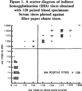
Figure 1. A scatter diagram of indirect
hemagglutination (IHA) titers obtained with 128 paired blood specimens:
Serum titers plotted against filter paper eluate titers.
I.2048 . .
11024
IHA POSITIVE TITERS P 1:128
eluate prepared from the same venous blood sample. Since a filter paper eluate in 0.2 ml of buffer is considered equivalent to a 1:s dilution of serum, filter paper titers below 8 cannot be detected in the IHA test. Nearly all filter paper eluate titers of 64 through 256 were one or two twofold dilu- tions below the corresponding serum titers. The differences in titers between paired specimens decreased, however, as the con- centration of antibody increased; so that filter paper titers above 256 showed increas-
ing agreement with serum titers. With a
titer 2128 taken to indicate a positive re- sponse, 55 specimens (43%) were positive by both methods, and 64 (50%) were negative
by both methods, resulting in 93% agree- ment between the methods. If a filter paper titer L 64 had been considered positive, the titers for only two specimens would have been in disagreement, both specimens being
negative by the filter paper method and
positive by the serum method.
Reproducibility of IHA Results
Comparative findings obtained by ti-
trating 44 filter paper eluates twice with T. cruzi antigen are shown in Table 1. Of the paired results, the titers of 34 specimens (77%) were the same or within one twofold dilution; eight specimens (189r) showed a difference of two twofold dilutions; only
two specimens (5%) showed differences
greater than two twofold dilutions. These results are within the range of acceptable laboratory variation.
Comparison of IHA and CF Tests
One hundred and fifty sera were tested against T. cruzi antigen with both of these methods (see Table 2). With CF titers of 8
or greater considered positive, 86 sera
(57%) yielded a positive CF response. With IHA titers of 128 or greater considered positive, 91 sera (61%) yielded a positive re- sponse. Overall, 121 of the 150 sera were positive or negative by both test methods,
resulting in 81% agreement between the
two techniques.
Table 1. Reproducibility of the results of indirect hemagglutination (IHA)
tests using filter paper eluates and T. MLzi ant&n.
Differences, in number of twofold dilutions, between the two titers obtained by testing the
same eluates twice against T. cruri antigen.
Total No. of eluates No. of twofold
dilution differences
0 1 2 L-2
No. of filter paper 19 15 8 2 44
blood eluates
344 PAHO BULLETIN l vol. XII, no. 4, 1978
Comparison of IHA and DA Tests
The titers obtained by testing 98 filter paper blood eluates with the IHA and DA techniques, using T. cruzi as antigen, are shown in Table 3. With titers of 128 or greater in both tests considered positive, only two of 42 specimens positive in the IHA test yielded negative results. Overall, 96 of the 98 sera were positive or negative by both test methods, so that there was 98% agreement between the two techniques.
Table 4. Comparison of complement fixation (CF) and direct agglutination (DA) results obtained
by testing 67 sera with T. c& antigen. CF testb
Total
+ -
DA testa + 42 9 51
- 6 10 16
Total 48 19 67
aPositive DA test: titer? 128. bpositive CF test: titer 2 8.
Com@arison of CF and DA Tests positive, and 51 of the 67 sera (76%) yielded
Sixty-seven sera were tested by the CF
positive results. Overall, 52 of the 67 sera
and DA methods, using T. cruzi as antigen were positive or negative by both test
(Table 4). With a CF titer of 8 or greater methods, resulting in 78% agreement be- considered positive, 48 of the 67 sera (7.2%)
tween the tWO techniques.
showed a positive CF response. In the DA Cross-Reactivity between T. cruzi, T. ran- tests, titers of 128 or greater were considered geli, and L. mexicana Antigens
Table 2. Comparison of indirect hemagglutination (IHA) and complement fixation (CF) results
obtained by testing 150 sera with
T. cnui antigen. CF testb
Total
+ -
IHA testa + 74 17 91
- 12 47 59
Total 86 64 150
apositive IHA test: titer 2 128. bositive CF test: titer 2 8.
Table 3. Comparison of indirect hemagglutination (IHA) and direct agglutination (DA) results
obtained by testing 98 filter paper eluates with T. cruri antigen.
DA testb
Total
+ -
IHA testa + 40 2 42
- 0 56 56
Total 40 58 98
Comparison of T. cruzi and T. rangeli
antigens: When tested by IHA, using T.
cruzi as antigen, sera from 27 Chilean pa-
tients with Chagas’ disease confirmed by
xenodiagnosis all yielded titers of 128 or greater. These sera were also tested with an
antigen prepared from T. rangeli. The
IHA titers to T. rangeli ranged from 2 to 128, with most reacting at titers of 16 and 32. Only one of the sera showed a titer of 128, which is considered the lower limit for a positive titer against T. rangeli in the IHA test.
Twelve sera from children in Panama
with parasitologically confirmed T. rangeli infections were tested by IHA, using both
T. cruzi and T. rangeli antigens; all 12 yielded negative results.
Sera from 41 subjects in Mhida, Mexico, that yielded negative titers with the T. cruzi antigen also showed negative titers with the T. rangeli antigen. The T. cruzi titers were 16 or less, and the T. rangeli titers were 8 or less.
with T. cruzi and T. rangeli antigens (Table 5). Some reactivity to T. rangeli antigen was found among the 70 sera posi- tive with T. crud antigen, the titers with T. rangeli ranging from 4 to 256. All of the titers obtained with the T. rangeli
antigen, however, were lower than those
obtained with the T. cruzi antigen, and
only four sera in this group (3y0) yielded positive results with both antigens. All 70 of the samples seronegative with T. cruzi antigen were also seronegative with T. ran- geli antigen .
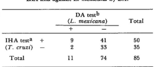
Comparison of T. cruzi and L. mexicana Antigens: Eighty-five of the sera collected in Oaxaca in 1969-1970 were tested with T.
cruzi antigen by IHA and with the L.
mexicana antigen by DA (Table 6). Nine
sera (11%) yielded positive titers with both T. cruri and L. mexicana (128 or higher), and 33 were found negative in both tests. Of the remaining sera, 41 yielded positive results with T. cruzi but negative results with L. mexicana, and two were negative
with T. cruzi but positive with L.
mexicana.
Discussion
The U.S. Center for Disease Control
(CDC) has used the IHA test in seroepide- miologic studies of parasitic diseases for a number of years because of the test’s effec-
tiveness in measuring the prevalence of
antibodies to several parasitic antigens in
Table 5. Comparison of indirect hemagglutination (IHA) results obtained by testing 140 Oaxaca sem
with T. crwi antigen and T. mngeli antigen. T. rangeli antigenb
Total
+ -
T. nuzi antigena + 4 66
- 0 70 7’0”
Total 4 136 140
aPositive IHA test (T. cruri): titer 2 128. bpositive IHA test (T. Tang.&‘): titer 2 128.
Table 6. Comparison of indirect hemagglutination (JHA) and direct agglutination (DA) results obtained
by testing 85 Oaxaca sera against T. crztzi by IEL4 and against L. mexicana by DA.
IHA testa + (T. cm?i) -
Total
DA testb (L. mexicana)
f -
9 41
2 33
11 74
Total 50 35 85 apositive IHA test (T. cruzi): titer 2 128. BPositive DA test (L. mexicana): titer 2 128.
large numbers of specimens. The IHA test has previously been used for seroepidemio- logic studies of Chagas’ disease in Brazil
(4), Colombia (Kagan, unpublished), and
Oaxaca, Mexico (1).
The use of filter papers greatly facilitates the collection and storage of blood samples in the field, because taking blood from a finger prick is much more acceptable to the
population than venipuncture, and because
transporting filter paper blood specimens is much easier than transporting sera, since filter papers do not require refrigeration. As already noted, in order to evaluate the filter paper method for field studies of Chagas’ disease, we compared IHA titers in pairs of sera and filter paper blood samples from 128 persons. Except at the highest antibody concentrations, where the titers were about equal, titers from the filter paper eluates tended to be one or two two- fold dilutions lower than those from the sera. Overall, however, the observed loss in sensitivity was only 7yo when titers of 128 or greater were considered positive. This indicates that collecting and transporting blood on filter papers can be a useful tech- nique-and one well-suited to procurement
of accurate results-when employed in
346 PAHO BULLETIN l vol. XII, no. 4, 1978
IHA V), CF (6, and indirect immuno-
fluorescence (7, 8, 9).
The reproducibility of the IHA titrations was high; 77.3% of the titers obtained were either the same or within one twofold dilu- tion, and 95.5% were within two twofold dilutions.
The IHA test for Chagas’ disease is con- sidered as sensitive or more sensitive than the CF test, the standard against which
newer tests are evaluated. However, the
IHA test is thought to give a larger (though still small) percentage of false positive re- actions (IO). In our first Oaxaca study (I), 73% of the results for 83 sera examined con- currently by both IHA and CF tests were in agreement: that is, the results of both tests were either negative or positive. In the study described here the overall agreement for 150 sera was 77%. Some of the results with specimens in these studies that yielded positive reactions by IHA but negative ones by CF may have been due to false positive reactions, or to the IHA test detecting anti- bodies that had persisted longer than those detected by CF.
The high correlation between the IHA
and DA test results was unexpected. The fact that all but two of the 42 sera with a positive IHA titer also yielded a positive DA titer suggests that the DA test may be a useful serologic procedure for detecting
antibodies to T. cruzi. However, the
finding that 18% of the sera with a positive DA titer were negative in the CF test sug- gests the possibility of false positive DA re- actions.
The specificity of the antibody detected in the Oaxaca sera with T. cruzi antigen
was evaluated by testing the sera with
antigens prepared from two other infectious agents, L. mexicana and T. rangeli. Dermal leishmaniasis (Chiclero’s ulcer of the ear)
caused by L. mexicana is found in some
parts of southern Mexico, although it has not been found in the Pacific coastal region of Oaxaca. T. rangeli infections have not
been reported from Mexico in man, ani-
mals, or vectors; but the parasite has been
found as far north as Guatemala in
Rhodniusprolixus (11). The distribution of T. rangeli is thought to be coterminous with the distribution of its principal vector, R. prolixus. Therefore, since R. prolixus has been in Oaxaca, T. rangeli may later be found in that area.
The data in this study support the evi- dence presented in the relatively few other studies concerning serologic cross-reactions between T. cruzi and T. rangeli (11, 12, 13, 14, 15). These studies indicate that T. ran-
geli infections in man do not elicit
antibodies that cross-react with T. cruzi; yet, conversely, infection in man with T. cruzi produces antibodies that do cross-react at low levels with T. rangeli. Our study of
12 sera from subjects with parasitologically
confirmed T. rangeli infections did not
detect any antibody to either T. rangeli or T. cruzi. However, one of 27 sera from subjects with parasitologically confirmed T.
cruzi infections yielded a titer of 128 against T. rangeli antigen, and sera from four sub- jects that yielded positive titers with T. cruzi antigen also yielded positive titers with T. rangeli antigen. To evaluate these cross-reactions further, cross-absorption techniques should be carried out with im- mune sera from animals infected with each of the parasites, as well as with sera from
people who have had naturally acquired
infections.
reaction with T. cruzi antigen, two yielded a positive response in the DA test with L.
mexicana. Because the presence of L.
mexicana infection in the area of Oaxaca where the sera had been collected cannot be ruled out, the reactions obtained from some or all of the 11 sera that showed positive titers with L. mexicana antigen may reflect the presence of antibodies to L. mexicana.
In the DA test for leishmaniasis with an antigen prepared from L. donovani, a titer of 32 or greater is considered positive (17). Further studies of sera from persons with
parasitologically confirmed L. mexicana
infections are needed so that the sensitivity and specificity of serologic tests for L. mexi- cana can be evaluated.
ACKNOWLEDGMENTS
The field studies reported here were con- ducted under a program for collaborative research between Benito Juarez University of Oaxaca and the University of California (San Francisco). Research was supported in part by grants from the National Institutes
of Health, U.S. Public Health Service-
Al-10051 (University of California Inter- national Center for Medical Research) and HD-06922.
The authors wish to thank Drs. Fernan-
do Galindo, Mario Perez Ramirez, and
Hugo Sarmiento DIaz of Benito Jugrez
University, and Gerard0 Varela of the
Institute of Health and Tropical Diseases in Mexico City for their help and encour- agement.
A comparative evaluation has been made of three serologic tests used for epidemiologic studies of Chagas’ disease in Oaxaca, Mexico. The results of this evaluation, concerned with the techniques of indirect hemagglutination
(IHA), direct agglutination (DA), and comple- ment fixation (CF), were as follows:
The reproducibility of IHA test results was demonstrated by testing the same sera twice by IHA against Trypanosoma crzlri antigen. The results of these tests showed 95c$ agreement. In addition, 81% agreement was obtained when blood samples were tested against T. cru.zi by
IHA and CF. Similar testing of samples by IHA and DA yielded 98% agreement, while 78%
agreement was obtained with samples tested by
DA and CF.
When T. rangeli antigen was used, sera tested by IHA showed few or no positive results. Cross- reactivity was found in 11% of 85 sera tested against Leishmaniu mexicana antigen by DA and against T. crwzi by IHA.
Sera from 128 subjects were tested against T.
cruzi antigen by IHA, and the results were compared to those obtained using eluates from filter paper strips containing biood specimens from the same subjects. Agreement between the
two sets of results was obtained in 93% of the
cases, indicating that collecting and transporting blood specimens on filter paper can serve as a useful and accurate technique in epidemiologic
studies concerned with IHA antibodies to T.
CTUZi.
REFERENCE3
(1) Goldsmith, R. S., I. G. Kagan, M. A. glutination test. Bull Pan Am Health Organ 6:
Reyes-Gonzalez, and J . Cedefio-Ferreira. Sero- 39-52, 1972.
348
iio-Ferreira. Epidemiologic studies of Chagas’ disease in Oaxaca, Mexico. Bull Pan Am Health
Organ 12(3): 236-250, 1978.
(3) Allain, D. S., andI. G. Kagan. An evalua- tion of the direct agglutination test for Chagas’ disease.] Purasitol 60: 179-184, 1974.
(4) Cuadrado, R. R., and I. G. Kagan. The prevalence of antibodies to parasitic diseases in sera of young army recruits from the United States and Brazil. Am J EPidemioE 86: 330-340, 1967.
(5) Neal, R. A., and R. A. Miles. Indirect haemagglutination test for Chagas’ disease, with a simple method for survey work. Rev Znst Med Trofi Sdo Paul0 12:325-332, 1970.
(6) Maekelt, G. A., and C. Colmenares de Alayon. Metodo sencillo para el envio de sueros Chagasicos desde las zonas rurales. Arch Venezo- lanes Med Trap Parasitol Med 3: 133-142, 1960.
(7) Loureiro de Souza, S., and M. E. Camar- go. The use of filter paper blood smears in a practical fluorescent test for American trypano- somiasis serodiagnosis. Rev Znst Med Trap S&o Paul0 8: 255-258, 1966.
(8) Alvarez, M., A. M. De Rissio, G.J.W. De Martini, L. Abram0 Orrego, and J. A. Cerisola. Recoleccion de sangre en papel para diagnostic0 de infeccidn chagasica por immunofluorescen- cia. Bol Chil Parasitol 26: 2-6, 1971.
(9) France, M. F., and L. G. Chamma. Tech- nical modification of indirect immunofluores- cent antibody test using filter paper blood eluates. Znt Arch Allergy Appl Zmmunol 44: 692-696, 1973.
PAHO BULLETIN . vol. XII, no. 4, 1978
(10) Cerisola, J. A., M. Alvarez, H. Lugones, and J. B. Rebosolan. Sensibilidad de las Reac- ciones serologicas para el diagndstico de la en- fermedad de Chagas. Bol Chil Parasitol 24: 2-8, 1969.
(II) World Health Organization. Chugas’
Disease: Report of a Study Group. WHO Tech- nical Report Series No. 202, Geneva, 1960.
(12) Brener, Z., and E. Chiari. Variacoes morfoldgicas observades em differentes amostras de Trypanosoma cruzi. Rev Znst Med TroP Scio Paul0 5: 220-224, 1963.
(I?) Gonzalez Cappa, S. M., and I. G. Kagan. Agar gel immunoelectrophoretic analy- sis of several strains of Trypanosoma cruzi. Exp Purusitol 25: 50-57, 1969.
(14) Kagan, I. G., L. Norman, and D. Allain. Studies on Trypanosoma cruzi isolated in the United States: A review. Rev Biol Trap 14:
55-73, 1966.
(Z5) Nussenzweig, V., L. M. Deane, and J, Kloetzel. Diversidade na constitucgo antigenica de amostras de Trypanosoma cruri isoladas do homem e de gambas. Rev Znst Med Trap Scio Paul0 4: 409-410, 1962.
(16) Pan American Health Organization.
Report of a Study Group on Chagas’ Disease.
PAHO Scientific Publication No. 195, Wash- ington, 1970.
(17) Allain, D. S., and I. G. Kagan. A direct agglutination test for leishmaniasis. Am J Tro$ Med Hyg 24: 232-236, 1975.
INTERNATIONAL UNION FOR
HEALTH EDUCATION