Evaluation of some oral postradiotherapy
sequelae in patients treated for head and
neck tumors
Avaliação de algumas seqüelas bucais
pós-radioterapia em pacientes tratados de
neoplasias de cabeça e pescoço
Abstract: The aim of this study was to evaluate the oral sequelae of radiotherapy in pa-tients treated between 1999 and 2003 for head and neck tumors. One-hundred papa-tients (24 women, 76 men) ranging in age from 30 to 83 years (mean 59.2 years) were examined. Time since radiotherapy ranged from 1 to 72 months (mean 28 months). The total mean radiation dose received by the patients was 5,955 cGy. The evaluation protocol included anamnesis, intraoral and extraoral examination, measurement of stimulated salivary low and salivary pH. Symptoms reported by the patients included dry mouth (68%), dyspha-gia (38%), and dysgeusia (30%). In 64% of the patients, the mean stimulated salivary low rate was less than 0.7 ml/min. The mean salivary pH was 6.97 (± 0.714). Stimulated salivary low increased with increasing postradiotherapy time (p < 0.05). The prevalence of mucositis was associated with higher radiation doses (p < 0.05), and the prevalence of atrophic candidiasis was related to a longer post-treatment period (p < 0.05). Two cases of recurrence of the primary tumor were detected during the study. The main effect of radiotheraphy in the head and neck region was a reduction of the salivary low rate, even though our study demonstrated that there was a modest late improvement of the salivary low.
Descriptors: Radiotherapy; Xerostomia; Saliva; Head and neck neoplasms.
Resumo: O objetivo deste estudo foi avaliar as seqüelas bucais provocadas pela radio-terapia em pacientes com neoplasias de cabeça e pescoço, tratados entre 1999 e 2003. Foram examinados 100 pacientes (24 mulheres e 76 homens), com idades entre 30 e 83 anos (média de 59,2 anos). O tempo desde a radioterapia variou de 1 a 72 meses (média de 28 meses). A média da dose total de radiação recebida pelos pacientes foi de 5.955 cGy. O protocolo de avaliação consistiu de anamnese, exame físico, aferição do luxo salivar estimulado e pHmetria da saliva. Os sintomas referidos foram boca seca (68%), disfagia (38%) e disgeusia (30%). Em 64% dos indivíduos o valor médio do luxo salivar estimula-do esteve abaixo de 0.7 ml/min. O pH médio da amostra foi de 6.97 (± 0.714). O luxo es-timulado e a ocorrência de candidíase atróica aumentaram conforme o aumento do tem-po pós-radioterapia (p < 0.05). A ocorrência de mucosite esteve associada a maiores doses de radiação (p < 0.05). Dois casos de segundo tumor primário foram diagnosticados. O principal efeito da radioterapia na região de cabeça e pescoço foi a redução do luxo sali-var, apesar de nosso estudo ter demonstrado que há uma melhora tardia do luxo salivar. Descritores: Radioterapia; Xerostomia; Saliva; Neoplasias de cabeça e pescoço. Cássia Maria Fischer Rubira(a)
Nádia Juliana Devides(b)
Liliane Torsani Úbeda(b)
Antônio Geraldo Bortolucci Jr(c)
José Roberto Lauris(d)
Izabel Regina Fischer Rubira-Bullen(d)
José Humberto Damante(e)
(a) PhD Student; (b)Undergraduate Students; (d)Associate Professors; (e)Professor and
Chairman – Stomatology Department, School of Dentistry of Bauru, University of Sao Paulo.
(c) DDS, Bucomaxillofacial surgeon, Amaral
Carvalho Hospital, Jaú, SP, Brazil.
Corresponding author:
José Humberto Damante
Faculdade de Odontologia de Bauru – Universidade de São Paulo (USP) Departamento de Estomatologia Alameda Octávio Pinheiro Brizola, 9-75, Bauru - SP - Brazil
CEP: 17012-901
E-mail: [email protected]
Introduction
The expression “head and neck tumors” com-prises a large number of tumors with different histo-logical characteristics arising from various anatomi-cal sites such as the lip semimucosa, oral mucosa, pharynx, larynx, cervical portion of the esophagus, paranasal sinuses, salivary glands, thyroid,
parathy-roid, and skin.11
Surgery, radiotherapy and chemotherapy ap-plied alone or in combination are used for cancer treatment. Radiotherapy is aimed at eliminating or reducing the number of neoplastic cells without ex-ceeding the tolerance of normal tissues, one of the most important factors for limiting the dose. The duration of radiotherapy, the ield to be irradiated and the radiation dose are factors that determine the extent and intensity of local sequelae. Immedi-ate (acute) complications observed at the beginning and during radiotherapy are mucositis, xerostomia,
dysgeusia, dysphagia, candidiasis and others.3 Late
occurrences such as osteoradionecrosis may be ex-pected.
The objective of the present study was to evalu-ate the postradiotherapy oral health status in pa-tients treated for head and neck tumors at the Ama-ral Carvalho Hospital, Jaú, SP, Brazil.
Material and Methods
A total of 1,104 records from patients treated for head and neck tumors at the Radiotherapy Ser-vice, Amaral Carvalho Hospital, Jaú, SP, Brazil, between 1999 and 2003, were analyzed. Only 115 patients accepted the invitation and 100 completed the evaluation process. All patients received detailed information about the study and signed an informed consent form. The study was approved by the Ethics Committee of the institution.
The patients were submitted to thorough clinical examination consisting of anamnesis and physical exams aimed at the detection of radiotherapy se-quelae. Complementary exams included sialometry, measurement of salivary pH and panoramic radiog-raphy.
Data regarding the primary tumor, types of treat-ment, radiation ield, and total radiation dose were collected from the hospital records. The patients
were classiied according to the use or not of medi-cations causing xerostomia (hypotensive, anxiolytic, antidepressive, diuretic, antidiabetic, anorexic,
anti-inlammatory, and antiparkinson drugs).12
For stimulated sialometry, the patients chewed a piece of surgical latex tube (1.0 x 0.5 cm, Aurilex, São Roque, SP, Brazil) attached to dental loss for 5 min, and saliva was collected into a transparent
mil-limeter-graded container.18 Two saliva samples were
obtained at an interval of 15 min. Patients present-ing a mean of both stimulated saliva low rates less
than 0.7 ml/min were classiied as xerostomic.7 The
patients were instructed not to eat anything at least one hour before the exam.
Salivary pH was measured by a portable appa-ratus (Sentron, model 1001, Roden, Drenthe, Hol-land) calibrated at the beginning of each day with two standard solutions (pH 7.0 and 4.0). The mea-surements were made immediately after sialometry. The mean value of the two pH measurements was obtained, which corresponded to the pH of each
subject.18
Results
The 100 patients studied were divided in 24 women and 76 men, with a proportion of approxi-mately 1:3. The mean age of the sample was 59.2 years (30-83 yrs).
Most of the patients (9 women, 58 men) had squamous cell carcinoma (SCC); basocellular carci-noma accounted for 8% of the neoplasms, and other tumors accounted for 25%. The most frequent site of tumors was the mouth (30%), distributed at the tongue (14%), gingiva (7%), mouth loor (5%) and lips (4%). The other sites were pharynx (20%), lar-ynx (14%), salivary glands (5%), thyroid (5%), na-sal cavity and paranana-sal sinus (3%), and other sites (23%).
The sample distribution, according to total ra-diation dose received, was 68% between 5,000 and 7,000 cGy and 20% between 7,001 and 8,000 cGy, meaning that 89% were treated with doses higher than 5,000 cGy. The mean total dose received by the patients was 5,955 cGy.
radio-therapy, was 28 months (1-72 months). Thirty nine percent had up to 12 months of postradiotherapy time, 15% up to 24, 12% up to 36, 15% up to 48 and 19% up to or more than 60 months.
Symptoms corresponding to possible sequelae of radiotherapy reported by the patients upon an-amnesis were dysgeusia in 30%, dysphagia in 38%, and xerostomia in 68%. Forty-three percent of the patients were completely edentulous. There were no complaints of trismus nor was it clinically detected.
No clinical or radiographic signs of osteoradio-necrosis were observed. High risk for caries and periodontal disease was detected in just one patient.
Two male patients (50 and 61 years) treated for SCC had indication for intraoral biopsy. In the irst case, the time since radiotherapy (6,300 cGy) was 23 months and the lesion was an erythematous asymptomatic macula located next to the surgical scar. In the second case, the time since radiotherapy (5,000 cGy) was 72 months and the lesion was a red spot with whitish points located in the left retromo-lar region. The microscopic results were of SCC.
The t-Student test was used to investigate
possi-ble differences between the sequelae groups (dysgeu-sia, dyspha(dysgeu-sia, dry mouth, mucositis, candidiasis) and the non-sequelae groups, regarding postradio-therapy time and treatment radiation dose.
The candidiasis group showed a statistically
sig-niicant higher postradiotherapy time (39.3 ± 19.2
months, p = 0.028) than that of the group without
candidiasis (25 ± 19.6 months). A signiicant
dif-ference was also found between the radiation dose
of the group with mucositis (7,520 ± 3,507 cGy;
p = 0.044) and that of the group without mucositis
(5,914 ± 3,507 cGy). There were no signiicant
dif-ferences between the other groups.
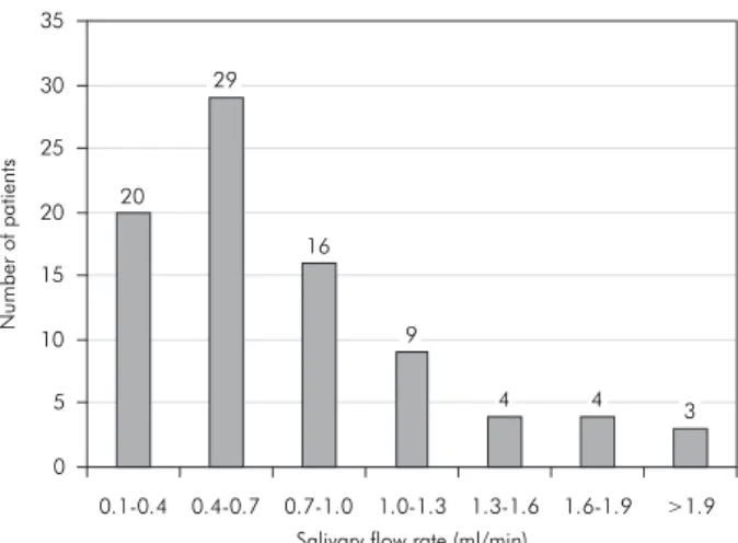
Stimulated salivary low was less than 0.7 ml/
min in 64% of the patients (hyposalivation).9 After
ive minutes, suficient saliva could not be collected for sialometry from 15 of these patients and their salivary low was therefore considered to be zero (Graph 1).
The sample was divided into three groups ac-cording to inclusion of the salivary glands in the ir-radiated ield. In group 1 (n = 39), only the face was included in the radiation ield, with most patients
be-ing treated for tumors of the mouth, salivary glands and maxillary sinus. In group 2 (n = 23), only the neck but not the face was included in the radiation ield. Most patients were treated for thyroid or vocal cord tumors. In group 3 (n = 38, patients with oro-pharyngeal cancer) the neck and face were included in the radiation ield. Backward stepwise multiple re-gression analysis was applied to determine the com-bined effect of the variables radiation ield, dose, use of xerostomic drugs and postradiotherapy time on salivary low. The variables showing statistical signiicance (p < 0.05) remained in the model. The results of multiple regression analysis are shown in Table 1. The only variable that showed no signii-cant correlation with salivary low was the radiation dose.
Salivary pH was measured in 93 patients, with a total of 186 results. The system permitted pH mea-surement with a single drop of saliva. It was
there-Graph 1 - Stimulated salivary flow rate in the irradiated
patients.
Salivary flow rate (ml/min)
N
u
mber
o
f
patients
0 5 10 15 20 25 30
20 29
16
9
4 4
3 35
0.1-0.4 0.4-0.7 0.7-1.0 1.0-1.3 1.3-1.6 1.6-1.9 >1.9
Table 1 - Results of multiple linear regression analysis:
ra-diation field in Group 1 and Group 3, use of xerostomic drugs and postradiotherapy time on salivary flow.
Independent Variables β p r
2
adjusted Radiation field (Face/Group 1) –0.3377 0.007
fore possible to measure pH in 12 out of the 15 pa-tients who were unable to perform sialometry. The
mean pH of the sample was 6.97 ± 0.71.
Discussion
Squamous cell carcinoma (SCC) was the most prevalent tumor in the present study (67%). Accord-ing to the literature, this tumor accounts for 90% of all malignant neoplasms of the mouth and
phar-ynx.1 The prevalence of oral cancer in our study
was higher among males (27%) than among females (8%), with a ratio of 3.3:1. The number of new cas-es of oral cancer for 2006 is cas-estimated to be 10,060 among men and 3,410 among women, with a ratio
of 2.9:1,8 which is similar to the ratio found in our
data.
Dysgeusia is deined as an altered sense of taste resulting from reduced salivary low and biochemi-cal alterations in the saliva. This condition frequent-ly occurs in irradiated patients, whose reduced sali-vary volume and low impair the physical contact
of foods with the taste papillae.9 These papillae are
also destroyed depending on the radiation doses.21
In the present study, 30% of the patients report-ed total loss of taste, with these patients having re-ceived doses higher than 5,000 cGy. Post-treatment time was up to 12 months in 16 patients and ranged from 17 to 72 months in the other 14 patients. Al-though some of the patients were within the period
of reestablishment (12 months),4 all received doses
close to 6,000 cGy, implying a possibly permanent
taste loss.13
Dysphagia is a dificulty in masticating and
swal-lowing foods.4 This condition is caused by radiation
ibrosis within the pharyngeal constrictor muscles.
Cintra et al.5 (2005) evaluated oropharyngeal
swal-lowing in patients treated for epidermoid carcinoma of the larynx and hypopharynx by chemotherapy in combination with radiotherapy (total dose of 7,040 cGy). Swallowing was regular in 54.8% of the sample studied (discrete to moderate dyspha-gia) and poor in 9.7% (severe dysphadyspha-gia). In our study, 38 subjects reported swallowing dificulties (dysphagia). They received a mean radiation dose of 6,063 cGy (4,000-10,000) and the radiation ield included the region of the oropharynx and neck.
Xerostomia is one of the most frequent sequelae of head and neck radiotherapy because the salivary glands are radiosensitive organs. A reduction in the quality and quantity of saliva also leads to an imbal-ance in the microbiota and consequent occurrence
of diseases.11,16,19
In the present study, most patients (89%) were irradiated with a dose higher than 5,000 cGy. This protocol involves a poor prognosis regarding the return of normal glandular function. How-ever, our results (Table 1) showed an increase in salivary secretion with increasing postradiotherapy
time (β = 0.2307; p = 0.020). Although 89% of the
sample received doses higher than 5,000 cGy and supposedly these doses cause irreversible damage
to saliva production,10 no statistic correlation was
observed between salivary low and total radiation dose (p > 0.05).
Groups 1 and 3 (Table 1), including patients whose major salivary glands were involved in the radiation ield, presented a signiicant reduction in salivary low (p = 0.007 and p = 0.008, respective-ly). Moreover, the patients using xerostomic medica-tions (Table 1) showed a signiicant inluence of this factor on the reduction of salivary low (p = 0.037).
Möller et al.14 (2004) observed a decrease in
pH during irradiation, with the lowest value being reached 3 months after the beginning of radiother-apy and increasing gradually during the subsequent months. However, salivary pH continued to be slightly acid (pH = 6.87) 12 months after treatment, and stimulated salivary low was reduced by 93% compared to the beginning of treatment. In the pres-ent study, the mean pH of the two stimulated saliva
samples was 6.971 ± 0.714. This salivary pH was
also classiied as slightly acid. Sixty four percent of the patients presented a salivary low of less than 0.7 ml/ml, which was in agreement with the study
by Möller et al.14 (2004) regarding the quantitative
and qualitative salivary alterations observed in irra-diated patients.
Mucositis generally occurs in the second week of
treatment with doses of 2,000 cGy or higher,6 and
affects about 80% of irradiated patients.14,20 Caielli
et al.4 (1995) demonstrated that mucositis persisted
of treatment, regressing within a few weeks. Dib et
al.6 (2000) observed the presence of mild mucositis
in the second week of treatment with a dose of up to 1,800 cGy, and of severe mucositis in the ifth week of treatment with a dose of 4,500 cGy.
Two cases of mucositis treated with a mean
ra-diation dose of 7,520 cGy (± 3.507; p = 0.044) were
observed in the present investigation. One patient was irradiated in the region of the palatine ton-sils and epiglottis and the other on the back of the tongue. Both patients were evaluated 2 months after radiotherapy. The low prevalence of mucositis (2%) observed in the present study might be explained by the small number of patients who complied with the study during the immediate post-treatment phases.
The most important cause of candidiasis in pa-tients undergoing radiotherapy is xerostomia. The
increased number of Candida spp. as a result of
radiotherapy may persist for several months after
treatment.2 We found a signiicant correlation
be-tween the frequency of candidiasis and higher mean postradiotherapy time (39.3 months; p = 0.028). Eleven patients presented chronic candidiasis, and only two of them had concluded radiotherapy less than 9 months before, a fact that could explain the observation of the chronic form.
Candidiasis is a consequence of low salivary low rates and slightly acid saliva (pH = 6.971), plus the use of old dentures. The large number of edentulous patients using dentures (43%) in the present sample
should be emphasized. Redding et al.17 (1999)
re-ported the presence of pseudomembranous candi-diasis in 90% of cases and persisting after the end of radiotherapy. In the present study only the chronic (atrophic) form was found.
The therapeutic measures available for the treat-ment of head and neck SCC have been questioned in view of the increasing number of patients over the
last decades that develop second primary tumors.10
Continuous exposure to the same carcinogens (ield
cancerization) might be one reason for the increased
probability of the occurrence of a second tumor.10
Ogata et al.15 (1997), retrospectively analyzing 125
patients with head and neck SCC, reported a 10.4% prevalence of second primary tumor, with the mean time of diagnosis of the second tumor being 36 months. In the present sample, 67% of the patients were treated for SCC, with the cancer occurring in the mouth in 26%. Two cases of second primary tu-mor were diagnosed 23 and 72 months after radio-therapy, respectively. These indings emphasize the importance of follow-up with patients treated for SCC of the mouth.
The frequency of osteoradionecrosis depends on the age of the patient, radiation dose, and the irra-diated mandibular volume. According to some
au-thors,10 the period of highest risk ranges from 4 to
12 months postradiotherapy, while others consider
the risk to be permanent.11 No case of
osteoradio-necrosis was observed in our study despite the high radiation doses applied and the high proportion of older patients evaluated. The pre-treatment preven-tive procedures adopted probably contributed to the low risk observed.
Conclusions
This study suggests that the effects of radiothera-py persist throughout the years and depend on a set of variables which include radiation ield, use of xero-stomic medication, radiation dose and postradiother-apy time. The main effect of radiotherpostradiother-apy in the head and neck region was a reduction of the salivary low rate, even though our study demonstrated that there was a modest late improvement of the salivary low.
Acknowledgments
We wish to thank the medical team, nurses and staff of the Amaral Carvalho Hospital, Jaú, SP, Bra-zil, for their direct or indirect participation which permitted the execution of this study.
References
1. Barnes L, Eveson JW, Reichart P, Sidransky D. Classification of tumors: pathology & genetics of head and neck tumors. Geneva: World Health Organization; 2005.
preva-lence, azole susceptibility profiles and response to antifungal treatment. Oral Microl Immunol. 2004;19(6):347-51. 3. Boraks S, Chilvarquer I, Panella J. Radiomucosite: contribuição
ao estudo dos efeitos das radiações ionizantes na mucosa bucal normal de pacientes portadores de carcinoma espinocelular submetidos a tratamento radioterápico.Rev Odontol UNICID. 2000;12(2):149-61.
4. Caielli C, Martha PM, Dib LL. Seqüelas orais da radioterapia: atuação da odontologia na prevenção do tratamento. Rev Bras Cancerol. 1995;41(4):231-41.
5. Cintra AB, Vale LP, Feher O, Nishimoto IN, Kowalski LP, Angelis EC. Deglutição após quimioterapia simultânea para carcinomas de laringe e hipofaringe. Rev Assoc Med Bras. 2005;51(2):93-9. 6. Dib LL, Gonçalves RCC, Kowalsky LP, Salvajoli JV. Abordagem
multidisciplinar das complicações orais da radioterapia. Rev Assoc Paul Cir Dent. 2000;54(5):391-6.
7. Ericsson Y, Hardwick L. Individual diagnosis, prognosis and counseling for caries prevention. Caries Res. 1978;12(Suppl 1):94-102.
8. INCA. Estimativa 2006: incidência de câncer no Brasil; 2006. [acesso 24 ago 2006]. Disponível em: http://portal.saude.sp.gov. br/resources/gestor/destaques/estimativa_incidencia_cancer-2006.pdf.
9. Joyston-Bechal S. Management of oral complications follow-ing radiotherapy. Dent Update. 1992;19(6):232-4, 236-8. 10. Kufe D, Pollock RE, Weichselbaum RR, Bast RC, Gansler TS,
Holland JF et al. Cancer Medicine 6, BC Becker. [book online] [cited 2005 Mar 15]. Available from: http://www.ncbi.nlm.nih. gov/books/bv.fcgi?call=bv.View..ShowTOC&rid=cmed6. TOC&deptH = 10.
11. Lopes MA, Coletta RD, Alves FA, Abbade N, Rossi A. Reco-nhecendo e controlando os efeitos colaterais da radioterapia. Rev Assoc Paul Cir Dent. 1998;52(3):241-4.
12. Marcucci G. Fundamentos de odontologia: estomatologia. Rio de Janeiro: Guanabara Koogan; 2005.
13. Meraw SJ, Reeve CM. Dental considerations and treatment of the oncology patient receiving radiation therapy. J Am Dent Assoc. 1998;129(7):201-5.
14. Möller P, Perrier M, Ozsahin M, Monnier P. A prospective study of salivary gland function in patients undergoing radio-therapy for squamous cell carcinoma of oropharynx. Oral Surg Oral Med Oral Pathol Oral Radiol Endod. 2004;97(2):173-89.
15. Ogata AC, Soares EWS, Soares GVS, Araki LT. Tumor segundo primário em pacientes operados com carcinoma espinocelular de cabeça e pescoço. Rev Bras Otorrinolaringol. 1997;63(6):583-6.
16. Pontes CB, Polizello ACM, Spadaro ACC. Clinical and bio-chemical evaluation of the saliva of patients with xerostomia induced by radiotherapy. Braz Oral Res. 2004;18(1):69-74. 17. Redding SW, Zellars RC, Kirkpatrick WR, Mcatee RK,
Ca-ceres MA, Fothergill AW et al. Epidemiology of oropharyn-geal Candida colonization and infection in patients receiv-ing radiation for head and neck cancer. J Oral Microbiol. 1999;37(12):3896-900.
18. Silva MA, Damante JH, Stipp AC, Tolentino MM, Carlotto PR, Fleury RN. Gastroesophageal reflux disease: new oral findings. Oral Surg Oral Med Oral Pathol Oral Radiol Endod. 2001;91(3):301-10.
19. Spolidorio DMP, Spolidorio LC, Barbeiro RH, Hofling JF, Ber-nardo WLC, Pavan S. Avaliação quantitativa de Streptococcus
do grupo mutans e Candidasp. e fatores salivares na cavidade bucal de pacientes submetidos à radioterapia. Pesqui Odontol Bras. 2001;15(4):354-8.
20. Vissink A,Jansma J, Spijkervet Kl, Burlage FR, Coppes RP. Oral sequelae of head and neck radiotherapy. Crit Rev Oral Biol Med. 2003;14(3):199-212.