Lucas da Fonseca Roberti GARCIA(a)
Alailson Domingos dos SANTOS(b)
João Carlos Silos MORAES(b)
Carlos Alberto de Souza COSTA(c)
(a)Universidade Estadual Paulista – UNESP,
Araçatuba School of Dentistry, Department of Restorative Dentistry, Araçatuba, SP, Brazil.
(b)Universidade Estadual Paulista – UNESP,
School of Engineering, Department of Physics and Chemistry, Ilha Solteira, SP, Brazil.
(c)Universidade Estadual Paulista – UNESP, Araraquara School of Dentistry, Department of Physiology and Pathology, Araraquara, SP, Brazil.
Cytotoxic effects of new MTA-based
cement formulations on fibroblast-like
MDPL-20 cells
Abstract: The present study aimed at evaluating the cytotoxic
effects of a novel cement called CER on periodontal ibroblast-like cells of mice (MDPL-20), in comparison with different formulations of Mineral Trioxide Aggregate (MTA), by means of the cell viability test (MTT) and cell morphology analysis. Thirty-two round-shaped samples were fabricated with the following cements: white MTA, white and gray CER and experimental white MTA. The samples were immersed in serum-free culture medium for 24 hours or 7 days (n = 16). The extracts (culture medium + components released from the cements) were applied for 24 hours to previously cultured cells (40.000 cells/cm2) in the wells of 24-well plates. Cells seeded
in complete culture medium were used as a negative control. Cell viability was assessed using the MTT assay. Two samples of each cement were used for cell morphology analysis by Scanning Electron Microscopy (SEM). The extracts obtained at the 7-day period presented higher cytotoxicity compared with the 24-hour period (p < 0.05). The gray CER obtained at 24 hours presented the highest cytotoxic effect, whereas the experimental white MTA presented the lowest, similar to the control (p > 0.05). However, at the 7-day period, the experimental white MTA presented no signiicant difference in comparison with the other cements (p > 0.05). At the 7-day period, CER cement presented cytotoxic effects on ibroblast-like cells, similar to different MTA formulations. However, the immersion period in the culture medium inluenced the cytotoxicity of the cements, which was greater for CER cement at 24 hours.
Keywords: Biocompatible Materials; Dental Materials; Endodontics;
Silicate Cement.
Introduction
Mineral Trioxide Aggregate (MTA) cement was initially used
as a root-end filling material and for root perforation treatment;1
however, due to its favorable attributes, it has also been used for
apexification,2 root resorption treatment,3 pulpotomy4 and pulp
capping in conservative procedures.5
Studies have reported that the success rate of pulp therapy using MTA
is higher than that using calcium hydroxide-based materials.6 Considering
that MTA cements are applied in direct contact with connective tissues,
Declaration of Interests: The authors certify that they have no commercial or associative interest that represents a conflict of interest in connection with the manuscript.
Corresponding Author: Carlos Alberto de Souza Costa E-mail: [email protected]
DOI: 10.1590/1807-3107BOR-2016.vol30.0028
Submitted: Jul 23, 2015
such as periodontal ligament and pulp, and that
moisture from the surrounding tissue also acts as
an activator of its bioactivity, the cytotoxic effects and the biological properties of this material have
been the focus of extensive research.7,8,9
The ability of tissue to regenerate and the antibacterial property of MTA-based cements
are related to ionic dissociation in calcium and
hydroxyl ions.10 The calcium ions released by the
cement react and produce calcite granules, when the cement comes into contact with the carbon
dioxide and carbonic acid from the cell catabolism.7
In addition, there is a tendency for fibronectin to accumulate. This is a glycoprotein found in the tissue, and synthesized by fibroblasts and endothelial cells.11 Glycoproteins allow adhesion, cell
differentiation and growth, leading to mineralized
tissue deposition.11
Despite the clinical success of MTA as a sealing
cement, it has some disadvantages, such as a sandy
consistency and a long setting time.12 MTA takes
approximately 180 minutes to set; however, its working
time it less than 4 minutes.12 A long exposure period
of cells to the cement before its complete hardening may compromise its biological property, leading to an inlammatory reaction, which may affect its physical characteristics.10
A novel experimental cement, called CER, was developed to improve some undesirable characteristics of MTA cement, such as poor handling from porosities
formed after manipulation,1 making it highly unstable,
high solubility,9 long setting time,10 and dental
staining.3,12 The name CER is the Portuguese acronym for
“cimento endodôntico rápido”, or “quick-setting endodontic
cement” in English. This cement is basically composed of clinker (raw material used in the manufacture of Portland cement), barium sulfate (radiopaciier), water and an emulsiier (proprietary) to increase the
handling characteristics of CER.12
Studies have reported that calcium and hydroxyl ions released by CER cement were similar to those
released by conventional MTA-based cements.12,13
In addition, CER presented greater ability to release calcium ions than MTA, when in contact with an aqueous environment. This feature could
accelerate tissue repair in endodontic therapy.12,13
The authors of these studies also reported that CER has a shorter setting time and improved
handling, in comparison with MTA, in addition to a coeficient of thermal expansion similar to that of dentin, thus making it more suitable for
sealing root perforations.12,13 However, in vitro and
in vivo studies are needed before a new dental material can be safely recommended for clinical practice, observing the research levels established by the associations and federations responsible for
biological test standardization.14
In vitro cytotoxicity protocols, performed to
compare materials and predict their safe clinical
application, have been considered the initial tests
needed to support further in vivo studies in animals.14 The aim of this in vitro study was to evaluate the
cytotoxic effects of CER cement on ibroblast-like MDPL-20 cells, in comparison with different MTA-based cement formulations, by means of the cell viability (MTT) test and cell morphology analysis. The null hypothesis tested was that there would be no difference in cytotoxicity promoted by the different cements tested.
Methodology
Cell culture
Periodontal ligament ibroblasts (MDPL-20) were cultivated in 75 cm2 sterilized plastic bottles (Costar
Corp. Cambridge, USA) in Dulbecco’s Modified Eagle’s Medium (DMEM - Sigma Chemical Co., St. Louis, USA) containing 10% fetal bovine serum (FBS, GIBCO, Grand Island, USA), 100 mg/mL of penicillin, 100 µg/mL of streptomycin, and 2 mol/L of glutamine (GIBCO), in a humidiied atmosphere
at 37oC, containing 5% CO
2 and 95% air. The cells
were sub-cultured every three days, at a density of 3 x 10⁴ cells/cm2,to obtain the number of cells required
to perform this in vitro study.
Cements extracts
MTA - Ângelus - powder with CER cement emulsiier). Since the emulsifier improves the handling
characteristics of CER cement,12 the cytotoxicity
of Experimental White MTA was also assessed. Complete DMEM (Ambion - Life Technologies, Grand Island, USA) was applied to the cell and used as the control. The composition, the powder/liquid
ratio and the setting time of the cements assessed
in this study are shown in Table 1.
The tested cements were manipulated according to the manufacturer’s recommendations to fabricate thirty-two standardized round-shaped samples (2 mm thick and 4 mm in diameter) of each dental material. After setting, the thirty-two samples of each cement were randomly distributed according to the periods (24 hours or 7 days) (n = 16) of DMEM immersion. These speciic periods of immersion were used to correlate the calcium and hydroxyl ion release promoted by the cements throughout their setting process with their potential cytotoxic effects. Since the setting time of the tested cements may extend from 24 hours (initial setting time) up to 7 days (inal setting time), the rate of calcium
and hydroxyl ions released from the cements may
range signiicantly, thus allowing for an increase in cement cytotoxicity.
The extracts were obtained from the tested cements by individually placing the round-shaped samples in the wells of 24-well plates containing 1.1 mL of DMEM culture medium without fetal bovine serum (DMEM-FBS), and incubating them for 24 hours or
7 days, at 37oC, 5% CO
2 and 95% air.
The ibroblast-like MDPL-20 cells were seeded
(40.000 cells/cm2) in the wells of 24-well sterile
acrylic plates (Costar Corp., Cambridge, USA) and
maintained in an incubator at 37ºC, 5% CO2 and 95%
air, for 48 hours. After this period, the DMEM of each well was aspirated, and an aliquot of 400 µL from the
extract of each cement and the negative group control
(DMEM only) was applied to the cells for 24 hours. The protocols performed in this study followed the
ISO 10993-5 (2009)recommendations.15
Cell viability assay (MTT)
Cell viability was assessed with the methyl tetrazolium colorimetric assay (MTT), which is characterized by the cytochemical demonstration of succinic dehydrogenase produced by the cells. After 24-hour exposure to the extracts or to the DMEM (negative control), the cultured ibroblasts were replaced by 900 mL of DMEM and 100 mL of MTT solution (5 mg/mL in phosphate buffered saline - PBS) (SIGMA Chemical Co., St. Louis, USA). After a 4-hour incubation period, the solution was replaced by 600 mL of isopropanol acidiied with hydrochloric acid (0.04 N HCl) to dissolve the formazan crystals. Then, three aliquots of 100 µL from each compartment were transferred to 96-well plates (Costar Corp.) and the cell viability was assessed proportionally to the speciic absorbance at 570 nm, in an ELISA reader (ELX 800 - Universal Microplate Reader, BioTek Instruments, Winooski, USA). The experiments were performed in triplicate, according
to ISO 10993-5 (2009) recommendations.15
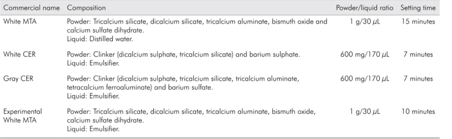
Table 1. Composition, powder/liquid ratio and setting time of cements evaluated in this study.
Commercial name Composition Powder/liquid ratio Setting time
White MTA Powder: Tricalcium silicate, dicalcium silicate, tricalcium aluminate, bismuth oxide and calcium sulfate dihydrate.
Liquid: Distilled water.
1 g/30 µL 15 minutes
White CER Powder: Clinker (dicalcium sulphate, tricalcium silicate) and barium sulphate. Liquid: Emulsifier.
600 mg/170 µL 7 minutes
Gray CER Powder: Clinker (dicalcium sulphate, tricalcium silicate, tricalcium aluminate, tetracalcium ferroaluminate) and barium sulfate.
Liquid: Emulsifier.
600 mg/170 µL 7 minutes
Experimental White MTA
Powder: Tricalcium silicate, dicalcium silicate, tricalcium aluminate, bismuth oxide, calcium sulfate dihydrate.
Liquid: Emulsifier.
Cell morphology (SEM)
Two representative specimens from each group were submitted to cell morphology analysis, using a Scanning Electron Microscope (SEM) (ZEISS DSM-940A model, Oberkochen, Germany). For this purpose, 12 mm diameter glass coverslips (Fisher Scientiic, Pittsburg, USA) were placed at the bottom of each well of a 24-well plate before seeding the ibroblasts. After 24-hour exposure of the cells to the extracts or to the DMEM, the solutions were aspirated and 1 mL of 2.5% buffered glutaraldehyde was applied to them. After 120 minutes, the ixative solution was aspirated and the cells were rinsed three times with 1 mL PBS (5 minutes each), and post-ixed with 1% osmium tetroxide solution for 60 minutes. Next, the cells were dehydrated by ascending exchanges of ethanol solutions (30%, 50%, 70%, 95% and 100%), and washed with solvent of low surface tension - 1,1,1,3,3,3-hexamethyldisilazane - (HMDS - Acros Organics, Fair Lawn, USA). The glass coverslips containing the attached cells were afixed to metal stubs and maintained overnight in a desiccator device. Afterwards, the samples were gold-sputtered, and cell morphology was carried out blindly by a single examiner.
Statistical analysis
The dataset for the viability of MDPL-20 cells did not present a normal distribution (Kolmogorov-Smirnov, p < 0.05). The data were then compiled and submitted to the non-parametric Kruskal-Wallis statistical test, complemented by the Mann-Whitney test (level of signiicance = 5%), using SPSS 19.0 software (SPSS Inc., Chicago, USA).
Results
Cell viability assay (MTT)
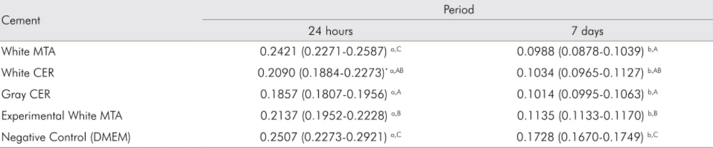
The cell viability data are shown in Table 2. There was a signiicant reduction in the production of the SDH enzyme by the cells after 7 days of contact with the extracts (p < 0.05), in all the experimental groups. At the 24-hour period, white MTA produced the lowest toxicity, statistically comparable to the control group (p > 0.05), whereas the white and gray
CER cements presented the highest cytotoxic effects
on the ibroblast-like MDPL-20 cells. However, at the 7-day period, white MTA presented a toxic potential similar to that of white and gray CER cements, with no statistical difference among them (p > 0.05).
Cell morphology (SEM)
The morphology of MDPL-20 cells can be observed in Figure.
Two representative specimens of each cement and of the control group were selected to assess the morphology of the ibroblasts that remained adhered to the glass substrate. In the negative control group, a number of cells remained adhered to the glass substrate during both periods evaluated, displaying numerous long and slender cytoplasmic processes, characteristic of typical fibroblasts (Figure E/F). Similar morphology was observed in the cells exposed to the extracts from the treated groups, irrespective of the analysis period. No cytoplasmic shrinkage was seen; however, a noteworthy decrease was observed in the number of cells attached to the glass substrate, for all treated groups, in comparison with the negative control group (Figure A-D).
Table 2. Mean values of succinate dehydrogenase enzyme (SDH) production by cells exposed to the cement extracts (24 hours or 7 days) or to DMEM.
Cement Period
24 hours 7 days
White MTA 0.2421 (0.2271-0.2587) a,C 0.0988 (0.0878-0.1039) b,A
White CER 0.2090 (0.1884-0.2273)* a,AB 0.1034 (0.0965-0.1127) b,AB
Gray CER 0.1857 (0.1807-0.1956) a,A 0.1014 (0.0995-0.1063) b,A
Experimental White MTA 0.2137 (0.1952-0.2228) a,B 0.1135 (0.1133-0.1170) b,B
Negative Control (DMEM) 0.2507 (0.2273-0.2921) a,C 0.1728 (0.1670-0.1749) b,C
*Median (interquartile range P25/P75).
Discussion
This study aimed at evaluating the cytotoxic effects of
a novel cement called CER on periodontal ibroblast-like cells, in comparison with different formulations of MTA. Based on the results obtained, it can be stated that the null hypothesis tested was partially accepted, since there was a signiicant difference in the cytotoxicity of the cements, but only at the 24-hour period.
Since MTA cement is recommended for diverse endodontic therapies, its biological properties have been evaluated by several authors using different cell lines, such as MDPC-23 and OD-21 pulp cells, periodontal ligament fibroblasts (MDPL-20) and
MG-63 osteoblasts.16,17 These studies have reported
a reduced toxic effect of the different types of MTA
cements available in the dental market.16,17
In general, the present study demonstrated greater cell viability at the 24-hour period, in comparison with the 7-day period. This may be explained by the longer exposure period of cells to the extracts obtained from the cements. Fibroblasts exposed for 24 hours to the extracts from conventional white MTA showed less cytotoxicity in comparison with the other cements. Despite the intense cytotoxicity caused by the gray version of CER cement at the 24-hour period, both gray and white versions presented a toxic potential
similar to that of the different MTA formulations
assessed at the 7-day period.
Figure. Representative SEM images of MDPL-20 cells exposed to the extracts from the experimental cements or the DMEM at 24-hour and 7-day periods. (A) White MTA: Only a few cells remained adhered to the glass substrate, in comparison with the negative control group, irrespective of the period of analysis. Note the typical morphology of fibroblasts - wide cytoplasm with several small cytoplasmic processes. (B) White CER: Significant decrease in the number of cells adhered to the glass substrate, in comparison with the negative control group. (C) Gray CER: Observe the similar morphology of cells exposed to the control extracts, despite the lower number of adhered cells. (D) Experimental White MTA: As found in the other experimental groups, a reduction was observed in the number of cells adhered to the glass substrate, in comparison with the negative control group, with no cell morphology alterations. (E) Negative control group: Note the greater number of cells adhered to the substrate than in the experimental groups. (F) High magnification of (A): the cells adhered to the glass exhibit long and slender cytoplasmic processes, typical of MDPL-20 fibroblasts, irrespective of the period of analysis.
A B C
The calcium and hydroxyl ion diffusion promoted
by the MTA cement increases the pH adjacent to the periodontal tissues, possibly acting on osteoclastic activity to promote alkalinization of the medium, which favors the healing process.17,18 It is known that
CER cement releases calcium and hydroxyl ions, and increases the pH of the medium in a manner similar
to that of MTA cement.13 After 24 hours, the calcium
and hydroxyl ions released by CER cement are greater than those released by MTA; however, following this
initial period, the values become similar.13
Despite the important role of calcium ion release
in the tissue repair process, the high rate of calcium
and hydroxyl ions released in the culture medium
probably resulted in irreversible damage to the cells,16,17
demonstrating that the association of cells to the
interaction period is significant,13 as can be seen
in the present study. Since the setting time of the tested cements may extend from 24 hours (initial setting time) up to 7 days (final setting time), the
rate of calcium and hydroxyl ions released from the
cements may vary signiicantly, thus allowing for
an increase in cement cytotoxicity.16,17 On the other
hand, a previous in vivo study18 reported a favorable
histological response promoted by CER and MTA at the 7-day period. However, it is worthwhile stressing
that in vitro conditions are not homeostatic, and that
there is no tissue elimination of toxic substances, such
as that reported for in vivo assays.19 In addition, a living host possesses defense mechanisms and a lymphatic
system that removes the toxic substances released.19
Several changes in the surface composition and the
morphology of MTA cement can lead to variations in its
biological response and may result in clinically relevant differences.20 The conlicting results observed in this
in vitro study for the different cements could be also related to the emulsion added, which probably modiied the chemical properties of the materials.12,13 With this
1. Torabinejad M, Parirokh M. Mineral trioxide aggregate: a comprehensive literature review - Part III: Clinical applications, drawbacks, and mechanism of action. J Endod. 2010;36(3):400-13. doi:10.1016/j.joen.2009.09.009
2. Floratos SG, Tsatsoulis IN, Kontakiotis EG. Apical barrier
formation after incomplete orthograde MTA apical plug
placement in teeth with open apex - report of two cases. Braz Dent J. 2013;24(2):163-6. doi:10.1590/0103-6440201302163
References
in mind, the chemical changes made in the cement composition were designed to yield dental materials with good handling characteristic and adequate
working time, associated with low cytotoxicity.21
The present study demonstrated that CER and the experimental MTA formulation not only presented
better physical and chemical properties than the conventional white MTA,12,13 but also exhibited greater
toxicity when applied to ibroblast-like MDPL-20 cells than the reference material, at the initial period. The cytotoxicity of the tested cements at the inal period of analysis was similar. Since time is a relevant factor in determining cement cytotoxicity, future studies are needed to analyze these new MTA-based cement formulations over longer periods of time.
Conclusion
According to the experimental conditions and the methodology employed in this in vitro study, it was concluded that CER cement presented cytotoxicity
similar to that of different MTA formulations at a 7-day period. However, the immersion period inluenced the cytotoxicity of the cements, which was greater for CER cement at 24 hours.
Acknowledgments
The authors acknowledge the contribution of the Center for Research Support/Electron Microscopy
(Núcleo de Apoio à Pesquisa em Microscopia Eletrônica
Aplicada a Agricultura - NAP/MEPA), Escola
Superior de Agricultura Luiz de Queiroz - ESALQ, Universidade de São Paulo - USP, coordinated by
Prof. Dr. Elliot W. Kitajima, of fundação para o
Desenvolvimento da UNESP - FUNDUNESP (grants # 0024/021/13-PROPe-CDC and 05/2014-PROPe/Unesp)
and of the Conselho Nacional de Desenvolvimento
3. Jacobovitz M, Lima RKP. Treatment of inflammatory internal root resorption with mineral trioxide aggregate: a case report. Int Endod J. 2008;41(10):905-12. doi:10.1111/j.1365-2591.2008.01412.x
4. Shahravan A, Jalali SP, Torabi M, Haghdoost AA, Gorjestani H. A histological study of pulp reaction to various water/powder ratios of white mineral trioxide aggregate as pulp-capping material in human teeth: a double-blinded, randomized controlled trial. Int Endod J. 2011;44(11):1029-33. doi:10.1111/j.1365-2591.2011.01916.x
5. Dantas RV, Conde MC, Sarmento HR, Zanchi CH, Tarquinio SB, Ogliari FA, et al. Novel experimental cements for use on the dentin-pulp complex. Braz Dent J. 2012;23(4):344-50. doi:10.1590/S0103-64402012000400006
6. Leye Benoist F, Gaye Ndiaye F, Kane AW, Benoist HM, Farge P. Evaluation of mineral trioxide aggregate (MTA) versus calcium hydroxide cement (Dycal®) in the formation of a dentine bridge: a randomised controlled trial. Int Dent J. 2012;62(1):33-9. doi:10.1111/j.1875-595X.2011.00084.x 7. Zhou HM, Du TF, Shen Y, Wang ZJ, Zheng YF, Haapasalo M.
In vitro cytotoxicity of calcium silicate-containing endodontic sealers. J Endod. 2015;41(1):56-61. doi:10.1016/j.joen.2014.09.012 8. Mestieri LB, Tanomaru-Filho M, Gomes-Cornélio AL, Salles
LP, Bernardi MI, Guerreiro-Tanomaru JM. Radiopacity and cytotoxicity of Portland cement associated with niobium oxide micro and nanoparticles. J Appl Oral Sci. 2014;22(6):554-9. doi:10.1590/1678-775720140209
9. Silva EJNL, Herrera DR, Rosa TP, Duque TM, Jacinto RC, Gomes BPFA, Zaia AA. Evaluation of cytotoxicity, antimicrobial activity and physicochemical properties of a calcium aluminate-based endodontic material. J Appl Oral Sci. 2014;22(1):61-7. doi: 10.1590/1678-775720130031
10. Ji DY, Wu HD, Hsieh SC, Teng NC, Chen CC, Ke ES, et al. Effects of a novel hydration accelerant on the biological and mechanical properties of white mineral trioxide aggregate. J Endod. 2011;37(6):851-5. doi:10.1016/j.joen.2011.03.015 11. Tjäderhane L, Palosaari H, Wahlgren J, Larmas M, Sorsa T,
Salo T. Human odontoblast culture method: the expression of collagen and matrix metalloproteinases (MMPs). Adv Dent Res. 2001 Aug;15(1):55-8. doi:10.1177/08959374010150011401 12. Santos AD, Araújo EB, Yukimitu K, Barbosa JC, Moraes
JCS. Setting time and thermal expansion of two endodontic
cements. Oral Surg Oral Med Oral Pathol Oral Radiol Endod. 2008;106(3):e77-9. doi:10.1016/j.tripleo.2008.04.021
13. Santos AD, Moraes JCS, Araújo EB, Yukimitu K, Valério Filho WV. Physico-chemical properties of MTA and a novel experimental cement. Int Endod J. 2005;38(7):443-7. doi:10.1111/j.1365-2591.2005.00963.x
14. Costa CAS, Hebling J, Scheffel DLS, Soares DGS, Basso FG, Ribeiro APD. Methods to evaluate and strategies to improve the biocompatibility of dental materials and operative techniques. Dent Mater. 2014;30(7):769-84. doi:10.1016/j.dental.2014.04.010
15. International Organization for Standardization. ISO 10993-5:1999 (E) - Biological evaluation of medical devices - Part 5: tests for in vitro cytotoxicity. 3rd ed. Géneve: ISO; 2009.
16. Paranjpe A, Zhang H, Johnson JD. Effects of mineral
trioxide aggregate on human dental pulp cells after
pulp-capping procedures. J Endod. 2010;36(6):1042-7. doi:10.1016/j.joen.2010.02.013
17. Samara, Sarri Y, Stravopodis D, Tzanetakis GN, Kontakiotis EG, Anastasiadou E. A comparative study of the effects of three root-end filling materials on proliferation and adherence of human periodontal ligament fibroblasts. J Endod. 2011;37(6):865-70. doi:10.1016/j.joen.2011.03.011 18. Gomes-Filho JE, Rodrigues G, Watanabe S, Bernabé PFE,
Lodi CS, Gomes AC, et al. Evaluation of the tissue reaction to fast endodontic cement (CER) and Angelus MTA. J Endod. 2009;35(10):1377-80. doi:10.1016/j.joen.2009.06.010
19. Wei W, Qi YP, Nikonov SY, Niu LN, Messer RL, Mao J, et al.
Effects of an experimental calcium aluminosilicate cement
on the viability of murine odontoblast-like cells. J Endod. 2012;38(7):936-42. doi:10.1016/j.joen.2012.03.020
20. DeAngelis L, Chockalingam R, Hamidi-Ravari A, Hay S, Lum V, Sathorn C, et al. In vitro assessment of
mineral trioxide aggregate setting in the presence
of interstitial fluid alone. J Endod. 2013;39(3):402-5. doi:10.1016/j.joen.2012.11.010