Artur Cunha VASCONCELOS(a)
Felipe NÖR(a)
Luise MEURER(b)
Gabriela SALVADORI(c)
Lélia Batista de SOUZA(d)
Pablo Agustin VARGAS(e)
Manoela Domingues MARTINS(a)
(a)Universidade Federal do Rio Grande do Sul – UFRGS, School of Dentistry, Department of Oral Pathology, Porto Alegre, RS, Brazil.
(b)Universidade Federal do Rio Grande do Sul – UFRGS, Hospital de Clínicas de Porto Alegre, Department of Pathology, Porto Alegre, RS, Brazil.
(c)University of Oslo – UiO, Faculty of Dentistry, Institute of Oral Biology, Oslo, Norway.
(d)Universidade Federal do Rio Grande do Norte - UFRN, Centro de Ciências da Saúde, Departamento de Odontologia, Natal, RN, Brazil.
(e) Universidade Estadual de
Campinas – UNICAMP, Piracicaba Dental School, Department of Oral Diagnosis, Piracicaba, SP, Brazil.
Clinicopathological analysis of salivary
gland tumors over a 15-year period
Abstract:Salivary gland tumors (SGT) are rare neoplasms that generate
interest due to their histopathological diversity and clinical behavior. The aims of the present study were to investigate clinicopathological aspects of SGTs diagnosed at a tertiary health center and compare the
indings with epidemiological data from different geographic locations.
Cases of tumor in the head and neck region at a single health center in the period between 1995 and 2010 were reviewed. Patient gender, age and ethnic group as well as anatomic location, histological type and clinical behavior of the tumor were recorded. Availability of complete information about these aspects was considered the inclusion criteria. Descriptive statistical analysis of the data was performed using the frequencies of categorical variables. Among the 2168 cases of tumors in the head and neck region, 243 (11.20%) cases were diagnosed in the salivary glands, 109 of which met the inclusion criteria: 85 (78%) benign tumors and 24 (22%) malignant tumors. Mean patient age was 46.47 years. The female gender accounted for 56 cases (51.4%) and the male gender accounted for 53 (48.3%). The major salivary glands were affected more (75.2%) than the minor glands. The most frequent benign and malignant SGTs were pleomorphic adenoma (81.2%) and adenoid cystic carcinoma (58.3%), respectively. In conclusion, pleomorphic adenoma and adenoid cystic carcinoma are the most frequent benign and malignant lesions, respectively. Comparing the present data with previous studies on SGTs, one may infer that some demographic characteristics and the predominance of malignant tumors vary in different geographic regions.
Keywords: Neoplasms; Salivary Glands; Adenoma, Pleomorphic;
Carcinoma, Adenoid Cystic; Carcinoma, Mucoepidermoid.
Introduction
Salivary glands tumors (SGTs) are uncommon neoplasms that pose a diagnostic challenge due to their complex histopathological features.1
Moreover, SGTs exhibit considerable variation with regard to clinical aspects, biology and clinical behavior.2,3,4
SGTs represent 3 to 6% of all tumors of the head and neck region, with an annual incidence throughout the world ranging from 0.05 to 2 cases per 100,000 individuals.5 Epidemiological data reveal different
frequencies of SGTs in distinct ethic groups and geographic locations, which
makes it dificult to establish global estimates.1,6,7,8,9,10,11,12,13,14,15,16 Although Declaration of Interests: The authors
certify that they have no commercial or associative interest that represents a conflict of interest in connection with the manuscript.
Corresponding Author: Manoela Domingues Martins E-mail: [email protected]
DOI: 10.1590/1807-3107BOR-2016.vol30.0002
Submitted: Sep 24, 2014
studies provide valuable knowledge, some data are contradictory. Local records are a useful strategy for the analysis of the distribution and particular
features of SGTs in a speciic population and the
establishment of appropriate treatment.
The aims of present study were to describe demographic and clinicopathological aspects of SGTs diagnosed at a tertiary health center and compare
the indings with epidemiological data from different
geographic locations.
Methodology
A retrospective analysis was carried out using archival data from the pathology laboratory of a tertiary health center located in southern Brazil
Hospital de Clínicas de Porto Alegre. This study received
approval from the Institutional Ethics Committee (protocol no. 130152/12).
In a 15-year period spanning from 1995 to 2010, 2168 cases of tumors in the head and neck region
were identiied, 243 of which were SGTs. Clinical
data regarding age, gender, clinical aspects, tumor localization, TNM Classification, treatment and follow-up information (clinical outcome and survival
time) were retrieved from medical iles. The follow-up period was deined as the time from diagnosis until
the last visit to the hospital. Availability of complete information about these aspects was considered the inclusion criteria and based on these only 109 were included. Slides stained with hematoxylin-eosin of all cases were reviewed based on the Histologic Typing of Salivary Gland Tumors established by the World Health Organization (WHO).17 Continuous
variables were expressed as mean, median and standard deviation values. Categorical variables were expressed as absolute number of cases and percentage values.
Results
Among the 109 cases of SGTs, 81 (74.3%) tumors were located in major salivary glands and 28 (25.7%) were located in minor salivary glands. The most frequent location among the major salivary glands was the parotid gland (66.9%), followed by the submandibular gland (7.3%). Among the minor glands, the most frequent location was the palate
(14.6%). No cases occurred in the sublingual gland (Figure and Table 1).
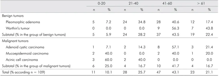
The male gender accounted for 53 cases (48.3%) and the female gender accounted for 56 (51.4%): male-to-female ratio of 0.9:1 for benign tumors and 1:1 for malignant tumors (Table 2). The majority of the tumors occurred in patients aged 41 to 60 years (median: 45 years). Mean patient age was 46.47 years (47.6 years for benign tumors and 42.3 years for malignant tumors). Young individuals (0-20 years-old) were the second group more affected by malignant tumors (25%) (Table 3).
Eighty-ive cases (78%) were classiied as benign
tumors and 24 (22%) were malignant tumors (benign-to-malignant ratio of 3.5:1). The proportions differed depending on the type of salivary gland analyzed. Among the 81 cases of SGT in the major glands, 67 (82.7%) were benign and 14 (17.3%) were malignant. Among the 28 cases in minor salivary glands, 18 (64.3%) were benign and 10 (35.7%) were malignant (Table 1). Among the 85 benign tumors, 68 (79.06%) were pleomorphic adenoma and 16 (18.6%) were cases of Warthin’s tumor. Among the 24 malignant
tumors, 14 (58.3%) were adenoid cystic carcinoma, ive (20.8%) were mucoepidermoid carcinoma and ive
(20.8%) were acinic cell carcinoma (Table 1).
Adenoid cystic carcinoma and mucoepidermoid carcinoma were classified based on the WHO Histologic Typing of Salivary Gland Tumors.17
Among the 14 cases of adenoid cystic carcinoma,
Number of SGT cases
80
Minor Gland Major Gland 70
50 60
30 40
20
10
0
Acinic cell carcinoma Mucoepicermoid
carcinoma Adenoid
cystic carcinoma Warthin’s
tumor Pleomorphic
adenoma
Table 1. Distribution of 109 salivary gland tumors according to anatomic location.
Parotid Submandibular Palate Other
n % n % n % n %
Benign tumors
Pleomorphic adenoma 48 69.6 3 4.3 11 15.9 7 10.1
Warthin’s tumor 16 100.0 0 0.0 0 0.0 0 0.0
Subtotal (% in the group of benign tumors) 64 75.3 3 3.5 11 12.9 7 8.2
Malignant tumors
Adenoid cystic carcinoma 3 21.4 3 21.4 4 28.6 4 28.6
Mucoepidermoid carcinoma 3 60.0 2 40.0 0 0.0 0 0.0
Acinic cell carcinoma 3 60.0 0 0.0 1 20.0 1 20.0
Subtotal (% in the group of malignant tumors) 9 37.5 5 20.8 5 20.8 5 20.8
Total (% according n = 109) 73 66.9 8 7.3 16 14.7 12 11.0
Table 2. Histologic and gender distribution of 109 salivary gland tumors.
n = 109 % % in the group of benign or malignant tumors
Gender
Male Female
n % n %
Benign tumors
Pleomorphic adenoma 69 63.3 81.2 31 28.4 38 34.9
Warthin’s tumor 16 14.7 18.8 10 9.2 6 5.5
Total 85 78 100 41 37.6 44 40.4
Malignant tumors
Adenoid cystic carcinoma 14 12.8 58.3 6 5.5 8 7.3
Mucoepidermoid carcinoma 5 4.6 20.8 3 2.7 2 1.8
Acinic cell carcinoma 5 4.6 20.8 3 2.7 2 1.8
Total 24 22 100 12 10.9 12 10.9
Table 3. Distribution of 109 benign and malignant salivary gland tumors according to age (in years).
0-20 21-40 41-60 > 61
n % n % n % n %
Benign tumors
Pleomorphic adenoma 5 7.2 24 34.8 28 40.6 12 17.4
Warthin’s tumor 0 0.0 0 0.0 9 56.3 7 43.8
Subtotal (% in the group of benign tumors) 5 5.9 24 28.2 37 43.5 19 22.4
Malignant tumors
Adenoid cystic carcinoma 1 7.1 2 14.3 8 57.1 3 21.4
Mucoepidermoid carcinoma 2 40.0 0 0.0 2 40.0 1 20.0
Acinic cell carcinoma 3 60.0 2 40.0 0 0.0 0 0.0
Subtotal (% in the group of malignant tumors) 6 25.0 4 16.7 10 41.7 4 16.7
seven (50.0%) cases were cribriform, ive (35.7%)
were tubular and two (14.3%) were solid. Among
the ive cases of mucoepidermoid carcinoma, four were classiied as low-grade and one was classiied
as intermediate-grade.
Regarding the clinical aspects, all benign tumors were nodules, whereas malignant tumors were nodules in 19 cases (11 cases of adenoid cystic carcinoma, four cases of mucoepidermoid carcinoma and four cases of acinic cell carcinoma) and ulcerated nodules in
ive cases (three cases of adenoid cystic carcinoma,
one case of mucoepidermoid carcinoma and one case of acinic cell carcinoma). Among the 24 malignant tumors, 11 (45.8%) cases were classified as TNM
stage I or II and 13 (54.2%) cases were classiied as
stage III or IV.
All patients underwent surgical treatment. Thirteen patients (11.92%) with malignant tumors also underwent radiation therapy. Among these patients, seven (53.8%) cases were adenoid cystic carcinoma, three (23.1%) were mucoepidermoid carcinoma and three (23.1%) were acinic cell carcinoma. The follow-up period ranged from four to 17 years (mean: 7.4 years). During the follow-up period, 19 patients (79.2%) of malignant lesions remained alive without recurrence and only one (4.2%) remained alive but with recurrence (adenoid cystic carcinoma). One patient (4.2%) died
due to an unspeciied cause and three (12.6%) died
due to the tumor (TNM stages III and IV): one case (4.2%) of acinic cell carcinoma and two cases (8.4%) of adenoid cystic carcinoma. Recurrence was observed in four cases (3.66%) of pleomorphic adenoma and four cases (3.66%) of adenoid cystic carcinoma.
Discussion
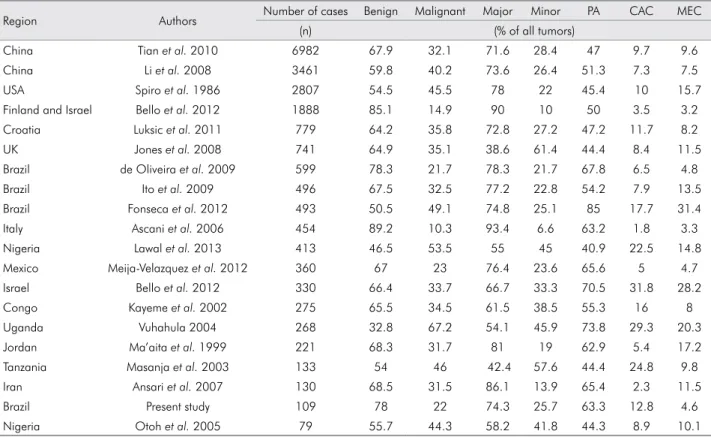
A morphologically diverse group of rare tumors arises from salivary glands. These tumors have different biological behaviors and their etiopathogenesis remains unknown.2,5 Epidemiologic studies conducted
in different parts of the world report differences in the incidence as well as frequency of histological types of SGTs (Table 4). The present study reviewed the demographic and clinicopathologic aspects of 109 cases of SGTs diagnosed at a tertiary health center
and the indings are in general agreement with data
reported in previous studies.1,8,10,12,15,16,18
According to the literature, the most common clinical presentation of SGT is a well-circumscribed nodular tumor with a normal overlying surface color. SGTs are usually asymptomatic and affect
females more, with peak incidence occurring the ifth
and seventh decades of life.10,15,17,19,20,21,22 The clinical
features that allow the differentiation of benign and malignant SGTs are the evolutionary course, pain and ulceration. Benign tumors tend to be insidious and slow growing, with an average course of three to six years and no adherence to surrounding tissues. Malignant lesions exhibit fast growth (often less than one year of evolution) and adhere to deep layers, with an ulcerated or telangiectatic surface. Pain is the most suggestive characteristic of malignancy.17 In the
present series, the majority of clinical characteristics observed were similar to those reported in previous studies. Patient age ranged from nine to 81 years old, with a mean of 46.47 years. The tumors were all nodules and 4.6% were ulcerated. The only difference in comparison to previous reports was the similar male-to-female ratio (0.94:1) with both benign and
malignant tumors. Although uncommon, Lukšić et al.15
and Tian et al.16 report similar indings.
Major salivary glands were affected more than minor glands, especially the parotid gland (66.9%), followed by palatine glands (14.7%). Similar results are described in the majority of studies on SGTs (Table 4), including those from Brazil (Table 5).1,6,7
The predominance of SGTs in major salivary glands may be explained by the fact that the present study was conducted at tertiary health center that receives most patients with parotid tumors. SGTs in minor salivary glands are usually diagnosed and treated at primary and secondary services, whereas only malignant SGTs in major salivary glands are referred to the hospital service.
Benign SGTs predominated in the major glands and comprised 78% of all salivary tumors, which is similar to rates reported in studies conducted in Iran,19
Congo,18 the United Kingdom,23 Jordan,24 Brazil6,7 and
China,16 in which benign tumors accounted for more
of malignant SGTs.11 In such cases, the uncommon
indings may be explained by the fact that the studies
were conducted at tertiary centers, which receive more severe cases of SGTs.
Pleomorphic adenoma is a benign neoplasm composed of epithelial and myoepithelial cells arranged in a large variety of morphological patterns, with areas of mesenchymal differentiation. In the present series, this tumor was the most frequent type of SGT, accounting for 63.3% of all tumors and 81.2% of benign tumors. The majority of such cases (73.9%) were located in major salivary glands. The frequency
of pleomorphic adenoma ranges from 40.9% to 73.8% of all STGs (Table 4).11,21 Studies involving large series
of SGTs report similar results, with pleomorphic adenoma representing 60% to 73% of parotid gland tumors as well as 40% to 60% of submandibular and minor salivary gland tumors.11,21 All cases of
pleomorphic adenoma reported in the present study were treated by surgery and recurrence was observed in four cases (3.66%).
Adenoid cystic carcinoma was the most prevalent malignant SGT (14 cases), representing 12.8% of all tumors and 58.3% of malignant tumors, followed by mucoepidermoid carcinoma and acinic cell carcinoma. The predominance of adenoid cystic carcinoma has been described in studies conducted in Congo, Tanzania, Croatia and Nigeria. In contrast, studies conducted in Iran, Brazil, Jordan, the USA, the United Kingdom and Italy report a greater frequency of mucoepidermoid carcinoma than adenoid cystic carcinoma, whereas similar distribution between these two types of SGTs has been reported in
China, Finland and Mexico (Table 4). These indings
Table 4. Distribution of salivary gland tumors in different countries.
Region Authors Number of cases Benign Malignant Major Minor PA CAC MEC
(n) (% of all tumors)
China Tian et al. 2010 6982 67.9 32.1 71.6 28.4 47 9.7 9.6
China Li et al. 2008 3461 59.8 40.2 73.6 26.4 51.3 7.3 7.5
USA Spiro et al. 1986 2807 54.5 45.5 78 22 45.4 10 15.7
Finland and Israel Bello et al. 2012 1888 85.1 14.9 90 10 50 3.5 3.2 Croatia Luksic et al. 2011 779 64.2 35.8 72.8 27.2 47.2 11.7 8.2
UK Jones et al. 2008 741 64.9 35.1 38.6 61.4 44.4 8.4 11.5
Brazil de Oliveira et al. 2009 599 78.3 21.7 78.3 21.7 67.8 6.5 4.8
Brazil Ito et al. 2009 496 67.5 32.5 77.2 22.8 54.2 7.9 13.5
Brazil Fonseca et al. 2012 493 50.5 49.1 74.8 25.1 85 17.7 31.4 Italy Ascani et al. 2006 454 89.2 10.3 93.4 6.6 63.2 1.8 3.3
Nigeria Lawal et al. 2013 413 46.5 53.5 55 45 40.9 22.5 14.8
Mexico Meija-Velazquez et al. 2012 360 67 23 76.4 23.6 65.6 5 4.7 Israel Bello et al. 2012 330 66.4 33.7 66.7 33.3 70.5 31.8 28.2
Congo Kayeme et al. 2002 275 65.5 34.5 61.5 38.5 55.3 16 8
Uganda Vuhahula 2004 268 32.8 67.2 54.1 45.9 73.8 29.3 20.3
Jordan Ma’aita et al. 1999 221 68.3 31.7 81 19 62.9 5.4 17.2
Tanzania Masanja et al. 2003 133 54 46 42.4 57.6 44.4 24.8 9.8 Iran Ansari et al. 2007 130 68.5 31.5 86.1 13.9 65.4 2.3 11.5
Brazil Present study 109 78 22 74.3 25.7 63.3 12.8 4.6
Nigeria Otoh et al. 2005 79 55.7 44.3 58.2 41.8 44.3 8.9 10.1
Table 5. Reports of salivary gland tumors in different states and regions in Brazil.
State/Region Authors Number of cases (n) Goiás/Middle-West de Oliveira et al. 2009 599 Paraná/South Ito et al. 2009 496 Paraná/South –
São Paulo/Southeast
Fonseca et al. 2012 493
suggest geographic variation in the frequency of malignant SGTs.
Adenoid cystic carcinoma exhibited the worst behavior among all SGTs analyzed. This tumor has a slow growth pattern with the early nerve and lymph node involvement. Advanced tumors require surgery and radiation therapy.25 In the present study,
adenoid cystic carcinoma recurred in four cases (28.6%) and death occurred in an additional two cases (14.28%). The patients in these cases were in TNM stages III or IV and received surgery plus radiation therapy. According to previous studies, the majority of patients with distant metastasis of an adenoid cystic carcinoma have tumor size of T3 or T4 and that surgery and radiation therapy achieve excellent results regarding both local and regional control.5,26,27 Another important aspect is the presence
of perineural invasion, which is a common inding in
adenoid cystic carcinoma that appears to contribute to a worse outcome.27 The mean follow-up period in
the present study was 7.4 years and a high survival was observed. However, previous studies indicate
that survival rates are usually high in the irst ive
years, whereas the cure rate over a 20-year period is much lower.5,8,26,27
Conclusion
In this retrospective study, major salivary glands were more affected than minor glands by neoplastic processes and pleomorphic adenoma and adenoid cystic carcinoma were the most frequent benign and malignant tumors, respectively, at a tertiary health center in southern Brazil. Analyzing the present
indings in light of data from previous studies on SGTs,
one may infer that some demographic characteristics (e.g., gender and age) and the predominance of malignant tumors (e.g., adenoid cystic carcinoma and mucoepidermoid carcinoma) vary in different geographic regions. Factors that influence SGT behavior and prognosis should be investigated further.
Acknowledgments
The authors are grateful to Flavia Rejane Giusti for technical support. This study was supported by the Postgraduate Research Group of the Hospital de
Clinicas de Porto Alegre (GPPG/FIPE: 12–0339). Luise
Meurer, Lelia Batista de Souza, Pablo Agustin Vargas and Manoela Domingues Martins are research fellows
of the Brazilian National Council of Scientiic and
Technological Development (Conselho Nacional de
Desenvolvimento Cientíico e Tecnológico - CNPq).
1. Oliveira FA, Duarte EC, Taveira CT, Maximo AA, Aquino EC, Alencar RC et al. Salivary gland tumor: a review of 599 cases in a Brazilian population. Head Neck Pathol. 2009;3(4):271-5. doi:10.1007/s12105-009-0139-9
2. Mariano FV, Silva SD, Chulan TC, Almeida OP, Kowalski LP. Clinicopathological factors are predictors of distant metastasis from major salivary gland carcinomas. Int J Oral Maxillofac Surg. 2011;40(5):504-9. doi:10.1016/j.ijom.2010.12.002 3. Eveson JW, Cawson RA. Salivary gland tumours: a review
of 2410 cases with particular reference to histological types, site, age and sex distribution. J Pathol. 1985;146(1):51-8. doi:10.1002/path.1711460106
4. Terhaard CH, Lubsen H, Van der Tweel I, Hilgers FJ, Eijkenboom WM, Marres HA et al. Salivary gland carcinoma: independent prognostic factors for locoregional control, distant metastases, and overall survival: results of the Dutch head and neck oncology cooperative group. Head Neck. 2004;26(8):681-93. doi:10.1002/hed.10400
5. Guzzo M, Locati LD, Prot t FJ, Gat ta G, McGurk M, Lic it ra L. Major a nd m i nor sa l iva r y g la nd
tumors. Crit Rev Oncol Hematol. 2010;74(2):134-8. doi:10.1016/j.critrevonc.2009.10.004
6. Ito FA, Ito K, Vargas PA, Almeida OP, Lopes MA. Salivary gland tumors in a Brazilian population: a retrospective study of 496 cases. Int J Oral Maxillofac Surg. 2005;34(5):533-6. doi:10.1016/j.ijom.2005.02.005
7. Fonseca FP, Carvalho MV, Almeida OP, Rangel AL, Ta k i zawa MC, Bueno AG et a l. Cli n icopat holog ic a nalysis of 493 cases of salivar y gla nd t umors i n a Sout her n Bra zi l ia n popu lat ion. Ora l Su rg Ora l Me d O ra l Pat hol O ra l R ad iol. 2012;114(2):230 -9. doi:10.1016/j.oooo.2012.04.008
8. Li LJ, Li Y, Wen YM, Liu H, Zhao HW. Clinical analysis of salivar y gla nd t umor cases i n West China in past 50 years. Oral Oncol. 2008;44(2):187-92. doi:10.1016/j.oraloncology.2007.01.016
9. Al-Khateeb TH, Ababneh KT. Salivary tumors in north Jordanians: a descriptive study. Oral Surg Oral Med Oral Pathol Oral Radiol Endod. 2007;103(5):e53-9. doi:10.1016/j.tripleo.2006.11.017
10. Bello IO, Salo T, Dayan D, Tervahauta E, Almangoush A, Schnaiderman-Shapiro A et al. Epithelial salivary gland tumors in two distant geographical locations, Finland (Helsinki and Oulu) and Israel (Tel Aviv): a 10-year retrospective comparative study of 2,218 cases. Head Neck Pathol. 2012 Jun;6(2):224-31. doi:10.1007/s12105-011-0316-5 11. Lawal AO, Adisa AO, Kolude B, Adeyemi BF, Olajide MA. A
review of 413 salivary gland tumours in the head and neck region. J Clin Exp Dent. 2013;5(5):e218-22. doi:10.4317/jced.51143 12. Mejia-Velazquez CP, Duran-Padilla MA, Gomez-Apo E,
Quezada-Rivera D, Gaitan-Cepeda LA. Tumors of the salivary gland in Mexicans. A retrospective study of 360 cases. Med Oral Patol Oral Cir Bucal. 2012;17(2):e183-9. doi:10.4317/medoral.17434
13. Spiro RH. Salivary neoplasms: overview of a 35-year experience with 2,807 patients. Head Neck Surg. 1986;8(3):177-84. doi:10.1002/hed.2890080309
14. Bjorndal K, Krogdahl A, Therkildsen MH, Overgaard J, Johansen J, Kristensen CA et al. Salivary gland carcinoma in Denmark 1990-2005: outcome and prognostic factors. Results of the Danish Head and Neck Cancer Group (DAHANCA). Oral Oncol. 2012;48(2):179-85. doi:10.1016/j.oraloncology.2011.09.005
15. Lukšić I1, Virag M, Manojlović S, Macan D. Salivary gland tumours: 25 years of experience from a single institution in Croatia. J Craniomaxillofac Surg. 2012;40(3):e75-81. doi:10.1016/j.jcms.2011.05.002
16. Tian Z, Li L, Wang L, Hu Y, Li J. Salivary gland neoplasms in oral and maxillofacial regions: a 23-year retrospective study of 6982 cases in an eastern Chinese population. Int J Oral Maxillofac Surg. 2010;39(3):235-42. doi:10.1016/j.ijom.2009.10.016
17. Barnes LE, Reichart P, Sidransky D. Patholoy and genetics of head and neck tumors. Lyon: IARC Press; 2005.
18. Kayembe MK, Kalengayi MM. Salivary gland tumours in Congo (Zaire). Odontostomatol Trop. 2002;25(99):19-22. 19. Ansari MH. Salivary gland tumors in an Iranian population:
a retrospective study of 130 cases. J Oral Maxillofac Surg. 2007;65(11):2187-94. doi:10.1016/j.joms.2006.11.025
20. Otoh EC, Joh n son NW, Olasoji H, Da n fillo IS, Adeleke OA. Salivary gland neoplasms in Maiduguri, north-eastern Nigeria. Oral Dis. 2005;11(6):386-91. doi:10.1111/j.1601-0825.2005.01137.x
21. Vuhahula EA. Salivary gland tumors in Uganda: clinical pathological study. Afr Health Sci. 2004;4(1):15-23.
22. Shishegar M, Ashraf MJ, Azarpira N, Khademi B, Hashemi B, Ashrafi A. Salivary gland tumors in maxillofacial region: a retrospective study of 130 cases in a southern Iranian population. Patholog Res Int. 2011;2011:934350. doi:10.4061/2011/934350
23. Jones AV, Craig GT, Speight PM, Franklin CD. The range and demographics of salivary gland tumours diagnosed in a UK population. Oral Oncol. 2008;44(4):407-17. doi:10.1016/j.oraloncology.2007.05.010
24. Ma’aita JK, Al-Kaisi N, Al-Tamimi S, Wraikat A. Salivary gland tumors in Jordan: a retrospective study of 221 patients. Croat Med J. 1999;40(4):539-42.
25. Triantafillidou K, Iordanidis F, Psomaderis K, Kalimeras E. Acinic cell carcinoma of minor salivary glands: a clinical and immunohistochemical study. J Oral Maxillofac Surg. 2010;68(10):2489-96. doi:10.1016/j.joms.2009.09.065
26. Seethala RR. An update on g rading of salivary gland carcinomas. Head Neck Pathol. 2009;3(1):69-77. doi:10.1007/s12105-009-0102-9