Arq Neuropsiquiat r 2004;62(4):1004-1007
Department of Magnetic Resonance Imaging, Clinica Radiologica Vila Rica, Brasilia DF - Brazil: 1Hospital Santa Lucia; 2)Clinica Radiologica Vila Rica; 3Hospit al Geral e Ort opedico; 4Fundaçao Hospit alar do Dist rit o Federal.
Received 19 M arch 2004, received in f inal f orm 9 June 2004. Accept ed 29 July 2004.
Dr. Sergio Lopes Viana - SQS 105 / Bloco D Apt o 506 / Asa Sul - 70344-040 Brasília DF - Brasil. E-mail: [email protected]
CAVERNOUS ANGIOM A OF THE CAVERNOUS SINUS
Imaging f indings
Jose Luiz Furt ado de M endonça
1,2, Sergio Lopes Viana
2,
M akot o M at sumine
2, Renat o Faria e Silva
3,
M aria Angelica de Carvalho Barbosa Viana
4, Flavia M endes de Oliveira Freit as
2ABSTRACT - Cavernous angiomas (cavernomas) of t he cavernous sinus are uncommon, w it h only scat t ered report s in lit erat ure. Unlike t heir parenchymat ous count erpart s, t hey are int ensely enhancing para-sellar masses, iso/hypoint ense in T1- and hyperint ense in T2-w eight ed images. Diff erent ial diagnosis w it h para-sellar meningiomas and schw annomas can be diff icult . We report t hree cases of t his condit ion, describing f indings of diff usion-w eight ed imaging in t his kind of lesion f or t he f irst t ime in lit erat ure.
KEY WORDS: cavernous angioma, cavernous sinus, magnet ic resonance imaging, comput ed t omography, angiography.
Angiom a cavernoso do seio cavernoso: achados aos exam es de im agem
RESUM O - Angiomas cavernosos (cavernomas) do seio cavernoso são lesões incomuns, com poucos relat os na lit erat ura especializada. Ao cont rário dos cavernomas parenquimat osos, são massas para-selares int en-sament e capt ant es, isoint ensas a hipoint ensas em T1 e hiperint ensas em T2 nas imagens de ressonância magnét ica. O diagnóst ico dif erencial com meningiomas e schw annomas para-selares pode ser dif ícil. Rela-t amos os achados de imagem de Rela-t rês casos desRela-t a enRela-t idade, descrevendo os achados da ressonância magné-t ica ponderada em dif usão nesmagné-t e magné-t ipo de lesão pela primeira vez na limagné-t eramagné-t ura.
PALAVRAS-CHAVE: angioma cavernoso, seio cavernoso, ressonância magnét ica, t omograf ia comput adori-zada, art eriograf ia.
Cavernous angioma of t he cavernous sinus is an ext remely rare condit ion, and it s diff erent ial diag-nosis w it h t umors, namely para-sellar meningiomas and schw annomas, is of t en diff icult . They are rich-ly vascularized lesions, made up of a net w ork of dilat ed, t hin-w alled vessels, and are responsible f or less t han one percent of all int racranial masses. The advent of magnet ic resonance imaging (M RI) rai-sed new diagnost ic perspect ives f or t his condit ion as w e report in t he st udy.
M ETHOD
This st udy w as approved by t he commit t ee of Et hics of Clinica Radiologia Vila Rica, Brasilia DF, Brazil.
Three f emale pat ient s, w it h 42-years-old (pat ient 1), 45-years-old (pat ient 2) and 37-years-old (pat ient 3), are, respectively, presented with complaints of right-sided cranial nerves def icit s (III nerve [pat ient s 2 and 3], V1 ner-ve [pat ient 2] and VI nerner-ve [pat ient s 1 and 2]) and
hea-daches. All of t hem underw ent magnet ic resonance ima-ging and cerebral digit al subt ract ion angiography (DSA). Pat ient s 2 and 3 w ere also submit t ed t o comput ed t o-mography (CT). In all pat ient s, M RI revealed w ell-delimi-t ed righell-delimi-t para-sellar lesions, unif ormly isoinell-delimi-t ense in T1-w eight ed images and hyperint ense in T2-, T2- and FLAIR-w eight ed images, bright ly enhancing af t er int ra-vascu-lar infusion of gadolinium. Diffusion-weighted sequence, perf ormed in pat ient 3, revealed an isoint ense mass.
RESULTS
angiogra-Arq Neuropsiquiat r 2004;62(4) 1005
phy (M RA) of cerebral vessels w it h 3D-TOF t echni-que, perf ormed in pat ient s 2 and 3, t he right -si-ded component s of t he circle of Willis w ere lat er-ally displaced by t he cavernous angiomas, w it hout any impairment of blood f low (Fig 2). DSA w as nor-mal in pat ient 1; pat ient s 2 and 3 had a supra-sel-lar and para-selsupra-sel-lar blush in lat e venous phase, once again w it hout impairment of int ernal carot id f low (Figs 6 and 7). Aneurysm w as excluded in all pat ienpat s. Operapat ive f indings w ere of w elldemarcapat -ed, hyperemic, reddish t o brow nish ext ra-dural pa-ra-sellar masses in all cases. These lesions w ere col-lapsible, readily ref illing af t er release of compres-sion. Punct ure yielded f reely f low ing blood. Planes of cleavage w it h surrounding neurovascular st ruc-t ures w ere noruc-t regarded as saf e, and none w as resect ed, given t he ant icipat ed surgical bleeding.
DISCUSSION
Cavernous angiomas are also known as caverno-mas and cavernous hemangiocaverno-mas. They are com-mon lesions of t he cerebral hemispheres, alt hough t hey can occur anyw here in t he cent ral nervous sys-t em. Exsys-t ra-axial cavernous angiomas are uncom-mon, and t he cavernous sinus is one locat ion in t his group. It is most common in w omen in t heir f if t h decade of lif e and in Japanese people, being rarely mult iple and heredit ary (specially in Hispanic-Ame-rican subject s)1-8. It is, in f act , a vascular malf
orma-t ion, w hich can behave like a real orma-t umor w hen iorma-t grows up to the point to compress neighbor structu-res. Exacerbat ion of sympt oms in pregnant w omen
Fig 1. Pat ient 1. Well-circumscribed right para-sellar and supra-sellar mass, slight ly hypoint ense in T1-w eight ed sequence (1A), show ing int ense enhancement af t er gadolinium inf usion (1B) and high signal int ensit y in T2-w eight ed image (1C). A small hemorrhagic f ocus is seen peripherally locat ed, due t o previ-ous biopsy. Lesion encircles right int ernal carot id art ery.
Fig 2. Patient 2. Right para-sellar and supra-sellar lesion iso/hypo-int ense in T1-w eight ed image (1A), bright ly enhancing af t er gadolinium infusion (1B and 1D) and hyperintense in T2-weight-ed image (1C). M RA (1E) show s displacT2-weight-ed right -sidT2-weight-ed vessels of t he circle of Willis, w hich have preserved f low. Right int ernal carot id art ery is seen w it hin lesion.
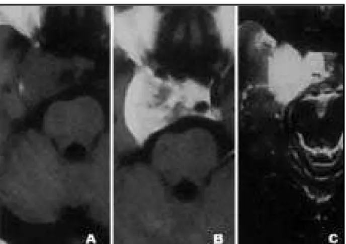
Fig 3. Pat ient 3. Right para-sellar and supra sellar mass show ing low t o int ermediat e signal int ensit y in T1-w eight ed image (1A), bright ly enhancing (1B), w it h high signal int ensit y in T2w eight -ed image (1C) and isointense in diffusion-weight-ed sequence (1D). Not e t he right int ernal carot id art ery encircled by t he mass.
have been report ed, improving af t er delivery2,3,9.
Present at ion complaint s most of t en consist of visu-al dist urbances (ret ro-orbit visu-al pain and headache accompanied of reduced ocular mot ricit y, pt osis, diplopia, exopht almos and impaired visual acuit y), mainly due t o compressive ef f ect or enclosure of neurovascular st ruct ures, namely t he cranial nerves passing t hrough t he cavernous sinus1-4,6,10. Seizures,
f acial numbness and neuralgia may also occur1,3,5.
1006 Arq Neuropsiquiat r 2004;62(4)
M acroscopically, t hey appear as w iny, w ell-cir-cumscribed, mult iloculat ed masses, richly vascu-larized, surrounded by a pseudocapsule. M eningio-mas and schw annoeningio-mas, t he most import ant hypo-t heses in diff erenhypo-t ial diagnosis are, unlike observed in our cases, compact ly solid masses, w it hout vascu-lar appearance, and do not yield abundant blood when punctured. Microscopically, they are a honey-comb of vascular spaces w hich lack muscular and elast ic component s, lined by a single endot helial layer, w it hout int ervening neuronal t issue5,6.
This ent it y appears as an iso/hyperdense mass on nonenhanced CT scans, enhancing int ensely af -t er inf usion of iodina-t ed media in mos-t cases3.
Cal-cification is an occasional finding, and it is most com-mon in meningiomas5,6,8. Erosion of t he sphenoid
bone can also be seen. DSA can be normal and show an avascular mass or a discrete to moderate tumoral blush, with feeding vessels originating from branch-es of t he ext ernal carot id or cavernous int ernal ca-rot id2-6,9,10. The int ernal carot id art ery is of t en
encir-cled by t he lesion in it s cavernous port ion, usual-ly maint aining it s normal caliber.
Unlike cerebral cavernous angiomas, their caver-nous sinus count erpart s do not have a pat hogno-monic appearance on M RI. Findings usually are of well-delimited para-sellar lesions, hypo or isointense in T1-w eight ed images and bright ly hyperint ense in T2-w eight ed images. A dumbbell-shaped mass can be seen, w it h a small supra-sellar component and most of lesion w it hin cavernous sinus1,4. M RI
allows superb evaluation of the relationships among t he cavernous angioma and t he surrounding st ruc-t ures1,2. Gradient -echo sequences may be usef ul t o
reveal hemorrhagic component. The mass was
isoin-Figs 4 and 5. Pat ient s 2 and 3. CT scans show right para-sellar and supra-sellar lesions, spont aneously hyperdense (4A and 5A), int ensely enhancing af t er inf usion of iodinat ed cont rast media (4B and 5B).
Fig 6. Pat ient 1. Select ive right carot id angiography show ing normally sized vessels, w it h adequat e cont rast f low. No mass ef f ect could be seen (6A and 6B).
Arq Neuropsiquiat r 2004;62(4) 1007
t ense in dif f usion-w eight ed images in our only pat ient in w hich it w as perf ormed, and, t o our kno-w ledge, t his paper is t he f irst so f ar t o report f ind-ings of t his t echnique in cavernous angioma of t he cavernous sinus. Enhancing pat t ern af t er gadolini-um inf usion in most cases is int ense and homoge-neous. The most import ant dif f erent ial diagnosis (and t he most common preoperat ive misdiagno-sis) is para-sellar meningioma. Although differentiat ion bedifferentiat w een differentiat hese differentiat w o condidifferentiat ions could be dif -f icult , a homogeneously enhancing mass enw rap-ping t he int ernal carot id art ery w it hout signif icant reduct ion of it s caliber should lead diagnosis t o-wards cavernous angioma. Other possible hypothsis in t his cont ext is para-sellar schw annoma. M e-ningiomas usually are isoint ense w it h gray mat t er bot h in T1- and T2-w eight ed images, w hile schw annomas t end t o have low er signal t han gray mat -t er in T1- and higher signal in T2-w eigh-t ed images. Enhancement is prominent bot h in meningiomas and schw annomas, but t ends t o be slight ly het ero-geneous1,3,6.
Alt hough parenchymal cavernomas are easily resect ed in most cases, surgical excision of caver-nous angiomas is often challenging because of abun-dant operative bleeding, due to the vascular nature of these lesions3-5,7,8,10. Predominantly vascular
mass-es and most ly organized onmass-es can be f ound9.
Des-pit e successf ul resect ions carried out , high mort ali-t y raali-t es sali-t ill remain, and conservaali-t ive ali-t reaali-t menali-t should be also considered. The possibilit y of a ca-vernous angioma must be kept in mind, because
of dif f erent surgical approaches possibly ut ilized. Radiot herapy may be a usef ul complement as t he primary t herapy or as a preoperat ive adjuvant2, 4.
In conclusion, no-demarcat ed, homogeneous-ly enhancing, avascular/hypovascular masses of t he middle f ossa, w hich are iso/hypoint ense in T1- and hyperint ense in T2-w eight ed images should raise t he hypot hesis of cavernous angiomas, despit e t he higher f requency of para-sellar meningioma. Because of t he high mort alit y w hen at t empt ing t o remove t hese lesions, radiologist s should alert sur-geons f or t his possibilit y.
REFERENCES
1. Sohn CH, Kim SP, Kim IM, Lee JH, Lee HK. Characteristic MR imag-ing findimag-ings of cavernous hemangiomas in the cavernous sinus. AJNR 2003;24:1148-1151.
2. Bristot R, Santoro A, Fantozzi L, Delfini R. Cavernoma of the cav-ernous sinus: case report. Surg Neurol 1997;48:160-163.
3. Momoshima S, Shiga H, Yuasa Y, Higuchi H, Kawase T, Toya S. MR find-ings in extracerebral cavernous angiomas of the middle cranial fossa: report of two cases and review of the literature. AJNR 1991;12:756-760. 4. Suzuki Y, Shibuya M, Baskaya MK, et al. Extracerebral cavernous angiomas of the cavernous sinus in the middle fossa. Surg Neurol 1996;45:123-132.
5. Gliemroth J, Missler U, Sephernia A. Cavernous angioma as a rare neuro-radiologic finding in the cavernous sinus. J Clin Neurosci 2000;7:542-560. 6. Tannouri F, Divano L, Caucheteur V, et al. Cavernous haemangioma in the cavernous sinus: case report and review of the literature. Neuroradiology 2001;43:317-320.
7. Meneses MS, Dallolmo VC, Kondageski C, Ramina R, Hunhevicz S, Pedrozo AA. Cirurgia estereotáxica guiada para angiomas cavernosos. Arq Neuropsiquiatr 2000;58:71-75.
8. Andrade GC, Prandini MN, Braga FM. Cavernoma gigante: relato de dois casos. Arq Neuropsiquiatr 2002;60:481-486.
9. Shi J, Hang C, Pan Y, Liu C, Zhang Z. Cavernous hemangiomas in the cavernous sinus. Neurosurgery 1999;45:1308-1312.