2018/2019
Cláudia Sofia Monteiro Pinto
Cardiovascular Effects of
Urocortin-2: Pathophysiological Mechanisms
and Therapeutic Potential
Mestrado Integrado em Medicina
Área: Ciências Médicas e da Saúde
Tipologia: Monografia
Trabalho efetuado sob a Orientação de:
Prof. Doutora Carmen Dulce da Silveira Brás Silva Ribeiro
E sob a Coorientação de:
Prof. Doutor Rui Miguel da Costa Adão
Trabalho organizado de acordo com as normas da revista
Cardiovascular Drugs and Therapy (ISSN: 1573-7241)
Cláudia Sofia Monteiro Pinto
Cardiovascular Effects of Urocortin-2:
Pathophysiological Mechanisms and
Therapeutic Potential
Projeto de Opção do 6º ano
Declaração de IntegridadeProjeto de Opção do 6º ano
Declaração de ReproduçãoA todos aqueles que me acompanharam nesta longa caminhada, contribuindo para o meu crescimento científico, profissional e pessoal.
Um especial obrigada à minha mãe, uma figura de destaque na ciência da vida, por fazer de mim aquilo que hoje me posso orgulhar de ser.
Como não poderia deixar de ser, agradeço à Professora Doutora Carmen Dulce da Silveira Brás Silva Ribeiro e ao Professor Doutor Rui Miguel da Costa Adão, por serem um exemplo não só na investigação, mas também enquanto pessoas, com os quais aprendi muito e me orgulho de ter tido a oportunidade de trabalhar ao longo dos últimos três anos.
Projeto de Opção do 6º ano
DedicatóriaCardiovascular Effects of Urocortin-2: Pathophysiological Mechanisms and Therapeutic Potential Cláudia Monteiro-Pinto1 (ORCID 0000-0001-6438-3248)
Rui Adão1 (ORCID 0000-0003-2203-436X)
Adelino F. Leite-Moreira1 (ORCID 0000-0001-7808-3596)
Carmen Brás-Silva1,2 (ORCID 0000-0003-1527-3776)
1- Department of Surgery and Physiology, Cardiovascular Research and Development Center - UnIC, Faculty of Medicine, University of Porto, Al. Prof. Hernâni Monteiro, 4200-319 Porto, Portugal
2- Faculty of Nutrition and Food Sciences, University of Porto, 4200-319 Porto, Portugal
Corresponding Author
Carmen Brás-Silva: Tel: +351 220 426 822; Fax: +351 225 513 646, E-mail: [email protected]
Acknowledgments
This work was supported by Portuguese Foundation for Science and Technology (FCT) through Grant UID/IC/00051/2013 and project IMPAcT‐ PTDC/MED‐FSL/31719/2017
Cardiovascular Effects of Urocortin-2: Pathophysiological Mechanisms and Therapeutic Potential
Abstract
Urocortin-2 (Ucn-2) is a peptide of the corticotrophin releasing factor-related family with several effects within the cardiovascular system. A variety of molecular mechanisms has been proposed to underlie some of these effects, although others remain mostly hypothetical. Growing interest in the cardiovascular properties of this peptide promoted several pre-clinical studies in the settings of heart failure and ischemia, as well as some experiments in the fields of systemic and pulmonary arterial hypertension. Most of these studies report promising results, with Ucn-2 showing therapeutic potential in these settings, and few clinical trials to date are trying to translate this potential to human cardiovascular disease. Ucn-2 also appears to have potential as a biomarker of diagnostic/prognostic relevance in cardiovascular disease, this being a recent field in the study of this peptide needing further corroboration. Regarding the increasing amount of evidence in Ucn-2 investigation, this work aims to make an updated review on its cardiovascular effects, molecular mechanisms of action and therapeutic potential, as well as to identify some research barriers and gaps in the study of this cardioprotective peptide.
CARDIOVASCULAR EFFECTS OF UROCORTIN-2: PATHOPHYSIOLOGICAL MECHANISMS AND THERAPEUTIC POTENTIAL
Urocortin-2 (Ucn-2) is one of the latest discovered peptides of the corticotrophin releasing factor (CRF)-related family. Until now, this family is composed of four members in vertebrates, coded by four distinct genes which appear to be highly conserved during evolution [1]. In 1955, CRF was discovered as the first mediator of stress response [2, 3], but it was only until 1995 that the first of the 3 urocortins (Ucns) known to date, urocortin-1 (Ucn-1), was isolated [4]. Growing interest on the pharmacological properties of Ucns leaded to the discovery of stresscopin and stresscopin-related peptide (SRP), homologues of urocortin-3 (Ucn-3) and Ucn-2, respectively [5].
This work focuses on Ucn-2, a 38-amino-acid (aa) peptide discovered in 2001 that stood out for its promising effects within the cardiovascular system, reported in multiple animal and human studies [6-8]. It aims to make an updated review of its cardiovascular effects, therapeutic potential, mechanisms of action and limitations in the current knowledge needing further elucidation.
Distribution and Structure
In both humans and animals, Ucn-2 is widely expressed throughout the body. It has a similar distribution to that of Ucn-1, being found in the cardiorespiratory, gastrointestinal, hematologic, endocrine, reproductive, urinary and central nervous systems, as well as in skeletal muscle, brown fat and skin [9-15].
Human Ucn-2 (hUcn-2) is coded by a gene with 621 base pairs (bp) from chromosome 3 and
shares 76% of homology with mouse Ucn-2 (mUcn-2) at the amino-acid level [16]. Ucn-2 gene transcription results in a precursor peptide that undergoes specific processing, giving rise to the bioactive peptide. Human SRP, a 43-aa peptide, was identified from the same gene as Ucn-2, but with different interpretation of the cleavage sites from the precursor peptide [11].
A particular characteristic of both SRP and hUcn-2 appears to be somewhat baffling, which is the lack of a proteolytic site that would allow for C-terminal processing. In fact, although ⍺-amidation of the terminal is commonly a requisite for generation of a bioactive peptide, hUcn-2, with a rather unusual C-terminal, does not appear to have a consensus amidation sequence [16, 9, 17]. Therefore, while the processing of the murine precursor peptide occurs with cleavage after the signal peptide, glycosylation and processing at the C-terminal (giving origin to an amidated protein), the human’s form only goes through the first two phases, with the C-terminus remaining unmodified [17]. The natural cleavage site for hUcn-2 appears to be after Leu14 [17, 18].
Functionally, both Ucn-2 N- and C- terminals seem to be responsible for its receptor selectivity [19]. The middle region, while not appearing to have an influence on selectivity for corticotrophin releasing factor receptor-2 (CRF-R2), ensures correct positioning of N- and C- terminals with the CRF-R2 and it also harvests the binding site for corticotrophin releasing factor-binding protein (CRF-BP) [20]. This region may arise as an important target since modifications in the hUcn-2 peptide that reduce its affinity to CRF-BP conceivably increase its pharmacological activity [20]. Ucn-2 exists as a highly glycosylated precursor, what may function as a way of increasing its bioavailability [17].
Receptors
Since its discovery, Ucn-2 was found to be a strong agonist of CRF-R2, for both ⍺ and βisoforms, with low or no affinity for corticotrophin releasing factor receptor-1 (CRF-R1) [9].
The CRF-R2 gene was assigned to human chromosome 7p21–p15 in 1997 [21]. In mouse, it was found to be located on the proximal end of chromosome 6, which is consistent with the high degree of homology of this region with human chromosome 7 [22]. The gene has 16 exons and depending on alternative splicing at the 5’ end gives rise to the three known variants of CRF-R2 identified in humans: CRHR2-⍺, CRHR2-β and CRHR2-𝛾 (mice do not possess the 𝛾 variant) [5].
Contrasting to what happens in rodents, the major CRF-R2 isoform in human heart and skeletal muscle is CRF-R2⍺, whereas CRF-R2β plays a minor role [23]. More specifically, CRF-R2⍺ mRNA is highly expressed in all four human cardiac chambers, whereas CRF-R2β mRNA expression predominates in left atrium showing general weak expression [24].
Both CRF-R1 and CRF-R2 belong to the B1 family of G-protein coupled receptors, possessing a N-terminal extracellular domain (ECD) linked to a 7-transmembrane helical bundle domain [25]. CRF-R2 corresponds to a splicing variant of CRF-R1, having an extra 29 aa inserted in the first intracellular loop (ICL) [26].
Peptide binding and receptor activation occur in two steps: primarily, the C-terminal portion of the peptide binds to the ECD, where disulfide bonds are critical for ligand recognition, and subsequently the N-terminal portion binds to the transmembrane domain initiating the intracellular signaling cascade [25]. This additional interaction happens specifically in the juxta-membrane domain of the receptor, which also contributes for its selectivity pattern, stabilizing affinity for Ucn-2 by about 30-fold in contrast with CRF-R1. The third ICL is believed to have a pivotal role in G-protein coupling and signal transduction [27, 28].
Cardiovascular Ucn-2 effects in physiological states 1) Cardiac effects
a) Left Ventricular Function i) Animal Studies
Bale, Hoshijima et al. [29] observed acute intravenous (iv) injection of Ucn-2 in mice resulted in potent positive inotropic effects, as shown by the rise in peak (+) dP/dt and increased slope of the left ventricular end-systolic pressure-volume relation (ESPVR) for any given heart rate (HR). Ucn-2 injection also produced a decline in maximum left ventricular (LV) pressure and accelerated isovolumic relaxation, therefore enhancing diastolic function. These improvements of inotropism and lusitropism leaded to a clear enhancement in ventricular function, as demonstrated by the rises in stroke volume (SV), LV ejection fraction (EF) and the average 27% increase in cardiac output (CO).
In wild-type (WT) mice isolated cardiomyocytes, Ucn-2 increased maximal velocity of shortening, relengthening, peak height and shortening, as well as amplitude and fall in mean constant of
Ca2+ transient decay (Tau), reinforcing its positive inotropic and lusitropic effects [30]. Similar
dose-dependent inotropic and lusitropic effects were further obtained in mouse ventricular myocytes [31], isolated rabbit myocytes [32], feline isolated left ventricular myocytes [33] and anaesthetized pig [34]. In the sheep normal heart, Ucn-2 iv injection caused a marked increase in CO, accompanied by a minor decrease in left atrial pressure (LAP) [35].
As shown by in vivo and ex vivo experiments performed by Gao, Lai et al.[36], Ucn-2 gene transfer in healthy mice may lead to potent, long lasting, positive inotropic and lusitropic effects, with improvements in ventricular function up to 7 months of gene transfer. In a comparative study of Ucn-2
versus Ucn-3 gene transfer effects, Giamouridis, Gao et al. [37] reported increases in ESPVR slope by 90%
(vs 63% with Ucn-3), CO by 65% (vs 50% with Ucn3) and a 31% drop in Tau (vs 24% with Ucn3) after obtaining chronically elevated levels of Ucn-2. Tsuda, Takefuji et al. [38] implanted healthy mice with osmotic pumps, supplying a sustained Ucn-2 infusion for 4 weeks. The results were conflicting with the previous reports, with the emergence of cardiac dysfunction. The use of osmotic pumps does not allow for exogenous Ucn-2 temperature control, which may affect the peptide stability. On the other hand, continuously elevated Ucn-2 plasma levels opposed to Ucn-2 given in pulses may produce deregulated activation of several signaling pathways, which may associate with the deleterious effects found in this study.
ii) Human Studies
There are few studies on Ucn-2 cardiovascular actions in healthy humans. Two of them were performed in vivo, through an iv infusion of Ucn-2. The results were overlapping, with dose-related improvements in echocardiographic markers of ventricular function and contractility [39, 40]. One must take into account these works lack loading and HR control, limiting direct conclusions of inotropism. To clarify whether the improvements in left ventricular function are the result of Ucn-2 direct positive inotropic properties in humans, in vitro studies are needed. Yang, Kockskämper et al. [31] adressed this necessity, directly objectifying Ucn-2 positive inotropic effects in humans, denoted through a 15% increase in twitch force in isolated atrial trabeculae.
iii) Signaling Pathways
Yang, Kockskämper et al. [32, 31] performed studies in isolated rabbit and mouse myocytes suggesting the beforehand mentioned effects induced by Ucn-2 are mediated by actions in Ca2+ dynamics.
In these works, Ucn-2 was shown to accelerate and increase the amplitude of Ca2+ transients (increasing
not only systolic i[Ca2+], but also diastolic i[Ca2+]), also with a marked acceleration of the decaying phase.
The authors point CRF-R2-activated Gs-cAMP-PKA signaling cascade as one of the main pathways mediating these effects. PKA activation ultimately leads to phosphorylation of known Ca2+ regulatory
proteins, such as L-type Ca2+ channels, ryanodine receptor channels and phospholamban. These
mechanisms increase L-type Ca2+ current and SERCA activity, with concomitant rise in sarcoplasmic
of the CaMKII pathway, which may be stimulated by an unclear PKA-independent mechanism, as well as by the rise in i[Ca2+] induced by PKA. The conclusion that arises is that CaMKII and PKA signaling
cascades work synergistically, regulating the phosphorylation of Ca2+ regulatory proteins, both being of
central importance in the inotropic and lusitropic effects produced by Ucn-2.
A posterior study by Chen, Wang et al. [30] in mouse isolated cardiac myocytes, while further acknowledging the important role played by cAMP-PKA pathway, stands out AMPK pathway as another relevant mediator of Ucn-2 effects on Ca2+ dynamics and inotropism. As the authors note, AMPK pathway
is stimulated by Ucn-2 treatment and has the potential to increase Ca2+ sensitivity and contractility of
cardiomyocytes through cardiac troponin I phosphorylation. The mechanisms of interaction and regulation between AMPK and PKA-signaling pathways are, however, still unclear.
Walther, Pluteanu et al. [41] demonstrated that there is an increase in nitric oxide (NO) production induced by Ucn-2 in rabbit isolated cardiomyocytes, and this effect is linked to its inotropic effects. In their work, at least two pathways were shown to be associated with endothelial NO synthase (eNOS) activation: cAMP-PKA and PI3K-PDK-Akt signaling cascades. As shown by the authors, these pathways do not appear to interact directly, but they converge in phosphorylation of eNOS at Ser1177, ultimately leading to increased NO production. As expected, this culminates in stimulation of soluble guanil cyclase, increasing intracellular levels of cGMP. In the same study, the authors found Ucn-2 also stimulated MEK1/2-ERK1/2 pathway, but it did not play a role in the eNOS-mediated NO signaling in cardiac myocytes.
b) Chronotropism
In vivo iv injection of Ucn-2 exerts a dose-dependent positive chronotropic effect in mice, rat,
sheep and humans [29, 42, 35, 39, 40]. Gao, Lai et al. [36], using AAV8.CBA.Ucn-2, performed in vivo studies reproducing the increase in HR seen in the previously mentioned works up to 7 months after Ucn-2 gene transfer. However, when UcnUcn-2 administration was performed in isolated perfused hearts, the studies revealed no group differences, indicating that chronotropic effects seen in vivo are probably of reflex origin other than an intrinsic effect of Ucn2 gene transfer [36]. Studies in conscious rats corroborate this conclusion, since sympathetic and parasympathetic blockage by propranolol plus atropine abolished the tachycardic effect of Ucn-2 administration [43, 44]. In sheep, Ucn-2 seems to have a direct chronotropic effect, independent of autonomic and baroreceptor activation [45]. In fact, in this species, Ucn-2 produces a sustained decrease in the sympathetic traffic to the heart while there is still increased HR, weakening the hypothesis of this effect arising only from baroreflex activation [45].
c) Cardiac Morphology and Electrical Activity
As reviewed bellow, when in the presence of a pathologic condition which may ultimately lead to hypertrophy, Ucn-2 appears to have an obvious anti-hypertrophic effect. However, the effects in the normal heart seem to be less clear, with little conformity when analyzing the effects in vitro and in vivo. Indeed, in
vitro works in rat neonatal isolated cardiomyocytes assuredly indicate towards a pro-hypertrophic effect of
cardiomyocyte size, protein to DNA ratio and the incorporation of [3H]-leucine, all markers of cardiac hypertrophy [46, 47]. On the other hand, in vivo studies accessing the effects of chronically elevated levels of Ucn-2 obtained through gene transfer seem to favor an anti-hypertrophic effect [36, 37]. Necropsy analysis indicated less hypertrophy in AAV8.CBA.Ucn2 transfected mice, with reduction in body weight-adjusted LV weight [36, 37]. Contrasting with these works, Tsuda, Takefuji et al.[38] evidenced a pro-hypertrophic effect after chronic Ucn-2 ministration in control mice, which is in line with the above mentioned emergence of cardiac disfunction found in the same study.
Yang, Kockskämper et al. [31], using both mouse ventricular myocytes and myocardium from right-atrium of patients undergoing cardiac surgery , showed an association of Ucn-2 with arrhythmogenic events. Noteworthy, spontaneous Ca2+ increases where observed and preceded the shortenings, pointing
out release from SR as the inherent mechanism. In vivo, only one study briefly mentions Ucn-2 effects in electrocardiographic parameters in healthy humans, finding no relation with arrhythmogenic events [39]. In other works testing Ucn-2 in healthy animals and humans there is no reference to electrocardiographic parameters. Nonetheless, there is no report of symptomatic arrhythmias and/or relevant electric events in the studies in which electrocardiographic monitoring was performed.
i) Signaling Pathways
Walther, Awad et al. [48] attempted to explain the mechanisms behind the pro-hypertrophic effect of Ucn-2 observed in vitro. The authors found Ucn-2 can disturb the equilibrium between transcription factors of maintenance (NFAT1c) and “maladptative” (NFAT3) hypertrophy. Indeed, they observed Ucn-2 induced nuclear export of NFATc1 to cytosol, while not altering the location of NFAT3. They also suggested the mechanism involved might be the Ucn-2-stimulated PI3K/Akt/eNOS/NO pathway. Activation of this pathway would ultimately lead to PKG1 activation by cGMP, also involving activation of other kinases (GSK3β, JNK, p38), resulting in phosphorylation, deactivation, and nuclear export of NFAT. A previous study in rat neonatal isolated cardiomyocytes also suggested the hypertrophic effect induced by Ucn-2 in the normal heart was promoted by PI3K/Akt pathway [46].
Chronic activation of Ucn-2 induced pathways, such as PKA, CAMKII and PI3K/Akt, may explain the pro-hypertrophic effects observed in vivo in control mice [38]. On the other hand, the apparent anti-hypertrophic effect observed after Ucn-2 gene transfer in vivo was suggested to be a result of renin– angiotensin–aldosterone system (RAAS) inhibition by Ucn-2 [36].
In the first study accessing the direct electrophysiological effect of Ucn-2 in action potential, Yang and Zhu [49] found Ucn-2 significantly prolonged action potential duration in a time and concentration-dependent manner in guinea pig isolated myocytes. The elongation of action potential induced by Ucn-2 may lead to changes in refractory period and this may be one of the explanations for the beforehand described arrhythmogenic effect of high Ucn-2 doses in vitro. The authors found this effect appeared to be mediated by PKA activation and Ca2+-dependent activation of Na+/Ca2+ exchange (NCX), as well as
increased IKr inhibition rate and reduced IKr current. The inhibition of IKr was not mediated by PKA signaling and the contribution of other Ucn-2 activated pathways was not evaluated yet. The authors also
noticed Ucn-2 induced a right shift in the threshold for IKs current activation and decelerated rate of IKs activation.
2) Vascular Effects a) Animal Studies
In vitro, increasing doses of Ucn-2 were shown to express a dose and time-dependent vasodilatory
effect, similar in potency to that of Ucn-3, both being more potent than CRF, but less potent than Ucn-1 [50].
In vivo, the effects tend to vary according to the species and vascular bed. In rats, several studies
have shown consistent dose-dependent decreases in blood pressure (BP) after Ucn-2 infusion [51, 42-44, 52]. In this specie, Ucn-2 appears to induce marked mesenteric vasodilatation, while it produces an inconsistent renal vasodilation, indicating less powerful coupling of CRF-R2 receptors in the renal vascular bed. In mice, Ucn-2 was found to be a potent vasodilator, with decreases in arterial elastance and resistance, as well as in systolic, diastolic and mean BP [29, 37]. In pig, intra-coronary infusion of Ucn-2 leaded to an approximate 15% increase in coronary blood flow [34]. Contrastingly, in healthy sheep, Ucn-2 iv injection resulted in a minor reduction in calculated total peripheral resistance (CTPR). On top of that, although an initial drop in mean arterial pressure (MAP) was found, the overall effect was elevation of MAP [35].
b) Human Studies
Wiley and Davenport [27] conducted in vitro studies with human internal mammary arteries showing a potent vasodilatory effect of Ucn-2, with approximately 61% reversal of endothelin-1 (ET-1)-induced vasoconstriction. More recently, in coronary arteries dissected from patients subjected to heart transplantation, Ucn-2 induced marked relaxation in both intact endothelium and endothelium-denuded arteries, with no significant differences between the two [53].
Venkatasubramanian, Griffiths et al. [54] performed intra-brachial infusions of Ucn-2 in healthy volunteers, resulting in a pronounced dose-dependent vasodilation in the infused arm’s arteries. No changes were noticed in blood flow in the non-infused arm or in systolic blood pressure (SBP). The authors also describe no signs of tachyphylaxis associated with locally induced vasodilatation. Similar effects were obtained in a posterior study after intra-brachial infusion of Ucn-2 in both healthy subjects and heart failure (HF) patients [55]. In the latter study, systemic iv delivery of Ucn-2 in healthy subjects leaded to significant falls in MAP and peripheral vascular resistance index, accompanied by rises in HR.
c) Endothelium-dependency and Signaling Pathways
The endothelium-dependency of Ucn-2 vasodilatory effect presumably varies according to the species and the vascular bed. In humans, in vitro studies in human internal mammary and coronary arteries indicate towards a potent endothelium-independent vasodilatory effect of Ucn-2, although there is also
evidence of eNOS activation contribution in vivo [27, 53, 54]. As to other species, Gardiner, March et al. [43] found no involvement of NO or prostanoids in Ucn-2-induced vasodilation in conscious rats. In pig, on the other hand, the increase in coronary blood flow induced by Ucn-2 appears to be NO-dependent, suggesting the dependence on endothelium [34].
As to the signaling pathways involved in vasodilation, cAMP-PKA signaling was shown to have an important role in Ucn-2 induced vasodilation, also with contribution of p38/MAPK pathway (speculated to lead to production of vasodilatory prostaglandins) [50]. In porcine isolated aortic endothelial cells, NO generation stimulated by Ucn-2 appears to be mediated by cAMP-PKA and CaMKII signaling pathways, as well as increased cytoplasmic [Ca2+] [56, 57]. As shown by Grossini et al. [57], Ucn-2 seems to raise
levels of cytoplasmic [Ca2+] through two mechanisms: influx from intracellular pools and extracellular
medium and membrane hyperpolarization as a consequence of K+ channels opening.
Therapeutic potential in cardiovascular disease 1. Heart Failure
a) Animal Studies
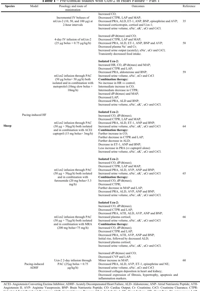
The first studies assessing Ucn-2 effects in failing hearts focused on documenting its acute effects in animal models of HF. In line with this, Bale, Hoshijima et al. [29] reported improvements in left ventricular diastolic and systolic functions in MLP-deficient cardiomyopathic mice after an iv bolus of Ucn-2. Rademaker, Cameron et al. [35] uncovered similar outcomes when using incremental iv boluses of mUcn-2 in an ovine model of HF. Moreover, they found a marked fall in CTPR and an overall reduction in MAP, contrasting with the findings from healthy sheep. A plausible explanation would be that in the HF model the arterial vasculature is in a pre-constricted state, which would enhance the vasodilatory properties of Ucn-2. The authors also reported important hormonal effects following Ucn-2 injection (detailed in Table I).
Rising interest in the therapeutic potential of Ucn-2 in this setting led a number of investigation groups to start exploring the effect of Ucn-2 given therapeutically. In a sheep model of chronic HF, a 4-day iv infusion of mUcn-2 presented with numerous favorable effects, improving cardiac function, hemodynamics, hormonal response and renal function [58]. On the other hand, potentially adverse effects of increased cortisol levels from secondary HPA axis stimulation and decreased food intake were only transient. In the same model of ovine HF using mUcn-2 administered through a pulmonary artery catheter, similar acute effects were observed in LV function, peripheral resistance, neurohormonal response and renal function [59]. Ucn-2 was also tested in the setting of acutely decompensated heart failure (ADHF) in ovines, resulting in a clear boost in cardiac function, suppression of potentially harmful neuro-hormonal response and amelioration of renal function [60]. The authors also documented lessened genetic expression of RAAS, as well as fibrosis, hypertrophy, apoptosis and inflammation markers. Histologically, Ucn-2 decreased collagen deposition in the heart and kidney.
Given that Ucn-2 has a rather short half-life, Lai, Gao et al. [61] made an effort to overcome this issue by performing an Ucn-2 gene transfer after induction of HF in mice. The gene transfer was attained by a single iv injection of a viral vector (AAV8) encoding Ucn-2 gene (AAV8.UCn2), giving rise to a 70-fold increase in LV Ucn-2 mRNA 5 weeks after injection. At this time, the authors reported an improvement in LV systolic and diastolic functions, with no group differences in MAP. The results also pointed to an anti-hypertrophic effect, with a reduced left ventricle/body weight ratio in treated animals. These findings also extended to Ucn-3 [62]. Ucn-2 gene transfer in Western-Diet (WD)-fed mice was also able to significantly improve glucose tolerance, with reduced fasting blood glucose and increased glucose disposal [63]. At the same time, it improved both diastolic and systolic LV functions and decreased liver fatty infiltration in mice with WD-induced diabetes-related LV dysfunction [63]. This highlights Ucn-2 as a potential therapeutic weapon in the setting diabetes-induced heart disease, targeting both glucose intolerance and cardiac dysfunction.
i. Ucn-2 in combination with established HF therapies
In order to prospect the realistic therapeutic potential of Ucn-2, Rademaker, Charles et al. performed a number of studies using Ucn-2 in combination with other drugs commonly used in the setting of HF, namely angiotensin converting enzyme inhibitor (ACEI) [64], loop diuretic [65], β-blocker [59] and mineralocorticoid receptor antagonist (MRA) [66]. All the works were performed in sheep with pacing-induced HF.
In the first study, the authors studied the effects of intra-arterial infusions of Ucn2 both isolated and in combination with the ACEI captopril [64]. They came to the conclusion that the addition of Ucn-2 to an ACEI may have beneficial effects on hemodynamic, hormonal and renal responses, to a greater extent than with ACEI treatment alone. Inclusively, in the matter of renal function, Ucn-2 co-treatment with ACEI appears to have the potential to neutralize some of the undesired effects observed with ACEI treatment alone, namely the reduction in creatinine clearance and urine output. Ucn-2 addition to ACEI was also able to neutralize the potentially adverse rise in plasma renin activity (PRA) induced by the latter.
Using the same regimen as in the previous work, Rademaker, Charles et al. [65] described Ucn-2 effects in combination with furosemide. The findings were compatible with an overall improvement of furosemide infusion effects. Specifically, the co-infusion resulted in enhanced renal function without additional K+ excretion, suppression of PRA and greater and more sustained decrease in aldosterone than
seen with both treatments alone. Given separately, Ucn-2 improved CO and hemodynamics, in accordance with previous studies in the same animal model of HF.
Employing a similar study protocol, the authors then accessed the effects of Ucn-2 in isolation and in addition to the 𝛽-blocker metoprolol [59]. The results were once again promising, with Ucn-2 avoiding the bradycardia, fall in CO and increase in CTPR induced by β-blocker therapy [59]. Ucn-2 may, therefore, have the appealing effect of increasing β-blocker tolerability, possibly allowing for an earlier and safest introduction in the setting of ADHF.
Lastly, the authors tested the effect of intra-arterial infusions of Ucn2, both isolated and in combination with a mineralocorticoid receptor antagonist (MRA) [66]. With combined therapy, the effects
of increased LV function, decreased LAP and CTPR were observed at a similar degree to those incited by Ucn-2 alone [66]. When added to a MRA, Ucn-2 also had the ability of improving the neuro-hormonal response and renal function, reducing PRA, angiotensin II and K+ levels [66]. Separate administration of
MRA increased plasma aldosterone, and Ucn-2 was capable of smoothing this response in co-therapy [66]. Overall, this study unveils possible beneficial effects of adding Ucn-2 to MRA therapy, with the particularly enticing effect of virtually inhibiting hyperkaliemia, a well-known dangerous side effect of MRA. Detailed information on the beforehand mentioned pre-clinical studies can be found on Table I.
b) Human studies
Davis, Pemberton et al. [67] studied the actions of iv hUcn-2 given to eight males suffering from congestive HF with reduced ejection fraction. They documented dose-dependent improvements in cardiac function and falls in SBP, DBP and MAP. They also noticed a slight reduction in urine volume and natriuresis, with no neurohormonal responses worth of consideration. This contradicts findings from ovine HF and is probably related to the deeper severity of disease and neurohormonal deregulation in this animal model. It seems likely that the natriuretic effect of Ucn-2 is only significant when there is an important background of RAAS activation and Na+ retention. Regarding side effects, Ucn-2 leaded to flushing in all
subjects, persisting within 2 hours after the infusion.
Chan, Frampton et al. [68] performed a double-blind, placebo-controlled clinical trial in patients admitted for ADHF, in which they studied the effect of Ucn-2 iv infusion as an adjunct to the conventional therapy initiated by the physician. Ucn-2 induced a prompt and marked hypotensive effect, as well as an increase in CO, although the latter was not sustained. The infusion transiently worsened renal function and increased PRA. Again, these findings diverge from those obtained in ovine HF. The emergence of reflex responses to the marked fall in BP and consequent decrease in renal perfusion may partially explain these discrepancies. The adverse effect of flushing was observed with Ucn-2 as previously documented, although it was not noticed by the patients. The peak plasma levels achieved in this study were twice the concentration recorded in the previous work in stable HF patients, remaining the question of the greater clinical applicability of this peptide in this scenario at a lower dose.
Stirrat, Venkatasubramanian et al. [55] conducted a double-blind, placebo-controlled cross-over study with iv infusions of either placebo or SNP, followed by either Ucn-2 or Ucn-3. They noticed increases in cardiac index, with concomitant falls in MAP and peripheral resistance index. Hypotension reaching sopping criteria occurred in one patient out of nine, once more rising the concern for important hypotension after Ucn-2 infusion in HF patients.
c) Signaling Pathways
Several mechanisms have been suggested to explain the positive inotropic and lusitropic effects of Ucn-2 in healthy hearts. These effects are also presumably activated after Ucn-2 administration in the setting of HF and justify, at least partly, the clear improvement in cardiac function observed in the several studies reviewed above. These mechanisms are explained in detail in the section “Left Ventricular Function” (subsection “Signaling pathways”).
Specifically in the failing heart, Lai, Gao et al. [61] documented effects in Ca2+ dynamics, reduced
phosphorylation of CAMKII, and increased cardiac myosin light chain kinase expression as potential mechanisms leading to the improvement in heart function induced by Ucn-2. In the matter of Ca2+
dynamics, the findings were in line with those previously described in non-failing isolated myocytes [32, 31]. Otherwise, reduced phosphorylation of CAMKII contrasts with findings from normal hearts, where activation of the CAMKII pathway appears to be one of the mechanisms mediating Ucn-2 cardiac effects [31].
As we can conclude from the pre-clinical studies exposed above, Ucn-2 appears to exhibit anti-hypertrophic properties in failing hearts. Walther, Awad et al. [48] proposed shifting of NFAT transcription factor isoforms balance towards the physiological state as one of the potential mechanisms mediating this effect. In their work in myocytes from rabbits with non-ischemic HF, Ucn-2 induced nuclear export of NFAT3, an isoform overexpressed in the nucleus in HF and associated with cardiac hypertrophy [48]. In
vivo, there is less knowledge regarding the mechanisms involved in the anti-hypertrophic effect of Ucn-2.
On one hand, the beneficial hormonal effects extensively evidenced from works in sheep HF may intuitively play a part. On the other hand, the significant improvement in hemodynamics alone could account for the reduction in cardiac hypertrophy and fibrosis in HF. Even so, despite having similar outcomes on hemodynamics when compared to Ucn-2, dobutamine was not able to mimic Ucn-2 effects of reducing cardiac fibrosis and hypertrophy markers, suggesting it possesses direct anti-hypertrophic properties [60].
Although this hypothesis was not tested in HF, the sustained reduction of sympathetic drive to the heart observed in healthy sheep may antagonize the harmful and potentially pro-arrhythmic sympathetic overdrive of cardiac dysfunction, arising as a potential additional mechanism explaining Ucn-2 beneficial effects in cardiac disease.
2. Ischemic Disease
The first study addressing Ucn-2 as a potential cardioprotective peptide was performed in neonatal rat ventricular myocytes submitted to hypoxia and reoxygenation [69]. The authors used SRP (1nM) administered prior to subjecting the cells to hypoxia or at the onset of reoxygenation, reporting an enhanced viability of the cardiomyocytes. Brar, Jonassen et al. [70] recorded similar findings in mouse cardiomyocytes. Ex vivo, Ucn-2 ministration from the onset of reperfusion significantly reduced infarct size in rat hearts subjected to ischemia/reperfusion (I/R) [70]. In this setting, Ucn2 treatment given before
ischemia was also shown to distinctly improve cardiac function, with slightly less marked results when Ucn-2 was given only during reperfusion [71]. Similar findings were obtained in mice isolated hearts [72].
In vivo, several studies in animal models of ischemia have evidenced Ucn-2 therapeutic properties
in this scenario. Liu, Yang et al. [73] observed a notable reduction in arrhythmias, allied to a 50% reduction in infarct size after iv Ucn-2 in a rat model of I/R. In male wistar rats submitted to left coronary artery ligation, an Ucn-2 iv injection before reperfusion was able to, not only limit infarct size and fibrosis, but also induce a sustained recovery of cardiac function in treated animals [74]. Likewise, an intraperitoneal Ucn-2 injection before induction of I/R injury in mice significantly restricted necrosis and improved contractile function in the previously ischemic area [72]. These results also applied to chronic Ucn-2 treatment, with the addition of left ventricle geometry improvement and significant attenuation of collagen 1 and β-myosin heavy chain gene expressions [75].
a) Signaling Pathways
Several mechanisms have been proposed to explain Ucn-2-mediated cardioprotection. One of these mechanisms is the phosphorylation of ERK1/2-P42,44, reported in works performed in both mouse [70] and rat [69] cardiomyocytes. The activation of this pathway seems to be mediated by MEK1 and Ras/Raf-1 kinase-dependent mechanisms, without intervention of PKA or cAMP [70]. Another suggested pathway is the attenuation of free-radical induced injury induced by Ucn-2 stimulation of anti-oxidative enzymes, such as superoxide dismutase and glutathione peroxidase [73]. Furthermore, Li, Qi et al. [72] highlighted the importance of Ucn2-CRFR2-PKCε -AMPK pathway to the pharmacological role of Ucn-2 in the setting of ischemia. They revealed the heart may secrete Ucn-2 in response to ischemia as an attempt to minimize cardiac damage, and AMPK also appears to be involved in this mechanism. Ucn-2 has also been shown to interfere with apoptotic pathways, inhibiting p38 and regulating Bcl-2 signaling towards an anti-apoptotic effect [71]. Specifically, Ucn-2 appears to induce Bcl-2 expression in the mitochondria, enhance Bim phosphorylation and decrease Bax phosphorylation, all of these actions translating into a pro-survival effect [71]. Ucn-2 has also been reported to prevent dysregulation in Ca2+ dynamics induced by
ischemia, and this may be an additional mechanism through which it exerts its cardioprotective actions [74].
3. Systemic Arterial Hypertension
Given its clear vasodilating properties and direct effects in the heart, the hypothesis of Ucn-2 as a plausible therapeutic option in systemic arterial hypertension became worth of test.
Dieterle, Meili-Butz et al. [76] were the first to explore this hypothesis with an experimental study in Dahl salt-sensitive rats, which they treated with intra-peritoneal (ip) hUcn-2 for five weeks. They found an immediate reduction of SBP, not accompanied by reflex tachycardia, as well as a sustained decrease in BP in treated animals. They also evidenced an apparent anti-hypertrophic effect of Ucn-2, together with acute positive inotropic effects. In addition to its BP-lowering properties, Ucn-2 produced clear enhancements in diastolic and systolic functions in spontaneously hypertensive rats, suggesting a potential
benefit in terms of LV function and geometry in arterial hypertension [77]. In the Langendorff-perfused heart at the stage of hypertension-induced severe HF, Ucn-2 perfusion (5nM during 20 minutes) induced positive inotropic and lusitropic effects, together with coronary blood flow improvement [78]. At the same time, it leaded to a significant reduction of monophasic action potential duration and LV conduction time, with an increase in ventricular fibrillation threshold.
a) Signaling Pathways
The mechanisms through which Ucn-2 exerts its vasodilating properties and their relative relevance seem to vary with the specie and the vascular bed involved, and they were reported in the section “Vascular Effects” (subsection “Endothelium dependency and Signaling Pathways”). Most of these works were performed in non-diseased vessels, and weather the same mechanisms apply under pathologic conditions remains a question of relevance.
Studies focusing on the signaling mechanisms mediating Ucn-2 effects in animal models of hypertension are rather scarce, and the existing ones address solely the pathways through which Ucn-2 may improve cardiac function in hypertension-induced HF. In this matter, Meili‐Butz, Bühler et al. [78] documented the improvement in contractile function seen acutely after Ucn-2 as a consequence of increased SR Ca2+ load. By its turn, Liu, Liu et al. [77] suggested Ucn-2 may relief the Ca2+ overload characterizing
SHR, probably through inhibition of current density through L-type Ca2+ channels, and this may be a
pathway through which it can improve LV geometry and function in this condition. 4. Pulmonary Arterial Hypertension
Recently, we reported Ucn-2 appears to display an overall favorable effect in Pulmonary Arterial Hypertension (PAH), accomplished by direct actions in both the pulmonary vasculature and right ventricle [79]. Indeed, Ucn-2 treated rats with monocrotaline-induced PAH exhibited higher percent of survival, as well as increased exercise tolerance and improved body weight. In addition, they presented with attenuated RV and pulmonary small arteries remodeling, improved biventricular systolic and diastolic functions and reduced levels of cardiac damage and fibrosis markers. In animals submitted to pulmonary artery binding, Ucn-2 was capable of undermining RV hypertrophy and fibrosis, indicating it exerts direct effects in the RV.
Details on pre-clinical studies using Ucn-2 in the conditions mentioned on 2., 3. and 4. can be found on Table III.
Ucn-2 potential as a biomarker in cardiovascular disease
A recent field in the study of Ucn-2 properties is focusing on its potential as a biomarker of diagnostic and prognostic relevance in cardiovascular disease. Although circulating levels of Ucn-2 in
healthy humans seem to be very low [39], a rising number of studies have found interesting associations between augmented Ucn-2 levels and conditions in the spectrum of cardiovascular disease.
Topal, Yağmur et al. [80] performed an observational study addressing this matter, gathering data from 86 hemodynamically stable outpatients. Ucn-2 serum levels in these patients were quantified by using Human Urocortin II ELISA Kit, with a detection range of 6.25-400 pg/mL. They found elevated levels of serum Ucn-2 in patients with mild to moderate systolic dysfunction, but not in severe ventricular dysfunction, indicating towards an increased secretion of Ucn-2 in early HF. There were no significant differences in Ucn-2 serum levels between patients with or without diastolic dysfunction. However, 73% of the patients in this group had only grade 1 diastolic dysfunction, remaining a question if Ucn-2 levels would remain unchanged in increasingly severe degrees of disease. In line with these findings, Tsuda, Takefuji et al. [38] found a median of 7.5-fold increase in Ucn-2 plasma levels in patients with non-ischemic dilated cardiomyopathy versus controls, again suggesting a possible relation between Ucn-2 levels and chronic HF. The only work acessing Ucn-2 plasma concentrations in patients with PAH found no significant differences in comparison with controls, although its mRNA levels were elevated in the RV of patients with RV failure when compared with non-failure patients [79].
Liew, Yandle et al. [18] published the first and single study to date using a high-sensitivity ELISA assay to measure a N-terminal pro-form of Ucn-2 (NT-proUcn-2) in human plasma. The authors extensively validated the assay, reporting a detection limit of 1.52 pmol/L (approximately 0.14 pg/mL) with an assay range of 4.3 – 102 pmol/L (1.17-27.8 pg/mL). Of relevance, this study shows increased NT-proUcn-2 plasma levels in patients suffering from HF, although this increase is rather modest when compared to the rise of NT-proBNP. Moreover, unlike plasma NT-proBNP, NT-proUcn-2 levels did not significantly differ between NYHA classes, but they showed an inverse relationship with 2-year mortality. This inverse relation is unique and somewhat intriguing since it is an uncommon finding among biomarkers in HF. Notwithstanding the lack of further corroboration and careful investigation, these works unveil a possible application of Ucn-2 as a prognostic marker in HF, which may proof substantially valuable.
In the field of vascular disease, Emeto, Moxon et al. [81] reported an association between aortic abdominal aneurism and Ucn-2 plasma levels, with a 4-fold rise in the prevalence of this condition in patients with Ucn-2 in the highest quartile.
It is relevant to notice that there is very little Ucn-2 assay validation to allow valid comparison between these studies and yield solid conclusions to wheather or not it can be used as a biomarker of clinic relevance in cardiovascular disease. Altough there is clear potential in the application of Ucn-2 as a biomarker for diagnosis and/or follow up in this conditions, further works are needed to corroborate previous findings and refine the situations in which this application may be of worth.
Future Directions and Concluding Remarks
Ucn-2 appears as a peptide of great potential in a multitude of conditions in the spectum of cardiovascular disease.
In fact, in vitro, ex vivo and in vivo works in numerous animal species as well as in humans show that Ucn-2 elicits potent positive inotropic and lusitropic effects, as shown by the improvement in both
echocardiographic and direct hemodynamic parameters of systolic and diastolic functions. A number of pre-clinical studies support its beneficial effects in several cardiovascular diseases which represent a great burden of disease worldwide. Therapeutically, it appears to directly improve cardiac function and exhibit intrinsic cardioprotective properties, having also important effects on the vasculature. Allied to these properties, it was shown to trigger favorable hormonal responses, down-regulating the deleterious hormonal response characterizing HF. It was also shown to positively interact with established mortality-reducing HF therapies.
These represent some of the advances in Ucn-2 investigation, but there were also some drawbacks when it comes to transitioning to clinical trials. In fact, there are few studies to date testing Ucn-2 ministration in human disease, all of them in the setting of HF. These studies report less encouraging results than those found in animal investigation, with the problem of hypotension ultimately leading to reduced renal perfusion and the resulting consequences. A plausible explanation could be that patients find themselves in less advanced stages of disease than the animal models. Additionally, the doses of Ucn-2 used appear to be inadequate to achieve the most advantageous balance between its direct cardiotropic effects and limiting vasodilating properties. Careful, evidenced-based, patient selection, combined with refined posology of ministration could contribute to overcoming the exposed problems and make progress in human applicability of this appealing peptide.
Although there seems to be sufficient evidence to encourage Ucn-2 investigation in human disease, there are still relevant questions left unanswered. For one, while we lack studies in physiological states to adress some of these questions, works in pathological conditions to clarify others are rather scarse. For example, there is some uncertainty when referring to Ucn-2 properties in terms of cardiac hypertrophy and arrhythmogenic potential under physiological conditions. At the same time, even though there is quite some evidence on the signaling pathways underlying Ucn-2 cardiovascular effects in the physiological state, most of these mechanisms were not tested under pathological conditions, and we do not know whether the same pathways apply in these situations. On the other hand, although we acknowledge Ucn-2 levels are elevated in human HF, the mechanisms promoting Ucn-2 secretion in this condition, as well as the cellular origin of the peptide remain unknown.
Lastly, although it is an appealing therapeutic option in the field of cardiovascular disease, there are still several gaps in Ucn-2 investigation. Given its potential, further works are needed to fill in these gaps, in order to take a step forward in achieving clinical applicability of Ucn-2.
Conflict of Interest The authors declare that they have no conflict of interest.
References
1. Chang CL, Hsu SYT. Ancient evolution of stress-regulating peptides in vertebrates. Peptides. 2004;25(10):1681-8.
2. Saffran M, Schally AV, Benfey B. Stimulation of the release of corticotropin from the adenohypophysis by a neurohypophysial factor. Endocrinology. 1955;57(4):439-44.
3. Saffran M, Schally AV. The release of corticotrophin by anterior pituitary tissue in vitro. Canadian Journal of Biochemistry and Physiology. 1955;33(3):408-15.
4. Vaughan J, Donaldson C, Bittencourt J, Perrin MH, Lewis K, Sutton S et al. Urocortin, a mammalian neuropeptide related to fish urotensin I and to corticotropin-releasing factor. Nature. 1995;378(6554):287. 5. Deussing JM, Chen A. The Corticotropin-Releasing Factor Family: Physiology of the Stress Response. Physiological reviews. 2018;98(4):2225-86.
6. Rademaker MT, Richards AM. Urocortins: actions in health and heart failure. Clinica Chimica Acta. 2017;474:76-87.
7. Adao R, Santos-Ribeiro D, Rademaker MT, Leite-Moreira AF, Bras-Silva C. Urocortin 2 in cardiovascular health and disease. Drug discovery today. 2015;20(7):906-14.
8. Onorati F, Chen-Scarabelli C, Knight R, Stephanou A, Mohanti B, Santini F et al. Targeting urocortin signaling pathways to enhance cardioprotection: is it time to move from bench to bedside? Cardiovascular drugs and therapy. 2013;27(5):451-63.
9. Hsu SY, Hsueh AJ. Human stresscopin and stresscopin-related peptide are selective ligands for the type 2 corticotropin-releasing hormone receptor. Nature medicine. 2001;7(5):605.
10. Chen A, Blount A, Vaughan J, Brar B, Vale W. Urocortin II gene is highly expressed in mouse skin and skeletal muscle tissues: localization, basal expression in corticotropin-releasing factor receptor (CRFR) 1-and CRFR2-null mice, and regulation by glucocorticoids. Endocrinology. 2004;145(5):2445-57. 11. Yamauchi N, Otagiri A, Nemoto T, Sekino A, Oono H, Kato I et al. Distribution of urocortin 2 in various tissues of the rat. Journal of neuroendocrinology. 2005;17(10):656-63.
12. Karteris E, Hillhouse EW, Grammatopoulos D. Urocortin II is expressed in human pregnant myometrial cells and regulates myosin light chain phosphorylation: potential role of the type-2 corticotropin-releasing hormone receptor in the control of myometrial contractility. Endocrinology. 2004;145(2):890-900. 13. Martinez V, Wang L, Million M, Rivier J, Taché Y. Urocortins and the regulation of gastrointestinal motor function and visceral pain. Peptides. 2004;25(10):1733-44.
14. Florio P, Torres PB, Torricelli M, Toti P, Vale W, Petraglia F. Human endometrium expresses urocortin II and III messenger RNA and peptides. Fertility and sterility. 2006;86(6):1766-70.
15. Pepels P, Spaanderman M, Bulten J, Smits P, Hermus A, Lotgering F et al. Placental urocortins and CRF in late gestation. Placenta. 2009;30(6):483-90.
16. Reyes T, Lewis K, Perrin M, Kunitake K, Vaughan J, Arias C et al. Urocortin II: a member of the corticotropin-releasing factor (CRF) neuropeptide family that is selectively bound by type 2 CRF receptors. Proceedings of the National Academy of Sciences. 2001;98(5):2843-8.
17. Vaughan JM, Donaldson CJ, Fischer WH, Perrin MH, Rivier JE, Sawchenko PE et al. Posttranslational processing of human and mouse urocortin 2: characterization and bioactivity of gene products. Endocrinology. 2013;154(4):1553-64.
18. Liew OW, Yandle TG, Chong JP, Ng YX, Frampton CM, Ng TP et al. High-sensitivity sandwich ELISA for plasma NT-proUcn2: plasma concentrations and relationship to mortality in heart failure. Clinical chemistry. 2016:clinchem. 2015.252932.
19. Mazur AW, Wang F, Tscheiner M, Donnelly E, Isfort RJ. Determinants of corticotropin releasing factor. Receptor selectivity of corticotropin releasing factor related peptides. Journal of medicinal chemistry. 2004;47(13):3450-4.
20. Isfort RJ, Wang F, Tscheiner M, Dolan E, Bauer MB, Lefever F et al. Modifications of the human urocortin 2 peptide that improve pharmacological properties. peptides. 2006;27(7):1806-13.
21. Meyer AH, Ullmer C, Schmuck K, Morel C, Wishart W, Lübbert H et al. Localization of the human CRF2 receptor to 7p21–p15 by radiation hybrid mapping and FISH analysis. Genomics. 1997;40(1):189-90.
22. Lesh JS, Burrows HL, Seasholtz AF, Camper SA. Mapping of the mouse corticotropin-releasing hormone receptor 2 gene (Crhr2) to Chromosome 6. Mammalian genome. 1997;8(12):944-5.
23. Valdenaire O, Giller T, Breu V, Gottowik J, Kilpatrick G. A new functional isoform of the human CRF2 receptor for corticotropin-releasing factor1. Biochimica et Biophysica Acta (BBA)-Gene Structure and Expression. 1997;1352(2):129-32.
24. Kimura Y, Takahashi K, Totsune K, Muramatsu Y, Kaneko C, Darnel AD et al. Expression of urocortin and corticotropin-releasing factor receptor subtypes in the human heart. The Journal of Clinical Endocrinology & Metabolism. 2002;87(1):340-6.
25. Pal K, Swaminathan K, Xu HE, Pioszak AA. Structural basis for hormone recognition by the Human CRFR2alpha G protein-coupled receptor. Journal of Biological Chemistry. 2010:jbc. M110. 186072. 26. Chen R, Lewis KA, Perrin MH, Vale WW. Expression cloning of a human corticotropin-releasing-factor receptor. Proceedings of the National Academy of Sciences. 1993;90(19):8967-71.
27. Wiley KE, Davenport AP. CRF2 receptors are highly expressed in the human cardiovascular system and their cognate ligands urocortins 2 and 3 are potent vasodilators. British journal of pharmacology. 2004;143(4):508-14.
28. Hillhouse EW, Grammatopoulos DK. The molecular mechanisms underlying the regulation of the biological activity of corticotropin-releasing hormone receptors: implications for physiology and pathophysiology. Endocrine Reviews. 2006;27(3):260-86.
29. Bale TL, Hoshijima M, Gu Y, Dalton N, Anderson KR, Lee K-F et al. The cardiovascular physiologic actions of urocortin II: acute effects in murine heart failure. Proceedings of the National Academy of Sciences. 2004;101(10):3697-702.
30. Chen S, Wang Z, Xu B, Mi X, Sun W, Quan N et al. The Modulation of Cardiac Contractile Function by the Pharmacological and Toxicological Effects of Urocortin2. Toxicological Sciences. 2015;148(2):581-93.
31. Yang LZ, Kockskämper J, Khan S, Suarez J, Walther S, Doleschal B et al. cAMP‐and Ca2+/calmodulin‐ dependent protein kinases mediate inotropic, lusitropic and arrhythmogenic effects of urocortin 2 in mouse ventricular myocytes. British journal of pharmacology. 2011;162(2):544-56.
32. Yang L-Z, Kockskämper J, Heinzel FR, Hauber M, Walther S, Spiess J et al. Urocortin II enhances contractility in rabbit ventricular myocytes via CRF2 receptor-mediated stimulation of protein kinase A. Cardiovascular research. 2006;69(2):402-11.
33. Makarewich CA, Troupes CD, Schumacher SM, Gross P, Koch WJ, Crandall DL et al. Comparative effects of urocortins and stresscopin on cardiac myocyte contractility. Journal of molecular and cellular cardiology. 2015;86:179-86.
34. Grossini E, Molinari C, Mary DA, Marino P, Vacca G. The effect of urocortin II administration on the coronary circulation and cardiac function in the anaesthetized pig is nitric-oxide-dependent. European journal of pharmacology. 2008;578(2-3):242-8.
35. Rademaker MT, Cameron VA, Charles CJ, Richards AM. Integrated hemodynamic, hormonal, and renal actions of urocortin 2 in normal and paced sheep: beneficial effects in heart failure. Circulation. 2005;112(23):3624-32.
36. Gao MH, Lai NC, Miyanohara A, Schilling JM, Suarez J, Tang T et al. Intravenous adeno-associated virus serotype 8 encoding urocortin-2 provides sustained augmentation of left ventricular function in mice. Human gene therapy. 2013;24(9):777-85.
37. Giamouridis D, Gao MH, Lai NC, Tan Z, Kim YC, Guo T et al. Effects of Urocortin 2 Versus Urocortin 3 Gene Transfer on Left Ventricular Function and Glucose Disposal. JACC: Basic to Translational Science. 2018;3(2):249-64.
38. Tsuda T, Takefuji M, Wettschureck N, Kotani K, Morimoto R, Okumura T et al. Corticotropin releasing hormone receptor 2 exacerbates chronic cardiac dysfunction. Journal of Experimental Medicine. 2017:jem. 20161924.
39. Davis ME, Pemberton CJ, Yandle TG, Fisher SF, Lainchbury JG, Frampton CM et al. Urocortin 2 infusion in healthy humans: hemodynamic, neurohormonal, and renal responses. Journal of the American College of Cardiology. 2007;49(4):461-71.
40. Chan WW, Charles CJ, Frampton CM, Richards AM, Crozier IG, Troughton RW et al. Human muscle sympathetic nerve responses to urocortin‐2 in health and stable heart failure. Clinical and Experimental Pharmacology and Physiology. 2015;42(9):888-95.
41. Walther S, Pluteanu F, Renz S, Nikonova Y, Maxwell JT, Yang L-Z et al. Urocortin 2 stimulates nitric oxide production in ventricular myocytes via Akt-and PKA-mediated phosphorylation of eNOS at serine 1177. American Journal of Physiology-Heart and Circulatory Physiology. 2014;307(5):H689-H700.
42. Mackay KB, Stiefel TH, Ling N, Foster AC. Effects of a selective agonist and antagonist of CRF2 receptors on cardiovascular function in the rat. European journal of pharmacology. 2003;469(1-3):111-5. 43. Gardiner SM, March JE, Kemp PA, Davenport AP, Wiley KE, Bennett T. Regional hemodynamic actions of selective corticotropin-releasing factor type 2 receptor ligands in conscious rats. Journal of Pharmacology and Experimental Therapeutics. 2005;312(1):53-60.
44. Gardiner S, March J, Kemp P, Bennett T. A comparison between the cardiovascular actions of urocortin 1 and urocortin 2 (stresscopin-related peptide) in conscious rats. Journal of Pharmacology and Experimental Therapeutics. 2007;321(1):221-6.
45. Charles CJ, Jardine DL, Rademaker MT, Richards AM. Urocortin 2 induces potent long-lasting inhibition of cardiac sympathetic drive despite baroreflex activation in conscious sheep. Journal of Endocrinology. 2010;204(2):181-9.
46. Chanalaris A, Lawrence KM, Townsend PA, Davidson S, Jashmidi Y, Stephanou A et al. Hypertrophic effects of urocortin homologous peptides are mediated via activation of the Akt pathway. Biochemical and biophysical research communications. 2005;328(2):442-8.
47. Ikeda K, Tojo K, Otsubo C, Udagawa T, Hosoya T, Tajima N et al. Effects of urocortin II on neonatal rat cardiac myocytes and non-myocytes. Peptides. 2005;26(12):2473-81.
48. Walther S, Awad S, Lonchyna VA, Blatter LA. NFAT transcription factor regulation by urocortin II in cardiac myocytes and heart failure. American Journal of Physiology-Heart and Circulatory Physiology. 2014;306(6):H856-H66.
49. Yang L-Z, Zhu Y-C. Urocortin2 prolongs action potential duration and modulates potassium currents in guinea pig myocytes and HEK293 cells. European journal of pharmacology. 2015;758:97-106. 50. Kageyama K, Furukawa K-I, Miki I, Terui K, Motomura S, Suda T. Vasodilative effects of urocortin II via protein kinase A and a mitogen-activated protein kinase in rat thoracic aorta. Journal of cardiovascular pharmacology. 2003;42(4):561-5.
51. Chen C-Y, Doong M-L, Rivier JE, Taché Y. Intravenous urocortin II decreases blood pressure through CRF2 receptor in rats. Regulatory peptides. 2003;113(1-3):125-30.
52. Akiba Y, Kaunitz JD, Million M. Peripheral corticotropin-releasing factor receptor type 2 activation increases colonic blood flow through nitric oxide pathway in rats. Digestive diseases and sciences. 2015;60(4):858-67.
53. Smani T, Calderon E, Rodriguez‐Moyano M, Dominguez‐Rodriguez A, Diaz I, Ordóñez A. Urocortin‐ 2 induces vasorelaxation of coronary arteries isolated from patients with heart failure. Clinical and Experimental Pharmacology and Physiology. 2011;38(1):71-6.
54. Venkatasubramanian S, Griffiths ME, McLean SG, Miller MR, Luo R, Lang NN et al. Vascular effects of urocortins 2 and 3 in healthy volunteers. Journal of the American Heart Association. 2013;2(1):e004267. 55. Stirrat CG, Venkatasubramanian S, Pawade T, Mitchell AJ, Shah AS, Lang NN et al. Cardiovascular effects of urocortin 2 and urocortin 3 in patients with chronic heart failure. British journal of clinical pharmacology. 2016;82(4):974-82.
56. Grossini E, Molinari C, Mary DA, Uberti F, Ribichini F, Caimmi PP et al. Urocortin II induces nitric oxide production through cAMP and Ca2+ related pathways in endothelial cells. Cellular Physiology and Biochemistry. 2009;23(1-3):087-96.
57. Grossini E, Caimmi PP, Molinari C, Mary DA, Uberti F, Vacca G. Modulation of calcium movements by urocortin II in endothelial cells. Cellular Physiology and Biochemistry. 2010;25(2-3):221-32.
58. Rademaker MT, Charles CJ, Ellmers LJ, Lewis LK, Nicholls MG, Richards AM. Prolonged urocortin 2 administration in experimental heart failure: sustained hemodynamic, endocrine, and renal effects. Hypertension. 2011:HYPERTENSIONAHA. 111.173203.
59. Rademaker MT, Charles CJ, Nicholls G, Richards M. Urocortin 2 sustains haemodynamic and renal function during introduction of beta-blockade in experimental heart failure. Journal of hypertension. 2011;29(9):1787-95.
60. Rademaker MT, Ellmers LJ, Charles CJ, Richards AM. Urocortin 2 protects heart and kidney structure and function in an ovine model of acute decompensated heart failure: Comparison with dobutamine. International journal of cardiology. 2015;197:56-65.
61. Lai NC, Gao MH, Giamouridis D, Suarez J, Miyanohara A, Parikh J et al. Intravenous AAV8 encoding urocortin-2 increases function of the failing heart in mice. Human gene therapy. 2015;26(6):347-56.
62. Giamouridis D, Gao MH, Lai NC, Tan Z, Kim YC, Guo T et al. Urocortin 3 Gene Transfer Increases Function of the Failing Murine Heart. Human gene therapy. 2019;30(1):10-20.
63. Kim YC, Giamouridis D, Lai NC, Guo T, Xia B, Fu Z et al. Urocortin 2 Gene Transfer Reduces the Adverse Effects of Western Diet on Cardiac Function in Mice. Human gene therapy. 2019(ja).
64. Rademaker MT, Charles CJ, Nicholls MG, Richards AM. Urocortin 2 combined with angiotensin-converting enzyme inhibition in experimental heart failure. Clinical Science. 2008;114(10):635-42. 65. Rademaker MT, Charles CJ, Nicholls MG, Richards AM. Urocortin 2 inhibits furosemide-induced activation of renin and enhances renal function and diuretic responsiveness in experimental heart failure. Circulation: Heart Failure. 2009:CIRCHEARTFAILURE. 109.861336.
66. Rademaker MT, Charles CJ, Nicholls MG, Richards AM. Interactions of enhanced urocortin 2 and mineralocorticoid receptor antagonism in experimental heart failure. Circulation: Heart Failure. 2013:CIRCHEARTFAILURE. 112.000205.
67. Davis ME, Pemberton CJ, Yandle TG, Fisher SF, Lainchbury JG, Frampton CM et al. Urocortin 2 infusion in human heart failure. European Heart Journal. 2007;28(21):2589-97.
68. Chan WW, Frampton CM, Crozier IG, Troughton RW, Richards AM. Urocortin-2 infusion in acute decompensated heart failure: findings from the UNICORN study (urocortin-2 in the treatment of acute heart failure as an adjunct over conventional therapy). JACC: Heart Failure. 2013;1(5):433-41.
69. Chanalaris A, Lawrence K, Stephanou A, Knight R, Hsu S, Hsueh A et al. Protective effects of the urocortin homologues stresscopin (SCP) and stresscopin-related peptide (SRP) against hypoxia/reoxygenation injury in rat neonatal cardiomyocytes. Journal of molecular and cellular cardiology. 2003;35(10):1295-305.
70. Brar BK, Jonassen AK, Egorina EM, Chen A, Negro A, Perrin MH et al. Urocortin-II and urocortin-III are cardioprotective against ischemia reperfusion injury: an essential endogenous cardioprotective role for corticotropin releasing factor receptor type 2 in the murine heart. Endocrinology. 2004;145(1):24-35. 71. Gao X-F, Zhou Y, Wang D-Y, Lew K-S, Richards AM, Wang P. Urocortin-2 suppression of p38-MAPK signaling as an additional mechanism for ischemic cardioprotection. Molecular and cellular biochemistry. 2015;398(1-2):135-46.
72. Li J, Qi D, Cheng H, Hu X, Miller EJ, Wu X et al. Urocortin 2 autocrine/paracrine and pharmacologic effects to activate AMP-activated protein kinase in the heart. Proceedings of the National Academy of Sciences. 2013:201312775.
73. Liu C-N, Yang C, Liu X-Y, Li S. In vivo protective effects of urocortin on ischemia-reperfusion injury in rat heart via free radical mechanisms. Canadian journal of physiology and pharmacology. 2005;83(6):459-65.
74. Domínguez-Rodríguez A, Mayoral González I, Avila-Medina J, Sánchez de Rojas-de Pedro E, Calderón-Sánchez E, Díaz I et al. Urocortin-2 prevents dysregulation of Ca2+ homeostasis and improves early cardiac remodeling after ischemia and reperfusion. Frontiers in physiology. 2018;9:813.
75. Ellmers LJ, Scott NJ, Cameron VA, Richards AM, Rademaker MT. Chronic urocortin 2 administration improves cardiac function and ameliorates cardiac remodeling after experimental myocardial infarction. Journal of cardiovascular pharmacology. 2015;65(3):269-75.
76. Dieterle T, Meili-Butz S, Bühler K, Morandi C, John D, Buser PT et al. Immediate and sustained blood pressure lowering by urocortin 2: a novel approach to antihypertensive therapy? Hypertension. 2009;53(4):739-44.
77. Liu C, Liu X, Yang J, Duan Y, Yao H, Li F et al. The effects of vasoactive peptide urocortin 2 on hemodynamics in spontaneous hypertensive rat and the role of L-type calcium channel and CRFR2. Pharmacological Reports. 2015;67(2):394-8.
78. Meili‐Butz S, Bühler K, John D, Buser P, Vale WW, Peterson KL et al. Acute effects of urocortin 2 on cardiac function and propensity for arrhythmias in an animal model of hypertension‐induced left ventricular hypertrophy and heart failure. European journal of heart failure. 2010;12(8):797-804.
79. Adão R, Mendes-Ferreira P, Santos-Ribeiro D, Maia-Rocha C, Pimentel LD, Monteiro-Pinto C et al. Urocortin-2 improves right ventricular function and attenuates pulmonary arterial hypertension. Cardiovascular research. 2018;114(8):1165-77.
80. Topal E, Yağmur J, Otlu B, Ataş H, Cansel M, Açikgöz N et al. Relationship of urocortin-2 with systolic and diastolic functions and coronary artery disease: an observational study. Anatolian Journal of Cardiology/Anadolu Kardiyoloji Dergisi. 2012;12(2).
81. Emeto TI, Moxon JV, Biros E, Rush CM, Clancy P, Woodward L et al. Urocortin 2 is associated with abdominal aortic aneurysm and mediates anti-proliferative effects on vascular smooth muscle cells via corticotrophin releasing factor receptor 2. Clinical Science. 2014;126(7):517-27.
Tables
Table I – Pre-clinical studies with Ucn-2 in Heart Failure – Part 1
Species Model Posology and route of
ministration Outcomes Reference
Sheep Pacing-induced HF Incremental IV boluses of mUcn-2 (10, 50, and 100 µg) at 2-hour intervals Increased CO;
Decreased CTPR, LAP and MAP;
Decreased PRA, ALD, ET-1, ANP, BNP, epinephrine and AVP; Increased corticotropin, cortisol and Ucn-1;
Increased urine volume, uNa+, uK+, uCr and CrCl.
35
4-day IV infusion of mUcn-2 (25 µg bolus + 0.75 µg/kg/h)
Increased dP/dt(max) and CO; Decreased CTPR, LAP and MAP;
Decreased PRA, ALD, ET-1, ANP, BNP and AVP; Decreased plasma Na+ and Cr;
Increased urine output (acutely), uNa+, uCr and CrCl; Transiently decreased food intake.
58
mUcn2 infusion through PAC (50 µg bolus+ 50 µg/h) both isolated and in combination with
metoprolol (10mg slow bolus + 10mg/h)
Isolated Ucn-2:
Increased HR, CO, dP/dt(max) and MAP; Decreased CTPR and LAP;
Decreased PRA, aldosterone and BNP; Increased urine volume, uNa+, uCr and CrCl Combination therapy:
No increase in HR vs control; Intermediate increase in CO; Intermediate decrease in CTPR; Increased dP/dt(max) and MAP; Decreased LAP;
Decreased PRA, ALD and BNP;
Increased urine volume, uNa+, uK+, uCr and CrCl.
59
mUcn2 infusion through PAC (50 µg + 50µg/h) both isolated and in combination with ACEI captopril (15 mg bolus+ 3mg/h)
Isolated Ucn-2: Increased CO, dP/dt(max); Decreased CTPR, LAP and MAP; Decreased PRA, ALD, ET-1, ANP and BNP; Increased urine volume, uNa+, uK+, uCr and CrCl Combination therapy:
Further increase in CO;
Further decrease in CTPR and LAP; Further decrease in ALD; Decrease in ET-1, ANP and BNP; Less increase in PRA (vs captopril alone) Increased urine volume, uNa+, uK+, uCr and CrCl.
64
mUcn2 infusion through PAC (50 µg + 50µg/h) both isolated and in combination with furosemide (20 mg bolus+3.3
mg/h)
Isolated Ucn-2: Increased CO, dP/dt(max); Decreased CTPR, LAP and MAP; Decreased PRA, ALD, AVP, ANP and BNP; Increased urine volume, uNa+, uK+, uCr and CrCl Combination therapy:
Increased CO, dP/dt(max); Decreased CTPR;
Further decrease in MAP and LAP; Decreased PRA, ALD, AVP, ANP and BNP; Increased urine volume, uNa+, uK+, uCr and CrCl.
65
mUcn2 infusion through PAC (50 µg + 75µg/h) both isolated and in combination with MRA
(200 mg bolus+75 mg/h)
Isolated Ucn-2: Increased CO, dP/dt(max); Decreased CTPR and LAP;
Decreased PRA, ATII, ALD, AVP, ANP and BNP; Increased plasma cortisol;
Increased urine volume, uNa+, uK+, uCr and CrCl Combination therapy:
Increased CO, dP/dt(max); Decreased CTPR and LAP;
Decreased PRA, ATII, AVP, ANP and BNP; Initial rise, followed by decreased ALD; Increased plasma cortisol;
Increased urine volume, uNa+, uK+, uCr and CrCl.
66
Pacing-induced ADHF
Ucn-2 2-day infusion through PAC (25μg bolus + 0.75
μg/kg/h)
Increased dP/dt(max) and CO; Decreased CVP and LAP; Minor increase in MAP;
Decreased PRA, ALD, AVP, ET-1, epinephrine and NE; Increased urine volume, uNa+, uCr and CrCl
Decreased collagen deposition in heart and kidney;
Decreased expression of fibrosis, hypertrophy, apoptosis and inflammation markers.
60
ACEI- Angiotensin Converting Enzime Inhibitor; ADHF- Acutely Decompensated Heart Failure; ALD- Aldosterone; ANP- Atrial Natriuretic Peptide; ATII- Angiotensin II; AVP- Arginine Vasopressin; BNP- Brain Natriuretic Peptide; CO- Cardiac Output; Cr- Creatinine; CrCl- Creatinine Clearance; CTPR- Calculated Total Peripheral Resistance; CVP- Central Venous Pressure; ET-1- Endothelin-1; HF- Heart Failure; HR- Heart Rate; IV- intravenous; LAP- Left Atrial Pressure; MAP- Mean Arterial Pressure; MRA- Mineralocorticoid Receptor Antagonist; NE- Norepinephrine; PAC- Pulmonary Artery Catheter; PRA- Plasma Renin Activity; Ucn-1- Urocortin-1; Ucn-2- Urocortin-2; uNa+-urinary Na+; uK+- urinary K+; uCr- urinary creatinine.