ORAL COMPLICATIONS OF CANCER TREATMENT IN PATIENTS WITH BREAST NEOPLASM – A RETROSPECTIVE OBSERVATIONAL STUDY IN
AN HOSPITAL SETTING.
Dissertação apresentada à Universidade Católica Portuguesa para obtenção do Grau de Mestre em
Medicina Dentária
Por
José Francisco da Silva Gonçalves
ORAL COMPLICATIONS OF CANCER TREATMENT IN PATIENTS WITH BREAST NEOPLASM – A RETROSPECTIVE OBSERVATIONAL STUDY IN
A HOSPITAL SETTING.
Dissertação apresentada à Universidade Católica Portuguesa para obtenção do Grau de Mestre em
Medicina Dentária
Por
José Francisco da Silva Gonçalves
Sob Orientação de : Prof. Doutor Rui Amaral Mendes
v
Resumo
A incidência global de cancro, particularmente cancro da cama, tem vindo a aumentar nas últimas décadas, com a cavidade oral a ser um dos locais afectados por efeitos adversos, nomeadamente osteoquimionecrose, devido ao uso de fármacos como os bifosfonatos e mais recentemente anticorpos monoclonais.
Objectivos: Analisar e caracterizar as complicações orais associadas ao tratamento oncológico em doentes com neoplasia da mama.
Materiais e Métodos: Um estudo observacional retrospectivo foi realizado. Os registos clínicos de 32 pacientes a receberem quimioterapia e radioterapia no Hospital Santos Silva(Centro Hospitalar de Vila Nova de Gaia /Espinho). De seguida todos os pacientes serão submetidos a examinação intra-oral e a um questionário. Tipo de Tumor, Estadio Tumoral , Esquemas Terapeuticos e manifestações orais serão determinados e a análise estatistica realizada usando SPSS 21.0.
Resultados : Foi obtida uma amostra de 32 pacientes, Carcinomas Invasivos foram os mais observados com uma incidência de 56.25% (n=18). Estadio IIIA foi o mais comum entre os vários estadios observados com uma incidência de 25% (n=8). FEC, 46.9% (n=15), foi o esquema terapêutico de quimioterapia mais utilizado. Disgeusia foi a manifestação oral mais frequente com uma incidência de 81.3%.
Conclusão: O conhecimento deste tipo de complicações decorrentes do tratamento do cancro da mama por parte dos pacientes é bastante reduzido e desvalorizadas pelos mesmos. É da nossa opinião que mais estudos sobre a ocorrência destas complicações durante o tratamento oncológico sejam realizados pelas entidades competentes de modo a permitirem o aumento da qualidade de vida do doente oncológico.
Palavras Chave : Complicações, Orais, Mama, Neoplasia, Cancro, Terapia, Xerostomia, Quimioterapia, Radioterapia, Mucosite
vii
Abstract
The overall incidence of cancer, particularly breast cancer, has increased substantially over the last decades, with the oral cavity being one of the locations where side effects usually occur, namely osteochemonecrosis, as a consequence of the use of drugs such as bisphosphonates and, more recently, monoclonal antibodies.
Purpose: To analyse and further characterise the main oral manifestations of cancer treatment associated with patients with breast neoplasm.
Material and methods: A retrospective observational study as carried out. Records from 32 patients undergoing chemotherapy will be retrieved from the Hospital Santos Silva (CHVNG/E) files and reviewed. Patients will subsequently be observed and a questionnaire applied. Tumour type, stage, therapeutic regimens, and oral manifestations will be determined and a statistical analysis performed using SPSS 21.0.
Results: A sample of 32 patients was obtained. Invasive Carcinoma were the most common breast neoplasm form with an incidence of 56.25% (n=18). Stage IIA was the most common with an incidence of 25 % (n=8). FEC, 46.9% (n=15), was the most employed therapeutic chemotherapy regimens. Dysgeusia was the most common oral complication observed with an incidence of 81.3% (n=26)
Conclusion: The awareness for oral complications during breast neoplasm between patients is very low and undervalued by them. More study on the subject are necessary so that the quality of life for the oncological patient can be improved significantly.
Keywords: Complications, oral, breast neoplasm, cancer therapy, xerostomia, chemotherapy, radiotherapy, mucositis
ix
Agradecimentos
Aos meus pais, José e Cândida e irmão António
por terem feito tudoo que está ao seu alcance para me ajudar nesta importante fase da minha vida! São o meu maior orgulho!
À Ana Sofia
Por ser a minha fonte de inspiração, alegria, amor e carinho mas especialmente por me encorajar a dar o meu melhor em tudo o que faço.sta dissertação não seria possível sem ti;
Aos meus avós Lúcia, Emília e António,
Não só por me avisarem vezes sem conta para fazer o meu “trabalhico”,para ser a melhor pessoa possível e para ajudar o próximo, mas também por todos os cafés que me pagaram e que ajudaram (e muito) nesta dissertação
À minha prima Joana e marido Paulo,
Que gentilmente me abriram os braços e ajudaram numa altura em que mais precisava;
Aos meus amigos Eduardo, João Pedro, Diogo, Rodrigo, Paulo e Hugo,
Por todos os momentos de descon(cen)tração e boa disposição que me proporcionaram.
Às minhas amigas Stefanie, Carolina e Branco,
Por todas as opiniões e sugestões que ajudaram na realização desta dissertação
À minha binómia Marlene,
Pela paciência demonstrada ao longo destes 3 anos.
Ao Professor Rui Amaral Mendes,
Pelo exemplo de vida que é, mostrando amor à profissão em tudo o que faz e agindo da forma mais correcta e lógica possível.
x
Ao Dr.Moreira Pinto, Enfermeira Irene e restante equipa do Serviço de Oncologia Médica do Centro Hospitalar Vila Nova de Gaia/Espinho
Agradeço do fundo do meu coração todo o apoio que me demonstraram na realização dos questionários e pelas respostas a todas as perguntas que lhes coloquei.
Aos doentes do Serviço de Oncologia,
Que partilharam comigo expriências de vida impressionantes e que, mesmo perante adversidades tremendas, são capazes de dar a outra face e ainda mostrar garra e alegria.
xi
INDEX
Resumo ... v Abstract ... vii Agradecimentos ... ix 1. Introduction: ... 4 2. Breast Cancer... 9 2.1. Definition ... 9 2.2. Anatomy ... 9 2.3. Classification ... 11 2.4. Diagnostic ... 132.4.1. Sentinel Node Biopsy ... 13
2.5. Breast Neoplasm Stages... 14
3. Treatment ... 18 3.1. Surgery ... 19 3.1.1. Lumpectomy ... 19 3.1.2. Total Mastectomy ... 19 3.2. Radiotherapy ... 20 3.3. Chemotherapy ... 23 3.4. Hormone Therapy ... 25 4. Oral Complications ... 28
4.1. Salivary Gland Hypofunction and Xerostomia ... 29
4.2. Dysgeusia and Smell Disorders (TSC) ... 31
4.3. Oral Mucositis ... 32
4.4. Dental Caries ... 35
4.5. Infections ... 36
xii
4.6.1. Inhibition of Osteoclastic Bone Resportion and Remodelling ... 38
4.6.2. Inflammation and Infection ... 38
4.6.3. Inhibition of Angiogenesis ... 38
4.6.4. Soft Tissue Toxicity and Immune Dysfunction... 39
5. Aim ... 42
6. Materials and Methods ... 45
7. Results ... 49
8. Discussion ... 61
9. Conclusion ... 69
10. Weaknesses and Limitations of the Study ... 73
11. Bibliography ... 76
12. APPENDIX ... a 12. Appendix ... c 12.1 Appendix A: Informed Consent ... c 12.2 Appendix B Online Questionnaire ... e 12.3 Image Index ... g 12.4 Table Index ... i 12.5 Graphic Index ... k
ORAL COMPLICATIONS OF CANCER TREATMENT IN PATIENTS WITH BREAST NEOPLASM – A RETROSPECTIVE OBSERVATIONAL STUDY IN
INTRODUCTION
4
1. Introduction:
Overall, all neoplasms start in the same way and breast neoplasms are not different: it begins when there is an out-of-control growth of abnormal cells2. These cells are not only different from normal cells, but, furthermore, continue to grow and form new abnormal cells. These cells can also disseminate through blood vessels and lymphatic tissue. Breast neoplasm is a malignant tumor that develops from the cells of the breast and there are two main types of breast neoplasm, the ductal (tubes that carry milk from the lobules to the nipple) and lobular (milk producing glands) carcinoma, they can metastasize through blood vessels near the breast or to nearby lymph nodes (axillary and supraclavicular p.e)2
. According to the latest statistical data, the worldwide cancer incidence is at 14,090,149 cases per year and breast neoplasm represents 11, 9% of those cases. In Portugal, breast neoplasm has the third biggest incidence rate (12.4%), a total of 67.61 cases in 2012 and a total number of 6.088 cases in the entire country3.
Currently the treatment for breast neoplasm consists of surgery, Chemotherapy, and Radiotherapy4. The type of treatment performed on a patient varies according to a different set of conditions that we will explore later, however, “Breast Conserving Surgery (BCS) followed by Radiotherapy is widely accepted as appropriate primary local management for most women with pre invasive and early-stage breast cancer”5. RT uses high energy beams to destroy abnormal cells and can be applied through external RT and braquitherapy, also known as internal radiation1, 6. CT for cancers outside the head and neck area includes a wide spectrum of classic, novel, and targeted chemotherapeutic medications, all of which can be highly bioactive and one of the areas that suffers the most from this therapy is the oral cavity.7
INTRODUCTION
5 Oral complications resulting from cancer therapies are underreported, under-recognized and undertreated8, 40 to 70 percent of cancer patients will experience oral side effects due to the malignancy or its treatment 9 It’s common practice that head and neck patients receive standardized oral health regimens but those are seldom routine for patients treated for other cancer diagnoses such as breast neoplasm7.
This may be caused by the fact that dentists are generally not part of the treatment teams enrolled in cancer patient’s management in health care facilities in Portugal.
Advancements in cancer therapy had an impact on previously recognized oral complications and are leading to newly recognized side effects. Many of these new advancements have no declaration regarding oral adverse effects which may help to explain some of the underreported cancer therapies7. There are a number of studies made regarding oral complications during cancer therapy though most the studies focus on single oral complications such as salivary gland hypofunction, xerostomia, caries, infections, mucositis, altered sense of taste and smell or bisphosphonate-related osteonecrosis of the jaws 8 Most of these complications contribute a great deal to the decrease of quality of life increasing the heightened morbidity associated with most cancer treatments especially chemotherapy being the likely cause of most of these oral complications 9 .
The data will be gathered by performing an oral examination on the patient and by applying a questionnaire that requires the cooperation of the patient and access to their medical file so that medical information (that the patient can’t provide correctly) can be interpreted. It is also important to understand if the medical personnel involved in the institution are aware of the oral complications that occur in their patients.
INTRODUCTION
6 This paper continues the work developed by a previous dissertation paper made in this University in 2010 that quantified Oral Complications during cancer therapy. From the statistical data referred previously, we know that breast neoplasm makes up most of the cancers being treated in Portugal.
Although the public awareness for the problem of Breast Neoplasm is high, the oral complications that come with the treatment aren’t known by most of the population at all, due to the lack of information being provided about this matter. Moreover, as Wong HM said: “Unfortunately, priority is often given to the more “life-threatening” condition that is cancer, and administering oral care has become an activity frequently neglected. Yet, it is the ethical and medical/legal responsibility of all health care practitioners, including professionals in the field of dentistry, to ensure that the oral health status of patients undergoing cancer therapy is thoroughly evaluated. ”4
BREAST CANCER
9
2. Breast Cancer
2.1. Definition
Cancer begins as an out-of-control growth of abnormal cells. These cells are not only different from normal cells, but, furthermore continue to grow and form new abnormal cells. These cells are also able to invade other tissues, essentially, cancer cells grow and invade other tissues. 1 “Cells become cancer cells because of damage to DNA. DNA is in every cell and directs all its actions. In a normal cell, when DNA gets damaged, the cell either repairs the damage or the cell dies. In cancer cells, the damaged DNA is not repaired, but the cell doesn’t die like it should. Instead, this cell goes on making new cells that the body does not need. These new cells will all have the same damaged DNA as the first cell does”2.
Breast Cancer is a malignant tumor that starts in the cells of the breast, although this type of cancer is more recurrent on women, men can also get it but in very insignificant numbers.1
2.2. Anatomy
The female breast has two important parts, the lobules where the milk-producing glands are located, the ducts that carry the milk to the nipple and the stroma that is composed of fatty tissue and connective tissue surrounding the ducts, lobules, blood vessels and lymphatic vessels..1
BREAST CANCER
10 Due
to the ability of cancer cells to migrate from one part of the body to another, it’s important to understand the lymph system that exists in the female breast. This lymph system vessels connect to those on the axillary nodes, internal mammary nodes and supraclavicular and infraclavicular nodes..2
Image 1: Anatomy of the Breast. Available on:
http://www.cancer.org/acs/groups/cid/ documents/webcontent/003090-pdf.pdf.
Image 2: Lymph nodes in relation to the breast. Available on: http://www.cancer.org/acs/groups/cid/ documents/webcontent/003090-pdf.pdf.
BREAST CANCER
11 2.3. Classification
Types of Breast Neoplasm
Breast cancers are classified according to their original location and their ability to migrate to other parts of the body and most of them are adenocarcinomas, which is a carcinoma that starts in glandular tissue, and in some cases a single tumour might be a mixture of invasive and in situ neoplasms.
This are the most common forms of breast cancer
Breast Neoplasms Carcinoma In Situ Lobular Carcinoma In Situ Ductal Carcinoma in Situ Invasive Ductal Neoplasm Medullar Neoplasm Tubular Carcinoma Metaplastic Tumours
BREAST CANCER
12 As demonstrated above, breast neoplasm can be divided into two main groups:
2.3.1.1. Carcinoma in Situ:
2.3.1.1.1.1. Lobular Carcinoma
Form of breast cancer limited to the breast lobules. This form of cancer rarely develops the ability to form metastases, however women who develop this form of cancer possess a higher risk of invasive cancer in both breasts. The invasive form of this carcinoma is harder to detect than invasive ductal carcinoma.1
2.3.1.1.1.2. Ductal Carcinoma
This is the most frequent form of breast neoplasm and it starts in the milk duct of the breast, breaking to the wall of the duct and growing in to the fatty tissue of the breast. Has a greater ability to spread through the lymphatic system and bloodstream.2
2.3.1.2. Invasive Carcinoma: 2.3.1.2.1. Invasive Ductal:
Represents eighty percent (80%) of all forms of invasive breast neoplasm, same origin of ductal carcinoma, however possesses the ability to metastasize. Clinically it may be presented as colloidal, medullar, tubular and metaplasic.2
2.3.1.3. Invasive Lobular
Ten to fifteen (10-15 %) of all forms of invasive breast neoplasm, same origin of Lobular, however possesses the ability to metastasize2
There are other rarer forms of breast neoplasm such as, inflammatory breast neoplasm, Paget disease of the nipple, Phyllodes Tumour, Angiosarcoma.
BREAST CANCER
13 2.4. Diagnostic
During the second half of the twentieth century, new methods of diagnostic became standard in the detection of all types of cancer. For the breast neoplasm was the mammogram that enabled the early detection therefore preventing the late detection of breast neoplasm.1
Currently the diagnostic of the breast neoplasm is made with some of the following methods:
● Mammogram;
● Sentinel Node Biopsy; ● MRI;
● Fine Needle Aspiration;
2.4.1. Sentinel Node Biopsy
Surgical procedure used to examine lymph nodes where no signs of neoplasm are present. A Radioactive dye is injected to lymphatic tissue vessels. This dye allows the surgeon to visualize the first dyed node and remove it; it’s then sent to the pathologist to be examined. If the tests come back negative, then there are no signs of neoplasm and no more need for surgery, however if it comes back positive, the preceding node dyed is removed. 1
BREAST CANCER
14 2.5. Breast Neoplasm Stages
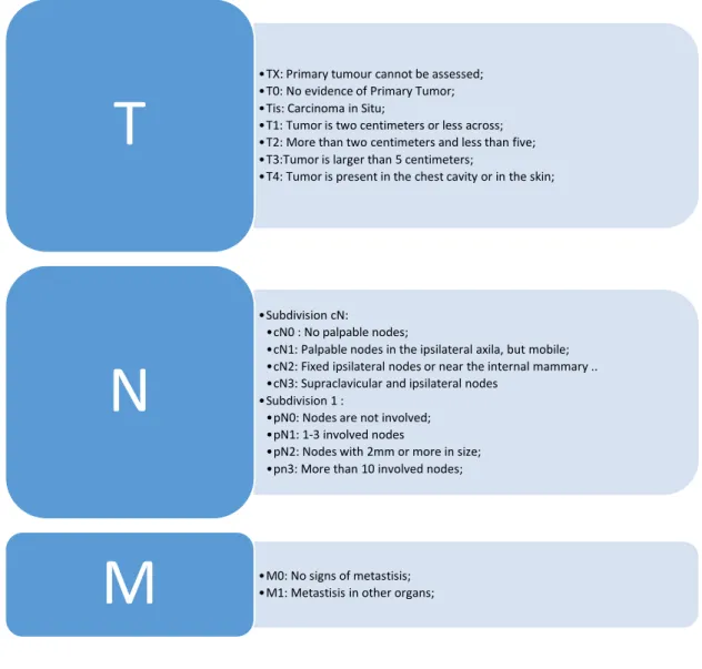
The American Joint Committee on Cancer (AJCC) TNM System is the standard system used to measure neoplasms stages.10
The TNM staging system classifies cancer by this criteria: T : Numbered 0 to 4 refers to the tumour size;
N : Numbered 0 to 3 indicates if the cancer has spread to lymph nodes; M : Numbered 0 to 1 indicates the presence of metastases;
Table 1: TNM staging system based on the article “NCC. Stages I and II Breast Cancer. National Cancer Society; 2014” 1
•TX: Primary tumour cannot be assessed; •T0: No evidence of Primary Tumor; •Tis: Carcinoma in Situ;
•T1: Tumor is two centimeters or less across; •T2: More than two centimeters and less than five; •T3:Tumor is larger than 5 centimeters;
•T4: Tumor is present in the chest cavity or in the skin;
T
•Subdivision cN:
•cN0 : No palpable nodes;
•cN1: Palpable nodes in the ipsilateral axila, but mobile; •cN2: Fixed ipsilateral nodes or near the internal mammary .. •cN3: Supraclavicular and ipsilateral nodes
•Subdivision 1 :
•pN0: Nodes are not involved; •pN1: 1-3 involved nodes
•pN2: Nodes with 2mm or more in size; •pn3: More than 10 involved nodes;
N
•M0: No signs of metastisis; •M1: Metastisis in other organs;
M
BREAST CANCER
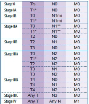
15 After the TNM staging system has been determined by histopathological and radiological diagnostics, we combine this information using stage grouping, which allows for a simple and quick understanding of the current stage of the cancer’s development 1.. The table below resumes those stages:
Table 2: Anatomic Stage/ Prognostic Groups extracted from the article “NCC. Stages I and II Breast Cancer. National Cancer Society; 2014” 1
TREATMENT
18
3. Treatment
The treatment for breast neoplasm consists of Surgery, Chemotherapy, and Radiotherapy 4. The type of treatment performed on a patient varies according to a different set of conditions that we will explore later, however, “Breast Conserving Surgery followed by Radiotherapy is widely accepted as appropriate primary local management for most women with pre-invasive and early-stage breast cancer”5. Radiotherapy uses high energy beams to destroy abnormal cells and can be applied through external Radiotherapy and braquitherapy also known as internal radiation (5, 6). Chemotherapy for cancers outside the head and neck area includes a wide spectrum of classic, novel, and targeted chemotherapeutic medications, all of which can be highly bioactive and one of the areas that suffers the most from this therapy is the oral cavity7.
Breast Neoplasm treatment can be divided into 1 : ● Noninvasive carcinomas ( LCIS and DCIS);
● Operable, loco regional invasive carcinoma associated with or without noninvasive tumors;
● Inoperable locoregional invasive carcinoma associated with or without noninvasive carcinoma;
TREATMENT
19 3.1. Surgery
Surgery is the most common form of treatment among noninvasive carcinomas and locoregional invasive carcinoma, however it is not a standalone treatment, since it's paired with other forms of treatment, radiotherapy being the most common.2
The type of surgery performed is related to the number of ganglions involved and the type of breast neoplasm diagnosed. Early onsets of breast neoplasm like DCIS and LCIS (Stage 0) are subjected to surgery as primary treatment due to their location and inability to metastasize. The most common procedures are Lumpectomy, Total Mastectomy, Surgical Axillary Staging, Lymph node surgery, Surgical Biopsy and Surgical Resection 1, 2, 11.
3.1.1. Lumpectomy
Removal of the breast lump however, being a type of Breast Conservative Surgery it does not require for the full removal of the breast but demands that a part safety margin of healthy tissue to be removed at the same time. 1, 11
3.1.2. Total Mastectomy
Removal of most or the totality of the breast leaving only the thoracic muscles. Most women prefer this form of surgery over Lumpectomy. During this cirurgical procedure, an Axillary Node Staging can be performed where the removed lymphatic tissue is analyzed to determine the stage of development of the breast neoplasm and the more lymph nodes retrieved, the better the staging of the breast neoplasm 1, 2, 11
TREATMENT
20 3.2. Radiotherapy
Radiotherapy uses a beam of high-energy rays to neutralize cancer cell, and it’s most commonly used in association with Surgery on early onsets of Breast Neoplasm due to its ability to eliminate the remaining cancer cells left behind after the surgery. 2
The patient can be subjected to two forms of Radiotherapy:
External Beam Radiation Therapy The most acknowledged form of RT, it uses a machine to emit radiation outside of the body;
Brachytherapy Also called interstitial radiation and makes use of a radioactive device placed besides the tumor bed after surgery. It may also be used as a boost for External Beam Radiation Therapy.
External Beam Radiation Therapy is administered accordingly to the patient’s previous surgery, stage of cancer and the location of the nodes diagnosed with cancer. There are three forms of External Beam Radiation Therapy that can be administered to the patient:11
TREATMENT
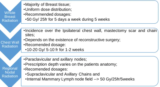
21 As described above, RT is administered accordingly to the stage of the breast In early stages of development (Stage 0) the Radiotherapy suggested by the National Comprehensive Cancer Network and Portugal´s Health Ministry is Whole Breast Radiotherapy. 11, 12
For the following stages (Stage I, IIA, IIB, T3, N1, M0) the Radiotherapy
suggested should be prescribed to the internal mammary lymph nodes that are clinically and pathologically positive for cancer cells, whole breast radiation + boost to tumor bed, infraclavicular region and supraclavicular area.11
For the more advanced stages (Stage IV, T4,M1), Radiotherapy functions
as a neo-adjuvant treatment as it is administered prior to CT to eliminate the residual cancer cells, the treatment consists of Chest Wall Radiation + Infraclavicular and Supraclavicular Regions. 11
Whole Breast Radiation
•Majority of Breast tissue; •Uniform dose distribution; •Recommended dosages:
•50 Gy/ 25fr for 5 days a week during 5 weeks
Chest Wall Radiation
•Incidence over the Ipsilateral chest wall, mastectomy scar and chain sites;
•Depends on the existence of reconstructive surgery; •Recomended dosage:
•10-20 Gy/ 5-10 fr for 1-2 weeks
Regional Nodal Radiation
•Paraclavicular and axillary nodes;
•Prescription depth varies on the patients anatomy; •Recomended dosages:
•Supraclavicular and Axillary Chains and
•Internal Mammary Lymph node field --> 50 Gy/25fr/5weeks
TREATMENT
22 Radiotherapy as greater results when associated with Chemotherapy as these treatments should be utilized when the patient is subjected to radiation in the internal mammary lymph node field in more advanced stages, but in early stages when we associate lumpectomy and whole breast radiation the recurrence rate of the breast neoplasm is reduced by 50%. 11
Oral complications caused by Radiotherapy are more frequent in irradiations to head and neck regions, however, they have little or none prevalence when the irradiated region is the chest wall, the breast, and lymph nodes.
TREATMENT
23 3.3. Chemotherapy
Chemotherapy is a neoadjuvant treatment used to make the chirurgical removal of tumors larger than 50mm easier by diminishing their size. It’s also a possible adjuvant treatment for early-stage breast neoplasm but always associated with surgery or radiotherapy. 1, 11, 13
The treatment received by the patient is dependent of certain hormonal receptors that are related to breast neoplasm, some of the main types of receptors are:
HER2; RH; Ki67;
Chemotherapy can be administered as a single agent or a chemotherapy regimen. Single agent is used on early stages and has fewer side effects, but the duration of the treatment is larger and the chemotherapy regimen that is administered to more aggressive neoplasm provides a faster treatment. However, the side effects are much more frequent and cause an increased morbidity. 2
Each of this methods to administer Chemotherapy is quantified by cycles of treatment followed by days of rest. The length of each cycle depends on the drug used and it can have the duration of fourteen, twenty-one and twenty-eight days.
TREATMENT
24 Each patient has a specific CT treatment based on the risk of recess, magnitude and benefit from applying adjuvant therapy, toxicity and comorbidity, but the two most important factors are the presence of some of the receptors mentioned above.
If three cycles of treatment of a single agent or a chemotherapy regimen do not provide a shrinkage of the tumor size, than another cycle of treatment with a different drug must be applied. In more advanced stages (Stage IV, M1), another drug is used after a single unsuccessful CT treatment.
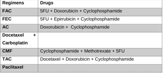
In the Serviço de Oncologia Médica do Centro Hospitalar de Gaia/Espinho some of the more common and recommended chemotherapy regimens by the Alto Comissariado da Saúde are 12:
Regimens Drugs
FAC 5FU + Doxorubicin + Cyclophosphamide FEC 5FU + Epirrubicin + Cyclophosphamide AC Doxorubicin + Cyclophosphamide Docetaxel +
Carboplatin
CMF Cyclophosphamide + Methotrexate + 5FU TAC Docetaxel + Doxorubicin + Cyclophosphamide Paclitaxel
TREATMENT
25 3.4. Hormone Therapy
Hormone therapy is another form of systemic treatment that is conditioned by the presence of hormonal receptors in the patient. Mostly used as an adjuvant therapy to reduce the risk of recurrence but also as a neoadjuvant treatment.2
Estrogen is naturally formed in the ovaries until menopause. This hormone promotes the growth of breast cancers that are hormone receptor-positive, so certain drugs are used to block the connection between estrogen and those receptors or by lowering estrogens levels in the blood stream. These drugs are:
Tamoxifen: Blocks estrogen receptors in breast neoplasm cells prevent them to signal the cell to grow and divide. However Tamoxifen can be perceived as estrogen in other parts of the body like the uterus and bone.14
Aromatase Inhibitors: These drugs are used in post-menopausal women that are receiving treatment for either early or advanced breast neoplasm. They block aromatase that is responsible for the production of estrogen in fat tissue. Those drugs are, Letrozole, Anastrozole, and Exemestane.14
ORAL COMPLICATIONS
28
4. Oral Complications
Oral complications resulting from cancer therapies are underreported, under-recognized and undertreated 8, 40 to 70 percent of cancer patients will experience oral side effects due to the malignancy or its treatment 9
. Advancements in cancer therapy caused an impact on previously documented oral complications and are leading to newly recognized side effects. Many of these new improvements have no substantiation as to their effects in the oral cavity which may help to explain some of the underreported cancer therapies and oral complications.7
The most reported oral complications related to cancers outside the head and neck region and specifically Breast Neoplasm are 4, 6-9, 14-16:
Salivary Gland Hypofunction and Xerostomia; Dysgeusia ;
Oral Mucositis; Dental Caries; Infections;
Drug-Induced osteonecrosis;
Most of these complications contribute a great deal to the decrease of quality of life increasing the heightened morbidity associated with most cancer treatments especially chemotherapy being the likely cause of most of these oral complications 7
ORAL COMPLICATIONS
29 4.1. Salivary Gland Hypofunction and Xerostomia
Saliva is a fluid released into the oral cavity that provides a crucial role in the maintenance of the teeth, digestion of food and at the same time providing antimicrobial activity preventing oral infections. It is also important for taste perception (that will be discussed later), formation of food bolus, swallowing and speech but the most important function of the saliva is that it provides calcium and phosphate for dental enamel integrity. 6
Saliva production is divided into two groups, the major salivary glands (parotid, submandibular and sublingual) that represent 90% off all saliva, the following 10% are produced by the minor salivary glands (17). However, they play an important role in maintaining optimal conditions in the oral cavity such as the lubrication of the oral mucosa and “the sensation of oral dryness may occur when a person’s normal unstimulated flow rate is reduced by about 45-50%”15.
This salivary gland hypofunction leads to the more commonly known xerostomia, which is defined as the feeling of dry mouth, which can result in the increase of oral infections and caries and also oral mucosal discomfort and pain. 15, 17
Due to these factors, patients with this symptoms refrain from their usual social activities and have a poorer general well-being.7
The decrease in saliva flow rates can be related to Chemotherapy as it decreases within a few days after the first treatment, with the possibility of returning to normal levels within one or two weeks. However Hyposalivation and xerostomia “(…) has been found during and 6 months following adjuvant moderate standard dose chemotherapy (…) for solid tumours (i.e., breast cancer) (…) it has been reported that patients having low salivary secretions before cancer treatment seem to be at higher risk of developing Hyposalivation in response to chemotherapy (…)”.15
ORAL COMPLICATIONS
30 The management of xerostomia is often made by the patient himself through the ingestion of water, however, in more severe cases, the clinician can prescribe the use of artificial saliva sprays, ice cubicles, mouth moisturizing gel and alcohol-free mouth rinses.6, 17
According to Jensen, “It is debatable if CT imposes salivary gland hypofunction, but acute changes in salivary flow rate and composition have been observed during cancer treatment. Nevertheless, from previous studies it is difficult to draw firm conclusions due to a mixture of cancer diagnoses, antineoplastic drugs and doses”15.
ORAL COMPLICATIONS
31 4.2. Dysgeusia and Smell Disorders (TSC)
Dysgeusia is a taste disorder that occurs frequently during cancer therapy. The frequency and severity of it depends largely on the type of treatment given to each patient. 6 About 50% to 75% of cancer patients will suffer from distorted or impaired ability to taste 8
Common causes of Dysgeusia are environmental factors (oral hygiene, oral infections), surgical interventions and medication. Hyposalivation can also lead to Dysgeusia as it reduces taste by limiting the delivery of tastings to the receptors.18
Chemotherapy affects taste by direct taste receptor stimulation due to secretion in saliva or via gingival crevicular fluid. This taste change may persist after treatment due to the damage to the taste buds.
It is a symptom that is sometimes overlooked by both medical practitioners and patients who fail to give it importance, but has a medium to high impact on the quality of life of the patient.
The management of Dysgeusia is not consensual. However, zinc supplementation has been shown to reduce the debilitating effects of Dysgeusia.8
ORAL COMPLICATIONS
32 4.3. Oral Mucositis
Oral mucositis has long been recognized as the most common debilitating side-effect of cancer therapies but only in recent years 16, 19-22, a pathobiological model for this disorder has been developed.23
According to Dossabhoy et al “Oral mucositis is a common adverse effect of cytotoxic chemotherapy and radiation, and it causes debilitating morbidity that may necessitate interruptions in cancer treatments” and is present in about 20% to 40% of patients receiving cytotoxic chemotherapy, a percentage that rises to 90% when combined with HTCT. 24
Mucositis is a form of mucosal barrier injury that describes a clinical condition characterized by oral erythema, ulceration and pain. Since the first proposed pathobiological method proposed by Sonis et al in 2004, more models have appeared but none of them provided a clear method to this pathology. 25
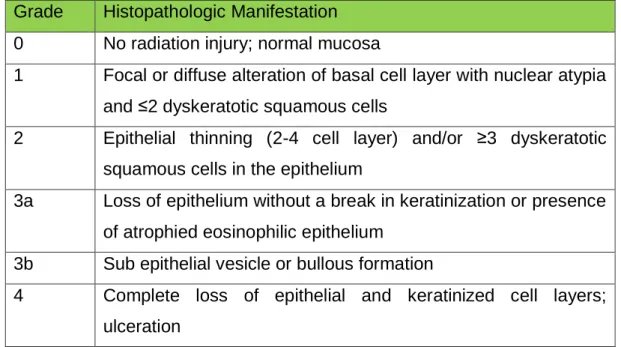
The most recent is a Histopathologic Grading Model proposed by Dossabhoy et al 24. This model categorizes the severity of disease on definite epithelial changes that begin as epithelial atypia and progress to erosion and ulceration.
ORAL COMPLICATIONS
33 The histopathologic criteria for staging oral mucositis are described in Table 1:
Grade Histopathologic Manifestation 0 No radiation injury; normal mucosa
1 Focal or diffuse alteration of basal cell layer with nuclear atypia and ≤2 dyskeratotic squamous cells
2 Epithelial thinning (2-4 cell layer) and/or ≥3 dyskeratotic squamous cells in the epithelium
3a Loss of epithelium without a break in keratinization or presence of atrophied eosinophilic epithelium
3b Sub epithelial vesicle or bullous formation
4 Complete loss of epithelial and keratinized cell layers; ulceration
Table 5: Histopathologic Grading Model proposed by Sunavala-Dossabhoy et al 24
The management of oral mucositis is based on the MASCC/ISSO Clinical Practice Guidelines for Oral Mucositis 23. These guidelines are constantly updated with the latest update being made in November 2014. According to these guidelines “(…) there was adequate positive evidence to support a suggestion in favour of using oral care protocols for the prevention of oral mucositis across all cancer treatment modalities”.19, 26
ORAL COMPLICATIONS
34 These guidelines are based on protocols that include a combination of tooth-brushing, flossing and more than one mouth rinses to maintain oral hygiene, Growth Factors and Cytokines, Anti-inflammatory agents, Antimicrobials, Coating agents, Anaesthetics, Analgesics, natural agents, Laser and other light therapy and Cryotherapy. 19
This guidelines are divided in three categories:
Recommendations in favour of an intervention; Suggestions in favour of an intervention;
Recommendations against an intervention;
Each of these categories integrates a set of clinical situations where the medical practitioner can apply these guidelines. These guidelines are available in the annex section.26
ORAL COMPLICATIONS
35 4.4. Dental Caries
Most of the dental complications diagnosed during cancer therapy can be attributed to fluctuations in the saliva's production and function.
Dental Caries are “(…) an infectious disease, post-eruptive, highly influenced by diet and it is mostly characterized by the centripetal progressive destruction of the mineralized tissues of teeth. “ (Pereira A, 1993).
This process occurs when certain forms of bacteria, present in the oral cavity, release certain acids during their metabolic process that lead to a demineralization of enamel, dentine and cement. This demineralization is thought to be mediated through decreased buffering capacity, the decreased availability of enamel substrates (Calcium, Phosphate and Fluoride), shifts in the oral flora and dietary changes. The enamel substrates mentioned before are distributed throughout the oral cavity by the saliva and are part of the remineralization process of teeth, however, if there is a deficit in the saliva production, these substrates are less frequent and are unable to contradict the demineralization lead by bacteria, which will lead to the continuous demineralization of dental tissues and to oral cavities. 6
We have shown that during cancer therapy salivary gland hypofunction and xerostomia are common oral complications, so we can expect these patients to have an increase in the number of carious lesions and according to Hong et al. the prevalence of dental caries in patients treated with chemotherapy and radiation is 21% 4
ORAL COMPLICATIONS
36 4.5. Infections
Chemotherapy and Radiotherapy negatively impact the patient’s immune system, making it more susceptible to opportunistic infections that increase morbidity and mortality among these patients
The most common infections occurring during cancer therapy are 4, 6, 8, 9, 15 : Bacterial Infections The oral floral is comprised of a wide variety
of bacteria that are controlled by the patient’s immune system, if this system is compromised, some of this bacteria may become pathogenic. Most of these infections focal points are in the oral mucosa and can be treated with a combination of penicillin and metronidazole.
Viral Infections The most frequent viral infection in the oral cavity is HSV. In most cases, this is due to stems from latent viral reactivation, however, the immunosuppression caused by chemotherapy as the main contributive factor establishes the prevalence near 40%. Oral Hairy Leucoplakia (OHL) can also be found in immunocompromised patients that are HIV negative. HSV and OHL treatment currently consists of acyclovir and valocyclovir. Candidiasis According to Lalla et al, “the prevalence of oral
fungal infections from all forms of cancer therapy was about 7.5% before treatment, 40% during treatment, and 30% after treatment”. However there is insufficient data to advise treatment of oral candidiasis with antifungal agents in patients undergoing cancer therapy, but in order to alleviate the symptoms, fluconazole can be prescribed. 19
ORAL COMPLICATIONS
37
4.6. Drug-Induced Osteonecrosis
Osteochemonecrosis is the necrosis of bone tissue induced by medication. It was first described “in 2003 by Marx RE, when he reported 36 cases of ONJ subsequent to the use of intravenous bisphosphonates (zoledronate and pamidronate) for the treatment of hypercalcemia related to multiple myeloma and metastatic breast cancer.” 27.
The American Association of Oral and Maxillofacial Surgeons (AAOMS) has since then released guidelines to help medical practitioners to detect and manage this type of oral complication. The most recent update to those guidelines was released in 2014 and it promoted the change in nomenclature from Bisphosphonate Related Osteonecrosis of the Jaw (BRONJ) to Medication Related Osteonecrosis of the Jaw (MRONJ). According to the AAOMS “The change is justified to accommodate the growing number of osteonecrosis cases involving the maxilla and mandible associated with antiresorptive (denosumab) and antiangiogenic therapies “ 28
The pathophysiology of this disease has yet to be determined, however there are some hypotheses, they are:
Inhibition of Osteoclastic Bone Resportion and Remodelling; Inflammation and Infection;
Inhibition of Angiogenesis;
ORAL COMPLICATIONS
38 4.6.1. Inhibition of Osteoclastic Bone Resportion and Remodelling
Bisphosphonates and antiresorptive drugs (ex. Denosumab), inhibit osteoclast differentiation and increase apoptosis, this causes a reduction in bone resorption and remodelling. This differentiation plays an important role in the healing and remodelling of skeletal sites, however, this form of osteonecrosis is more frequent in the jaws. This is due to the site’s increased remodelling rate, which makes it more vulnerable to side effects of Bisphosphonates.29-31
4.6.2. Inflammation and Infection
Inflammation and infections have always been an important component of Osteonecrosis of the Jaw, with some species of bacteria being more predominant, especially Actinomyces, as some studies shown this form of bacteria in necrotic bone removed from patients with Osteonecrosis of the Jaw.
The presence of this form of bacteria and others “(…) prompted studies to evaluate the possibility of a complex biofilm on exposed bone. These studies have identified bacteria in combination with fungi and viruses (…)” 28
4.6.3. Inhibition of Angiogenesis
Angiogenesis is the process that is responsible for the formation of new blood vessels, this process allows a tumour to grow and invade other regions of the human body. This takes results from the binding of Vascular Endothelial Growth Factor (VEGF) to receptors on endothelial cells.28
Some drugs like zoledronic acid have been shown to decrease angiogenesis, this fact associated with the very definition of osteonecrosis (interruption in vascular supply or avascular necrosis) makes the inhibition of angiogenesis one of the most promising hypotheses to ONJ pathophysiology.28, 29, 32
ORAL COMPLICATIONS
39 4.6.4. Soft Tissue Toxicity and Immune Dysfunction
Bisphosphonates primarily target osteoclasts and bind to the hydroxyapatite in bone tissue, however, there have been reports of soft tissue toxicity. A variety of cells has exhibited signs of increased apoptosis or decreased proliferation after being exposed to BPs in vitro.28
Other possible explanation is Immune Dysfunction. During animal tests, Osteonecrosis of the Jaw could only be induced when Bisphosphonates were combined with steroids or chemotherapy.28
Bisphosphonates are only used during the treatment of breast cancer in specific situations such as bone metastases. According to the AAOMS, the risk of developing Osteonecrosis of the Jaw during chemotherapy ranges from 0 to 0.019% or 1.9 cases per 10,000 patients with cancer but when the patient is exposed to Bisphosphonates this risk can reach 6.7%. When Breast Neoplasm is considered a study by Brufsky et al, shown that 3,8% of patients with Breast Neoplasm developed Osteonecrosis of the Jaw after long term treatment.28
AIM
42
5. Aim
The aim of this paper was to quantify and analyse oral complications that are present during cancer therapy applied to patients with Breast Neoplasm and ascertain if these patients have knowledge of this type of complications and if they are receiving treatment for them. This paper continues the work developed by a previous dissertation paper made in this University in 2010 that quantified Oral Complications during cancer therapy. From the statistical data referred previously we know that breast neoplasm makes up most of the cancer´s being treated in Portugal.
This aim was addressed by:
Conducting clinical examinations on each patients during their treatment at the Serviço de Oncologia Médica do Centro Hospitalar de Vila Nova de Gaia/Espinho;
Each patient signed an informed consent where they agreed to share their medical records, personal data and submit to an oral examination but their identities are confidential.
MATERIALS AND METHODS
45
6. Materials and Methods
1. Structure of the Study
This study consisted of an observational study. A questionnaire and an oral examination were performed to the patients being submitted to chemotherapy at Hospital Santos Silva (CHVNG/E).
An informed consent declaration was made to ensure the patient's data confidentiality and their rights, this declaration was presented to the patient at the time of the enquiry. An online questionnaire was used to gather the data, this questionnaire is available at Appendix B
The sample for this study was 32 patients observed between the 5th of June and the 17th of July.
Statistical Treatment
SPPSS 20 was used to make the necessary statistical analyses for this study. A significance level of α=0.05.
The variables in the study were:
Variables Type Scale Observations
Age Quantifiable Nominal Organized by
classes
Gender Qualitative Nominal Female = 0
Male = 1 Smoking Habits Qualitative Nominal Yes = 0
No = 1 Alcoholic Habits Qualitative Nominal Yes = 0
No = 1 Beginning of
Treatment
Qualitative Nominal Organized by classes Type of Tumour Qualitative Nominal 6 types were
taken into considerations Tumoral Stage Qualitative Nominal 9 stages were
taken into considerations Biological Markers Qualitative Nominal Multiple
Response with dichotomy Lymph node
involvement
Qualitative Nominal Yes = 0 No = 1
MATERIALS AND METHODS
46 Type of Treatment Qualitative Nominal 3 types
Radiotherapy Regimens
Qualitative Nominal 6 regimens evaluated as dichotomy Chemotherapy
Regimens
Qualitative Nominal 8 regimens evaluated as dichotomy Hormonal Therapy Qualitative Nominal Tamoxifen =0
AI`s = 1 Prosthodontics Qualitative Nominal 3 devices
evaluated as dichotomy
Caries Qualitative Nominal Yes = 0
No = 1 Oral Mucositis Qualitative Nominal Yes = 0
No = 1 Management of Oral
Mucositis
Qualitative Nominal 4 types of
management as dichotomy Osteochemonecrosis Qualitative Nominal Yes = 0
No = 1 Taste Changes
(Dysgeusia)
Qualitative Nominal 4 changes evaluated as dichotomy Smell Changes Qualitative Nominal 4 changes
evaluated as dichotomy
Medication Qualitative Nominal 5 changes
evaluated as dichotomy
Xerostomia Qualitative Nominal Yes = 0
No = 1 Xerostomia
Management
Qualitative Nominal 5 types of
management as dichotomy
RESULTS
49
7. Results
The present study was conducted in patients during their Chemotherapy sessions in Santos Silva Hospital – number of cases 32 patients.
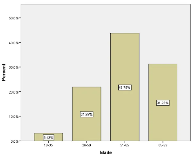
The mean age of patients which participated in the questionnaire was 57,75 years old ± 9,762 years, the youngest was 33 years old and the oldest 74 years old (Table 6). To distribute the number of cases for age group, we may find the most frequent age group, 43,75% (n=14), which is the group with ages between 51 and 75 years (graphic 1).
Statistics
N Minimum Maximum Mean Std.
Deviation
Kurtosis
Statistic Statistic Statistic Statistic Statistic Statistic Std. Error
Ages 32 40 74 57.75 9.762 -1.032 .809
Valid N (listwise) 32
Table 6: Descriptive analysis the of participants age (n=32)
RESULTS
50 In this study, we consulted the medical records of the patients to determine the beginning of their treatment. We separated them in three categories: less than a year, less than three year and more than five years. 75% (n=24) started the treatment less than a year prior to this study and 12.5% (n=4) had started it in less than three and five years.
Frequency Percent Valid < 1 year 24 75.0 < 3 years 4 12.5 < 5 years 4 12.5 Total 32 100.0
Table 7: Beginning of Treatment
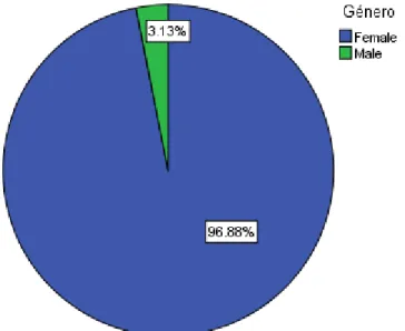
From all the participants 96, 88% (n=31) are females and 3, 13% (n=1) are male (Graphic 2).
Graphic 2: Distribution by gender.
The Ductal Invasive Carcinoma and the Lobular Invasive Carcinoma are the two most frequent types of tumours. (Table 8).
RESULTS
51
N Percent
in Situ Lobular Carcinoma 3 9.4
in Situ Ductal Carcinoma 4 12.5
Invasive Carcinoma Ductal 9 28.1
Invasive Carcinoma Lobular 9 28.1
Inflammatory Tumour 1 3.1
Other 6 18.8
Total 32 100.0
Table 8: Types of Tumour observed in the study
The CHVNG/E gives to patient’s treatment to all stages of Breast Cancer. As demonstrated 25, 00% (n=8) of the patients have Stage IIIA tumors and only 6, 25% (n=2) showed IA Stage tumour.
RESULTS
52 The patients at the CHVNG/E were being treated with Chemotherapy and Hormonal Therapy, however some have been submitted to Radiotherapy. This form of treatment isn’t administered at this facility. All of the patients (n=32) were submitted to Chemotherapy. 18% (n=6) were submitted to Radiotherapy and only 9.4% (n=3) were submitted to Hormonal Therapy
Responses Percent of Cases N Percent Patients submitted to Chemotherapy 32 78.0% 100.0% Radiotherapy 6 14.6% 18.8% Hormonal Therapy 3 7.3% 9.4% Total 41 100.0% 128.1%
Table 9: Type of Treatment used
N Percent Gangli on Yes 22 68.8 No 10 31.3 Total 32 100.0
Table 10: Ganglion Involvement.
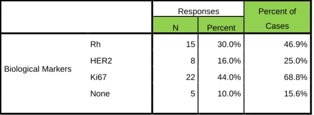
Table 11: Incidence of the Biological Markers observed.
Responses Percent of Cases N Percent Biological Markers Rh 15 30.0% 46.9% HER2 8 16.0% 25.0% Ki67 22 44.0% 68.8% None 5 10.0% 15.6% .
RESULTS
53 According to the guidelines made by the Portuguese Ministry of Health (12), there are some Therapeutic Regimens that each Hospital chooses to employ. In regards to Chemotherapy (table 12), Radiotherapy (table 13) and Hormonal Therapy (table 14) the CHVNG/E used this Therapeutic Regimens for the treatment of Breast Neoplasm.
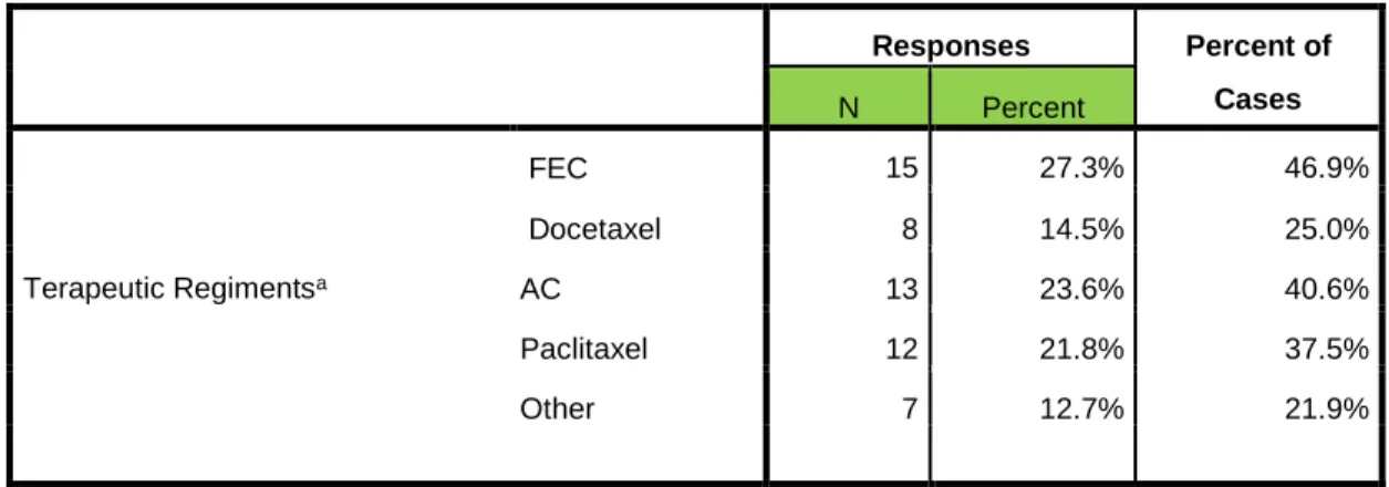
Responses Percent of Cases N Percent Terapeutic Regimentsa FEC 15 27.3% 46.9% Docetaxel 8 14.5% 25.0% AC 13 23.6% 40.6% Paclitaxel 12 21.8% 37.5% Other 7 12.7% 21.9%
Table 12: Distribution of the therapeutic regiment (n=32)
Responses Percent of Cases N Percent Submitted to 50Gy/ 25fr/ 5 weeks 5 15.6% 15.6% 45-50Gy/ 35fr / 5 weeks 1 3.1% 3.1% No Radiotherapy 26 81.3% 81.3% Total 32 100.0% 100.0%
Table 13: Radiotherapy Regimens
N Percent of Cases HT Tamoxifen AI`s 2 0 6.3 0 N/R 30 93.8 Total 32 100.0
RESULTS
54 According to the results obtained, only 6.3 %( n=2) didn’t shown any signs of Oral Complications. In table 8, the frequency of each oral complications surveyed is demonstrated in the remaining 93.7% (n=30).
Responses Percent of Cases N Percent Oral Complications Caries 20 20.4% 62.5% Signs of Mucositis 13 13.3% 40.6% Dysgeusia 26 26.5% 81.3% Smell Disorders 21 21.4% 65.6% Xerostomia 18 18.4% 56.3% Total 98 100.0% 306.3%
Table 15: Oral Complications among the patients
In the following tables, the results for crosstabs of some of the oral complications are presented, in order to observe possible correlations between them. Caries Total Yes No Xerostomia Yes 11 7 18 No 9 5 14 Total 20 12 32
RESULTS
55 In this table, we explore the relation between the tumoral breast neoplasm stages and the therapeutic regimen used.
Chemotherapy Regimens Total
FEC Docetaxel AC Paclitaxe
l Other Tumoral Stage Stage IA 0 0 1 1 0 2 Stage IB 5 3 0 0 1 5 Stage IIA 1 1 3 2 1 4 Stage IIB 0 0 3 2 0 3 Stage IIIA 3 2 4 4 2 8 Stage IIIB 2 0 0 0 0 2 Stage IIIC 2 0 1 2 1 3 Stage IV 2 2 1 1 2 5 Total 15 8 13 12 7 32
Table 17: Chemotherapy Regimens used according to the tumoral stages
The following table shows a cross tabulation of the oral complications associated with each chemotherapy regimen
Chemotherapy Regimens
FEC Docetaxel AC Paclitaxel Other
Oral Complications Caries 10 5 9 10 2 Dysgeusia 11 5 11 10 5 Smell Disorders 6 2 11 12 5 Xerostomia 7 3 9 7 5 Mucositis 5 3 7 8 2
RESULTS
56 In this graphic, the types of Mucositis encountered in this study are shown:
Graphic 4: Types of Oral Mucositis Observed
This table below demonstrates the presence of the several types of Dysgeusia (Table 19) and Smell Disorders (Table 20) observed in this study.
Responses Percent of Cases N Percent Dysgeusia Salty 14 24.6% 43.8% Sour 16 28.1% 50.0% Sweet 12 21.1% 37.5% Bittersweet 7 12.3% 21.9% No Changes 8 14.0% 25.0%
RESULTS 57 Responses Percent of Cases N Percent Smell Disorders Perfume 9 20.0% 28.1% Body Scent 14 31.1% 43.8% Cooking 5 11.1% 15.6% Hospital Scent 6 13.3% 18.8% No Changes 11 24.4% 34.4%
Table 20: Forms of Smell Disorders observed
These were the most commonly prescribed medications to the patients in this study: Responses Percent of Cases N Percent Medication Anti-depressants 10 19.6% 31.3% Anti-Histaminic 6 11.8% 18.8% Anti-Psychotics 8 15.7% 25.0% Analgesics 3 5.9% 9.4% Other 19 37.3% 59.4% No Medication 5 9.8% 15.6% Total 51 100.0% 159.4%
RESULTS
58 This table shows how the patients who complaint of Xerostomia controlled it:
Responses Percent of Cases N Percent Xerostomia Management No Xerostomia 14 41.2% 43.8% Ice Sickle 1 2.9% 3.1% Spray 1 2.9% 3.1% Gel 1 2.9% 3.1% Mouthwash 1 2.9% 3.1% Water 16 47.1% 50.0% Total 34 100.0% 106.3%
DISCUSSION
61
8. Discussion
A sample of 32 patients was obtained from the CHVNG/E after the personal interviews that took place between June and July 2015 in Vila Nova de Gaia, Portugal.
The mean age of the participant patients was 57,75 years old ± 9,762 years, the youngest was 33 years old and the oldest 74 years old (Table 6), and the most frequent age group was the 51-65 with a percentage of 43.75% . This data isn’t consistent with the projections of the Instituto Português de Oncologia (IPO) for the Northern Region of Portugal that estimated the percentage of breast neoplasm for a similar group age of 25.30%33. This difference can be explained by the low number of participant in this study and for the fact that this report isn’t circumscribed to the Porto District but as stated to all Northern Districts of Portugal which can influence the difference observed.
Studies made have shown that the incidence of breast neoplasm in 2015 is expected to be in the 129.5 for every 100.000 persons3
Relatively to gender, out of the 32 patients, 96.88% (n=31) were women and 3.13% (n=1) were men. The prevalence of a male breast neoplasm in Europe is 1 in 100 000´34, being uncommon it was unexpected to encounter this patient, however this can be attributed to the fact that this facility (CHVNG/E) is one of the biggest health facilities in the Northern Region of Portugal attending 490.954 people in 201435, which means that the probability of finding a male breast neoplasm patient was possible but unlikely.
The types of breast neoplasm observed are described in Table 8, the most common forms of breast neoplasm are the Invasive forms ( Lobular and Ductal) both with the same percentage 28.1% ( n=9) . This percentage is higher than that observed in the United States where the prevalence for the Invasive Lobular Carcinoma is 10% and where the Invasive Ductal Carcinoma represents 80% of all forms of invasive neoplasm (in this sample that percentage is reduced to 50%)2 ; this numbers differ maybe due to
DISCUSSION
62 demographical reasons and different social habits. 3.1% (n=1) of those neoplasm was an Inflammatory Tumour, which is consistent with the prevalence observed in other studies (1-3%).
Breast Neoplasm Stage was also registered in this study, as shown by Graphic 3, the most prominent stage found was the IIIA with 25% (n=8) of all patients followed by Stage IB and Stage IV both representing 15.63% (n=5). The data collected shows different results from a similar study conducted in Great Britain3 where the most frequent stages were Stage I (37.0%) and II (33.4%), as for Stage III (8.5%) and IV (5.6%) they were less observed. This discrepancy may be attributed to differences in screening methods and quality of public health care providers, however in Portugal there has been an improvement in this better and the public has become more aware of this condition.
All of the patients interviewed for this study were submitted to chemotherapy, however 65, 63% (n=21) were submitted exclusively to chemotherapy as 18, 8% (n=6) were also submitted to Radiotherapy and 9.4% (n=3) to Hormonal Therapy. Although Radiotherapy is widely used in the treatment of Breast Neoplasm, the reduced number of patients being submitted to it in this sample can be explained by the fact that such treatments aren´t performed at the CHVNG/E but in the IPO, so patients come to this hospital to receive chemotherapy and after the end of their cycle they are sent to the IPO to continue treatment.
Chemotherapy treatment is based in many factors such as the neoplasm stage, ganglion involvement and the presence of certain biological markers, to try to understand this process of selection, those factors were evaluated in the questionnaire. Ganglion involvement was present in 68.8% (n=22) of the patients and the tumoral related biological markers the most frequent were present in 84.38% (n=27) with the presence of HER2 (25%) and Ki67 (68.5%). Rh was included in this group to its presence in the clinical records of the patients. HER2 is one of the most important markers, has its presence represents the presence of invasive cancer, which makes it very important for a
DISCUSSION
63 correct diagnosis, “In breast cancer, immunohistochemical assessment of the proportion of cells staining for the nuclear antigen Ki67 has become the most widely used method for comparing proliferation between tumour samples” 36making it essential for a correct diagnosis.
Based on the factors mentioned above, there were several therapeutic regimens used in CHVNG/E, according to Table 12, the most used regimen were FEC with 46.9% (n=15), AC with 40.6% (n=13) and Paclitaxel with 37.5% (n=12). The FEC regimen is one of the most used in the treatment of Breast Neoplasm7, 15, 18, and according to some studies is associated with oral complication during chemotherapy treatment. Research about the frequency of use of these therapeutic regimens is almost none, it is our recommendation that this subject should be more investigated. Few patients were submitted to radiotherapy, whoever among these patients (n=6), 83,3% (n=5) were submitted to 50Gy/25fr for 5 weeks, this preference might be explained by the need for a quick and effective destruction of cancer cells in more aggressive forms of neoplasm. As for Hormonal Therapy it was the less often form of treatment (n=2) and in both cases Tamoxifen was used. In table 18 we can see the use of each therapeutic regimen according to breast neoplasm stage, FEC was used with Stage IB in 15.65% (n=5) followed by AC and Paclitaxel with 12.5% (n=4).
The main objective of this dissertation was to assess the presence of oral complications during the treatment phase for the Breast Neoplasm and some unexpected values were found and some were expected. In table 19 we can observe the presence of those oral complications in relation to the Therapeutic Regimens. The Regimens more associated with oral complications are AC and Paclitaxel with 47 complications recorder for each of them.
Xerostomia is one of the most well-known oral complications in cancer therapy, however it´s primarily associated with poor oral hygiene, medication and certain diseases. Salivary Gland Hypofunction couldn´t be determined for it is subject to a laboratorial examination of the saliva and that wasn´t done in this study, however the empirical manifestation of it, Xerostomia, was observed in 56.3% (n=18), Jensen15 explored this subject in great extent and observed that 64% of his patients complained of xerostomia, this numbers are slightly maybe
DISCUSSION
64 due to the fact that his study lasted for six months, we can speculate that if this thesis made an observational study of the same duration perhaps the values could be more similar. For the patients that complained of xerostomia we asked them how did they eased their symptoms, 50% (n=16) chose water and didn’t report those symptoms to their doctors or nurses, there was only one case where the patient was prescribed the Spray as well as the Gel and the Ice Sickle by the doctors at the CHVNG/E. This raises an important topic, the lack of proper attention to the debilitating condition that is xerostomia in Portuguese Hospital, according to the nurses at the Serviço de Oncologia Médica do CHVNG/E, they are aware that this forms of management exist, however few doctors prescribe them for most of the patients often choose to conceal their condition.
Taste and Smell Changes (Dysgeusia and Smell Disorders) were the most frequent oral complication detected in the questionnaires, 81.3% (n=26) for Dysgeusia and Smell Disorders account for 65.6% (n=21). This results are consistent to those observed by Bernhardson 18 that stated that in line with other studies in which 46-77% of patients undergoing chemotherapy reported TCs and 35-87% reported SCs. The results of our questionnaire are similar to these conclusions except for the higher number of Dysgeusia cases (TCs), this might be attributed to the use of different therapeutic regimens for this study abridged all types of cancer and also to demographic factors (Bernhardson conducted the study in Sweden). In this study we also evaluated which smells and tastes were lacking in the patients. As observed in the Tables 20 and 21 several tastes and smells were evaluated; in Dysgeusia the most frequent change was in sourly foods with 50% (n=16) followed by Salty and Sweet foods, 43.8% (n=14) and 37.5% respectively; in Smell Disorders the most reported change was with Body Scent 43.8% (n=14) followed by Perfume and Hospital Scent, 34.8% (n=14) and 18.8%(n=6) respectively. Using the study by Bernhardson as a comparison we can detect some differences, in his study the most reported Taste Change was Salty (41%) in our study it was Sour food but with only two cases more than Salty, this could be attributed to differences in gastronomical habits of each country, as for Smell Changes Perfume and Cooking were the most common but in our study this were the least reported,