Faculdade de Ciências
Departamento de Biologia Vegetal
Role of diet-derived retinoids in natural intraepithelial
lymphocytes
Bruno Miguel Belchior Raposo
Dissertação em Biologia Molecular e Genética
Dissertação orientada pela Doutora Manuela Ferreira e pela Professora
Doutora Margarida Telhada
2
Resumo
Uma grande maioria das células do sistema imunitário reside nas mucosas, onde se distribuem por diversas estruturas linfóides ou se estabelecem em contacto directo com o epitélio, como é o caso de linfócitos intraepiteliais (IEL). Os IEL são células T que constituem a primeira linha celular presente na mucosa, desencadeando respostas contra patogéneos, e promovendo a reparação e manutenção do epitélio, enquanto convivem com organismos comensais.
As mucosas epiteliais beneficiam da presença de uma população abundante de células citolíticas, os IEL ‘naturais’, CD8αβ- e CD4- (DN), que podem ser TCRαβ+ ou
TCRγδ+ e que se formam a partir de precursores pré-determinados no timo. A grande
maioria destas células expressa o homodímero CD8αα na superfície. O epitélio possui ainda IEL ‘induzidos’ que se formam a partir de células T convencionais por activação na periferia na presença de antigénios exógenos.
Os IEL naturais desenvolvem-se principalmente num período perinatal, colonizando o epitélio antes do nascimento, sendo estes as primeiras células T a estabelecerem-se no intestino. Deste modo, os IEL naturais desempenham um papel fundamental na imunidade de recém-nascidos, uma área da Saúde Pública de extrema importância mas ainda pouco explorada.
Os IEL naturais iniciam um programa transcripcional no timo caracterizado pela aquisição de moléculas típicas de células Natural Killer (NK) e por um reportório de TCRs auto-reactivo. As células precursoras de IEL naturais podem também adquirir receptores específicos do epitélio intestinal e migrar directamente para a mucosa entérica. Este programa genético é iniciado após o encontro com auto-antigénios no timo, no entanto, são ainda desconhecidos os mecanismos moleculares que regulam e determinam a geração de IEL naturais no timo assim como as suas funções específicas.
Nós e outros autores mostrámos anteriormente que componentes da dieta são críticos para a regulação de múltiplas funções imunitárias. Nomeadamente, o nosso laboratório demonstrou que os níveis de vitamina A na dieta materna têm um impacto na formação dos órgãos linfóides determinando a performance do sistema imunitário da descendência. O metabolito da vitamina A, ácido retinóico (RA), é um componente obtido através da dieta, e tem um papel essencial no sistema imunitário. O RA está envolvido na regulação de diversas respostas imunitárias, produção de células T reguladoras (Treg), na produção de IgA, sendo também fundamental para o tropismo
3 de certas células T e B para o intestino. O RA liga-se a receptores no interior das células, RARs e RXRs, funcionando como factores de transcrição que se ligam a sequências específicas no DNA (RAREs) regulando assim a expressão genética. No entanto, o papel do RA no desenvolvimento e função dos IEL naturais é ainda desconhecido.
Neste trabalho, o nosso objectivo foi determinar o papel do RA no desenvolvimento e função dos IEL naturais na defesa imunitária intestinal. Desta forma investigámos 3 pontos principais:
1. Papel do RA no desenvolvimento de IEL naturais;
2. Potenciais genes alvo do RA nas IEL naturais e mecanismo molecular; 3. Papel do RA na função de IEL naturais na defesa imunitária entérica. 1. Papel do RA no desenvolvimento de IEL naturais.
Investigámos o impacto da modulação dos sinais de RA no desenvolvimento e estabelecimento dos IEL naturais no epitélio intestinal. Para isso, recorremos a uma linha de ratinho geneticamente modificada que apresenta uma forma truncada no aminoácido 403 do receptor RARα introduzida no locus ROSA-26 com um codão STOP flanqueado por locais loxP (Rara403). Esta forma funciona como dominante negativo inibindo todos os RARs endógenos. Estes ratinhos Rara403 foram cruzados com distintas linhas Cre, nomeadamente CD2Cre e CD4Cre para induzir a expressão dominante negativa em diversas fases de desenvolvimento dos IEL naturais.
2. Potenciais genes alvo do RA nas IEL naturais e mecanismo molecular.
Para identificar possíveis eixos de sinalização que liguem RA aos mecanismos sinalizadores de IEL naturais, examinámos primeiramente a expressão da maquinaria de sinalização do RA, incluindo RARs e RXRs, a fim de determinar como os precursores e IEL naturais processam os sinais de retinóides. Foi pesquisado também o papel de sinais de RA como potenciais mecanismos moleculares de regulação do desenvolvimento dos IEL naturais. Precursores e IEL naturais estimulados na presença ou ausência de RA ou inibidores do mesmo foram purificados e a expressão de genes relevantes para os IEL naturais foi analisada.
4 3. Papel do RA na função de IEL naturais na defesa imunitária entérica.
Finalmente explorámos o papel do RA na função de IEL naturais na defesa contra parasitas entéricos. Os ratinhos CD2CreRara403 foram infectados com o parasita entérico natural, Eimeria vermiformis e o impacto na defesa imunitária do intestino foi examinada.
A análise de ratinhos CD2CreRara403 e CD4CreRara403 mostrou que a ausência de sinalização por RA nas populações de IEL naturais resulta numa redução drástica das populações IEL naturais TCRαβ+ e TCRγδ+ no epitélio intestinal e na alteração do seu
fenótipo activado.
Estes resultados demonstram que a sinalização pelo RA tem um papel indispensável no estabelecimento do compartimento de IEL naturais no intestino.
Os nossos resultados indicam que a manutenção, sobrevivência e expansão dos IEL naturais na periferia não são regulados por sinais de RA.
A análise do timo de ratinhos CD2CreRara403 e CD4CreRara403 demonstrou que as populações de IEL naturais precursores não estavam alteradas.
A transferência intravenosa de precursores tímicos DNTCRαβ+ de CD2CreRara403 para ratinhos imunocomprometidos demonstrou porém que os precursores tímicos de IEL naturais com disrupção da sinalização de RA não têm capacidade de colonizar especificamente o epitélio intestinal, distribuindo-se em oposição inespecificamente por outros órgãos periféricos.
Estes resultados indicam que o programa genético de precursores tímicos que carecem de sinais de RA está efectivamente alterado, sugerindo um papel fundamental dos retinóides em gerar sinais instrutivos para a programação e estabelecimento de IEL naturais intestinais.
A análise de ratinhos CD2CreRara403 e CD4CreRara403 mostrou que os níveis expressão de CCR9 e α4β7 estão significativamente reduzidos em precursores tímicos de IEL naturais, indicando que os sinais de RA são críticos para a expressão de receptores de migração para o epitélio intestinal em precursores tímicos de IEL naturais.
Em conformidade, estudos in vitro demonstraram que os precursores tímicos DNTCRαβ+ estimulados com RA apresentam maiores níveis de expressão de itga4 e
5 indicam que sinais de RA regulam a expressão dos marcadores de residência do intestino.
Notavelmente, a análise de ratinhos CD2CreRara403 infectados com Eimeria vermiformis, um parasita natural entérico, apresentaram uma maior perda de peso e notável dificuldade em recuperar. Estes dados sugerem que a ausência de sinalização por RA em IEL naturais compromete a defesa imunitária da mucosa intestinal contra parasitas entéricos.
Colectivamente, os nossos resultados indicam que o RA desempenha um papel crucial instrutivo no desenvolvimento de IEL naturais, medeia etapas cruciais no estabelecimento de IEL naturais no intestino e exerce funções essenciais na protecção entérica.
Assim, estes resultados sugerem que retinoídes derivados da dieta desempenham funções fundamentais na formação do compartimento intestinal de IEL naturais e consequentemente na capacidade do organismo se defender contra patogéneos intestinais.
Estes conhecimentos podem incentivar novas abordagens clínicas e terapias antimicrobianas para combater doenças inflamatórias entéricas e doenças infecciosas que são uma das maiores preocupações para a Saúde Pública.
Palavras-chave: Linfócitos intraepiteliais naturais, ácido retinóico, mucosa intestinal,
6
Summary
Intraepithelial lymphocytes (IEL) are the first cellular line of mucosal defense against invasive pathogens. Natural IEL develop from pre-committed thymic precursors and are the first antigen-experienced T cells colonizing the gut. However the factors that shape their development and function remain largely elusive.
Retinoic acid (RA), a diet-derived micronutrient obtained from the metabolism of vitamin A, has several immune regulatory functions and we have recently shown that maternal exposure to dietary retinoids impacts in innate lymphoid cell development and has long-term immune sequels in the offspring.
In this study, we aimed to determine the role of RA signaling in the development and function of natural IEL.
Here we show that the establishment of intestinal natural IEL depends on diet-derived retinoids, which fully condition enteric immune defense.
Taking advantage of loss of function genetic models we found that the disruption of RA signaling results in a drastic ablation of intestinal natural IEL in the small intestine and alteration of their activated phenotype.
While thymic precursor cell number was intact, competitive chimeras assays showed that thymic progenitors (DNTCRαβ+) lacking RA signaling were incapable of colonizing
the intestinal epithelium. Notably, these thymocytes presented downregulation of gut-homing markers, such as α4β7. Accordingly, in vitro assays revealed that RA induced upregulation of itga4 and itgb7 expression by IEL natural thymic precursors, whereas RAR inhibitors caused their downregulation.
We also show that mice, bearing natural IELs with disrupted RA signaling, when infected with Eimeria vermiformis, a natural enteric parasite, suffer higher disease impact relative to their littermate controls, indicating breakdown of their intestinal immune barrier.
Our results indicate that diet-derived retinoids mediate critical steps in the establishment of the enteric natural IEL compartment, likely shaping precursors for the acquisition of a complete natural IEL program beforehand in the thymus and consequently, controlling intestinal immune defense.
Key-words: Natural intraepithelial lymphocytes, retinoic acid, intestinal mucosa,
7
Acknowledgments
Quero começar por deixar um agradecimento muito especial às pessoas mais importantes para mim: à minha Mãe, Avó e Irmãos por toda a confiança, carinho, força e apoio incondicional que me deram sempre, sem vocês nada disto teria sido possível. Um grande obrigado ao meu Avô, Avó Mila e Tio, pelo apoio incondicional durante todo este tempo.
Obrigado aos meus amigos, nomeadamente ao Jota e à Catarina, por terem paciência para me aturar, mesmo naqueles dias em que estou mais em baixo, e por toda a força que me transmitiram.
Obrigado Henrique por me ter permitido fazer parte do seu excelente laboratório. Um grande obrigado à Manuela, por toda a orientação, simpatia, paciência e todo o conhecimento quer teórico como prático que me transmitiu durante todo o ano.
Obrigado à Sílvia por todos os conselhos que me foi dando e por me ‘chatear’ todos os dias só porque quer o meu bem.
Obrigado ao Hélder pelas discussões futebolísticas, por toda a ajuda e pelas centenas de DNAs que me foi dando para me ‘entreter’.
Obrigado ao Diogo por estar sempre disponível para ajudar, por saber tudo e mais alguma coisa e pelo seu humor característico.
Obrigado à Rita por ser ela mesma e animar o laboratório diariamente com a sua boa disposição e energia positiva.
Obrigado ao Carlos por me ter ajudado nos primeiros passos no laboratório e me ter ensinado uma enorme quantidade de coisas.
Obrigado a todas as restantes pessoas do laboratório que me ajudaram e que promovem um bom ambiente e um dia-a-dia mais animado e produtivo.
Obrigado a todas as pessoas do biotério, da citometria de fluxo, do bioimaging e da histologia por toda a ajuda disponibilizada.
8
Table of Contents
Introduction ... 10 1. General Aspects ... 10 2. T cell development ... 11 3. Mucosal Immunity ... 11 3.1. Gut homeostasis ... 11 3.1.1. Epithelial Cells ... 12 3.1.2. Paneth Cells ... 13 3.1.3. Mucus layer ... 13 3.1.4. Antimicrobial Peptides ... 14 3.1.5. GALT ... 14 3.2. Intraepithelial lymphocytes ... 15 4. Retinoic Acid ... 18 4.1. Mechanism of RA activity ... 18 4.2. Immunologic function ... 19 Methods: ... 20 Mouse strains: ... 20 Genotyping: ... 20 Thymus analysis: ... 20In vitro RA stimulation assay: ... 20
Quantitative Real-time PCR:... 21
Intraepithelial (IEL) and lamina propria (LP) cell suspension preparation: ... 21
Competitive chimeras: ... 22
Lung analysis: ... 22
Statistics: ... 23
Results ... 24
1. RA receptors are expressed in intestinal natural IEL and in their thymic precursors. ... 24
2. RA signaling controls intestinal natural IEL compartment ... 25
3. Regulation of natural IEL by RA signals ... 27
4. RA signals are dispensable for natural IEL precursor generation. ... 29
5. RA signaling deficient IEL thymic precursors fail to colonize the gut intraepithelial compartment ... 31
6. RA controls gut-homing molecules in the thymus ... 32
9
Discussion ... 35
Annex 1 ... 38
S1. CD2Cre is expressed in all IEL and cre recombination starts at DN stage ... 38
Annex 2 ... 39
S2. CD4Cre is expressed in all IEL and cre recombination starts at DP stage ... 39
Annex 3 ... 40
S3. Induced intraepithelial lymphocytes (IEL) in lymphocyte cell-autonomous RA signaling-disrupted mice. ... 40
Annex 4 ... 41
S4. T cell development in thymus of lymphocyte cell-autonomous RA signaling-disrupted mice. ... 41
Annex 5 ... 42
Mediums: ... 42
Real-time PCR probes (Applied Biosystems): ... 42
Annex 6 ... 43
Genotyping primers: ... 43
Antibodies Mix: ... 43
References ... 44
10
Introduction
1. General Aspects
The defense of the organism against potential pathogens and microorganisms is carried out by specific immune cell types, which can respond in a rapid and protective manner.
Immune responses involve cellular and molecular defense mechanisms that protect the host in a non-specific manner (innate) or involve antigen-specific responses (adaptive). Adaptive responses improve as successive encounters with the same pathogen leads to stronger and quicker responses. The main characteristics of the adaptive response are therefore specificity and memory [1].
Immune cells are produced by a process called hematopoiesis [2]. A unique hematopoietic stem cell (HSC) has the ability to give rise to all lymphoid cells, including Natural killer (NK), T, B cells and all myeloid cells including monocytes, macrophages, granulocytes and dendritic cells [2].
Immune cells migrate from primary lymphoid organs, where they are produced, to secondary lymphoid organs (SLOs) present in specific places through the lymphatic system where they interact with regulatory cells, antigen presenting cells and foreign antigens and pathogens that may invade the body [3]. Spleen, lymph nodes and organized lymphoid tissues within mucosal surfaces, such as Peyer’s patches, tonsils, bronchial, nasal and gut-associated lymphoid tissues are integrated in this category [3]. T and B lymphocytes are the main effectors of adaptive immunity. Naïve lymphocytes with a restricted specificity to a certain antigen are extremely rare – in the order of 1 in 106 – thus initiation of primary immune responses would be very inefficient in the wide context of all peripheral body tissues. Within SLOs naïve lymphocytes interact with antigen-presenting cells (APCs) coming from the periphery, maximizing the chance to encounter their complementary epitope. After this encounter they become proliferative and activated [1].
Most immune cells reside in mucosal tissues (MALT), where they can interact with invading pathogens and foreign antigens and mount an immune response. In the intestine they reside in gut-associated lymphoid tissue (GALT) where they contribute to host defense against pathogens [4].
11
2. T cell development
The thymus is the primary lymphoid organ where occurs the T cell development. Bone marrow derived CD4-CD8- (DN) precursor cells home in the thymus where they differentiate into T cells interacting with non-hematopoietic stromal cells. DN progenitors progress through four distinct stages: CD44+ CD25- (DN1), CD44+ CD25+ (DN2) in the cortex, and transit to the subcapsular zone as CD44- CD25+ (DN3) and finally CD44- CD25- (DN4) [5-7]. At DN3 stage, the rearrangement of the T cell receptor (TCR) β,γ,δ loci is initiated [5-7]. Cells that successfully rearrange TCRγ or TCRδ, will express TCRγδ and proceed to the γδ lineage pathway [5-7]. Alternatively, cells that rearrange TCRβ, express a pre-TCR form, resulting from the association of the TCRβ with an invariant pTα chain [5-7]. These cells undergo β selection and TCRα rearrangement, thus committing to the αβ T cell lineage [5-7].
At this stage, cells expand and become double positive (DP) for the co-receptors CD4 and CD8 [5, 8]. DP thymocytes expressing a surface TCR that engages at an intermediate affinity and/or avidity for self-peptide Major Histocompatibility Complex (MHC) MHC class I or II complexes downregulate one of the co-receptors and differentiate into CD8+ or CD4+ single positive (SP) thymocytes, respectively [5, 9]. These cells repress RAG expression and migrate to the medulla where they differentiate into mature T cells that may then leave the thymus. In contrast, thymocytes that express T cell receptors with high affinity for self-ligands are negatively selected, in such way that self-reactive cells are deleted [5, 10].
Interestingly, cells with a high self-reactive TCR can also be selected by agonist selection. Examples of these cells are NK-T cells, CD8αα expressing intraepithelial lymphocytes and regulatory T cells [11-13].
3. Mucosal Immunity
Body surfaces function as epithelial barriers against harmful pathogens and simultaneously interact with beneficial symbiotic microorganisms. Mucosal homeostasis is crucial for a healthy life, and imbalances of this equilibrium can result in serious diseases or even dead.
3.1. Gut homeostasis
The intestine is constantly exposed to pathogens that may endanger life. The intestinal mucosa comprises diverse components, including the single layer of intestinal epithelial cells and the immune system, whose balanced interplay ensures gut defense and
12 homeostasis. The immune system presents diverse specialized enteric components, which guarantee immune defense. Nevertheless, intraepithelial lymphocytes (IEL) constitute the first line of cellular immune defense impeding the entrance and spreading of pathogens.
3.1.1. Epithelial Cells
Intestinal epithelial cells are the main physical barrier to the entry of pathogens in the organism. Epithelial cells are connected by specialized molecular structures such as tight junctions that avoid paracellular traffic [14]. In addition, they have in their apical side microvillar extensions that prevent bacteria adhesion and invasion [14]. Epithelial cells are in direct contact with IEL and may contact lamina propria lymphocytes. This allows epithelial cells to modulate inflammatory and immunologic responses by antigen-specific responses or innate responses. Interestingly, epithelial cell express all the machinery necessary to process and present antigens. They are also able to distinguish between pathogenic or beneficial bacteria and mount immune response or promote tolerance, respectively [15].
Epithelial cells are also equipped with several receptors that can recognize specific molecules of bacteria and trigger a response. Examples of these receptors are Toll-like receptors (TLRs), NOD-like receptors (NLRs) and G Protein-coupled receptors (GPCRs). In addition, these cells produce antimicrobial peptides, such as defensins, cathelicidins, and calprotectins that can induce the formation of pores in bacteria cell walls, leading to their death [15].
TLRs are innate pattern-recognition receptors that have the capacity to recognize products derived from microbes. Most TLRs are expressed in the cell surface, but a minority is present in endosomal compartments. Stimulation of TLRs can activate NF-kB leading to the initiation of an immune response [16]. NLRs recognize intracellular ligands and bacterial signals and are important in the development of intestinal lymphoid tissues [16]. NLRs can mount specific immune responses like Th2 responses and after sensing of pathogenic bacteria can initiate the NF-kB pathway and increase the production of inflammatory cytokines. It has been reported that sensing of bacterial signals by NLRs is essential for the development of intestinal lymphoid tissues [17-19]. GPCRs are intracellular and respond to ligands produced by bacteria like butyrate [20]. The stimulation of these receptors can start an immune response and induce differentiation of T helper 17 (Th17) cells [20].
13
3.1.2. Paneth Cells
Paneth cells (PC) are specialized types of epithelial cells located below the epithelial stem cells in the base of small-intestinal crypts. These cells are important producers of antimicrobial peptides that preserve sterility of the crypt and protect the epithelial stem cell niche [21-26]. They originate from multipotent stem cells located at the interface of the villus and the crypt [21-26]. During their maturation and differentiation PCs move downwards to the bottom of the crypt and acquire apical cytoplasmic granules that contain antimicrobial peptides that can be released into the crypt lumen [21-26]. Examples of these antimicrobial peptides are defensins, cathelicidins and lysozyme [21-26].
PCs have the ability to produce several cytokines and mediators of inflammation like TNF-α as well as to present antigens as they express CD1 in their surface [27-30]. They express Fas ligand and can induce apoptosis of surrounding lymphocytes expressing Fas in their basolateral membrane [31, 32]. They can even secrete IgA in the secretory granules to defend against invading microorganisms [33].
In addition, PCs produce human trefoil factor and epidermal growth factor (EGF) that promote cell migration and growth of these cells during their maturation and renewal [34, 35].
3.1.3. Mucus layer
The mucus layer is formed by glycoprotein-polysaccharides, called glycocalyx, that forms a non-cellular barrier at the epithelial cell surface impairing the entrance of microorganisms [4]. This barrier is continuously produced by goblet cells present in the intestinal epithelium [4]. This mucus layer is constituted by heavily glycosylated mucin proteins and other protective molecules that help in tissue restitution and repair [4]. The mucus layer is divided in two distinct layers: an inner layer, small and stratified, which is firmly adherent to epithelial cells and an outer layer, large and nonattached [36-38]. These layers are formed by a net-like polimer of Mucin (Muc) 2, the main mucin present in the intestine [36-38]. Whereas the outer layer is the location where commensal bacteria live, the inner layer does not allow bacteria to penetrate due to its high compactness [36-38].
Microbe specific molecules, such as microbe-associated molecular pattern (MAMP), recognized by specific receptors in the host, can lead to an increase of mucin production by goblet cells and so the reconstitution of the mucus layer [39].
14
3.1.4. Antimicrobial Peptides
Antimicrobial peptides (AMPs) are produced by enterocytes and Paneth cells. These molecules may interact with the membrane of bacteria, leading to displacement of lipids, alteration of the membrane structure or can even enter the cell and affect bacteria metabolism. There are several types of AMPs, including defensins, cathelicidins, among others [40].
Defensins (type α and type β) are effective against a wide range of microorganisms and act by inducing the permeabilization of the membrane. Some defensins functions as opsonins or increase adherence to epithelial surfaces [41-43].
Cathelicidins have a diverse range of antimicrobial activity and can either kill directly microorganisms through membrane permeabilization or bind the endotoxin and block the biological effect of the infection [44] .
Lysozymes are mostly effective against Gram-positive bacteria, and act by catalyzing the hydrolysis of the components of the peptidoglycan, the major constituent of bacteria cell wall, leading to their lysis [45, 46].
3.1.5. GALT
Gut-associated lymphoid tissues (GALT) are lymphoid structures associated with the gut mucosa that comprise Peyer’s patches, cryptopatches, mesenteric lymph nodes and isolated lymphoid follicles (ILFs). These structures regulate lymphocyte function and contribute to the control of inflammatory or tolerant immune responses [47].
These lymphoid structures are sites of organized lymphoid cells that develop during embryonic life, fully assembling just after birth [3]. While Peyer’s patches can form at variable number along the anti-mesenteric side of the mid-intestine, mesenteric lymph nodes always develop in the same region of the mesentery. Yet, cryptopatches are
randomly located in the intestinal lamina propria [48-50]. Mesenteric lymph nodes (mLNs) develop in vascular junctions in the mesentery being
structures surrounded by a capsule and connected to lymphatic vessels, which allow the exchange of antigens and antigen-presenting cells [51-53]. mLNs have organized areas of naïve T and B cells permitting their encounter with peripheral antigens promoting their activation [51-53].
Peyer Patches (PPs) are constituted by several B cell follicles with germinal centers, which are flanked by small T cell areas [51, 52, 54]. Specialized epithelial cells called
15 microfold cells (M cells) are present in PPs [55-58]. These cells are specialized in the uptake of bacteria and foreign antigens to the interior of the PPs where they will contact with antigen-presenting cells and then trigger humoral responses mainly by IgA production [55-58].
Cryptopatches are structures of clustered group 3 innate lymphoid cells, which normally evolve to ILF as microbiota colonizes the intestine [54, 59].
ILFs are constituted by B cells with no clear T cell zone that have a germinal center, reflecting the capacity of humoral immune activation [60]. ILFs are involved in T cell independent IgA class-switching in mice, promoting humoral responses [51, 52, 61, 62].
3.2. Intraepithelial lymphocytes
Intraepithelial lymphocytes (IEL) comprise a population of T cells found in the epithelial layer of mucosal linings, for example in the gastrointestinal tract. In the gut IEL are in direct contact with enterocytes and due to their location, these cells constitute the first line of defense against invading pathogens [63, 64]. IEL promote repair and regeneration of the epithelium through secretion of several growth factors and mediate the death of pathogens through the secretion of antimicrobial peptides, avoiding their entry and spreading [63, 64].
According to the mechanism by which IEL are activated and to the cognate antigens recognized by these cells, there are two major IEL subsets:
- Natural IEL – Natural IEL directly differentiate from pre-committed thymic precursors acquiring their activated phenotype during their development in the thymus in the presence of self-antigens. Natural IEL are either TCRαβ or TCRγδ and do not express CD4 or CD8αβ in their surface. The majority of IEL express the homodimer CD8αα, although some lymphocytes are CD8αα-
[65] .
- Induced IEL – Induced IEL are the progeny of conventional CD4+ or CD8αβ+ T cells that express the TCRαβ, MHC-class II or MHC-class I
restricted, respectively. These cells that are selected in the thymus and activated in response to non-self-antigens in the periphery, such as in mesenteric lymph nodes or Peyer’s patches [65].
16 Induced IEL are scarce early in life increasing with age in response to peripheral antigens. In contrast, natural IEL are the first type of antigen-experienced T cells to colonize the gut, where the first natural IEL appear even before birth [66].
These natural intraepithelial lymphocytes represent an exclusive T cell lineage with diverse MHC restriction that have the capacity to sense an antigen repertoire totally different from the ones of CD4+ or CD8αβ+ T cells, which can reveal an important role in protecting mucosal sites against other types of aggressions [67]. These cells activate a different transcriptional program which leads to their specific characteristics similar to innate cells [67, 68].
IEL are antigen-experienced T cells which have an effector capacity, produce cytokines or mediate death of infected targeted cells after antigen encountering and they express diverse activation markers such as the likes of CD44, PD-1, CD8αα and CD69 [69-71]. They express high levels of cytotoxic Granzymes (GzmA and GzmB are the most abundant) which can induce apoptosis after entering target cells. IELs also express FasL which that binds Fas inside cells inducing apoptosis [68, 72-75]. These lymphocytes also have potential to produce the cytokines Interferon-γ, 2, 4 and IL-17, immunoregulatory chemokines such as RANTES and molecules associated with innate cell fuctions, in particular NK cells [68, 72-75].
Development of the natural TCRαβ+ IEL occurs in the thymus, where CD4+CD8β+
double positive (DP) cells acquire the expression of the homodimer CD8αα+
, becoming CD4+CD8β+CD8αα+ triple-positive (TP) cells [11]. CD8αα expressing cells but not CD8αβ are strong ligands for the thymic leukemia antigen (TL) tetramer, a non-classic MHC class I molecule, abundantly expressed in intestinal epithelial cells [76]. These TP thymocytes undergo an alternative positive selection, called agonist selection and differentiate into double-negative TCRαβ+ (DNTCRαβ+) by downregulating the
expression of CD4, CD8αβ and CD8αα while expressing TCRαβ. DNTCRαβ+ cells are
the progenitors of the natural TCRαβ+ IEL [11, 13]. DNTCRαβ+ cells express high
levels of CD69 and PD-1, indicating high TCR signaling, that might provide survival signals to these cells and can migrate directly to the gut epithelium [11, 63, 70, 77].
17 The selective expression of chemokine receptors, chemokines and adhesion molecules have an important role in T cell homing. Important gut homing receptors expressed in lymphocytes are α4β7, CCR9 and αEβ7, which in contact with their ligands in the small intestinal epithelial cells results in their entry in the epithelium [78-80]. The integrin αEβ7 is expressed on more than 90% of intestinal IEL and plays a role in the recruitment and expansion of TCRαβ IEL [81]. This integrin promote adhesion of T cells to the intestine through binding to E-cadherin, which is expressed selectively on epithelial cells [81]. CCR9 is a chemokine receptor functionally and selectively expressed in small-intestine lymphocytes which interacts with his ligand CCL25 (TECK), constitutively expressed in small-intestine epithelial cells, leading to the recruitment of circulating lymphocytes to the intestine [82-84]. The majority of CCR9+ cells co-express α4β7 integrin, which is also involved in the migration of lymphocytes to the gut [82-84]. This integrin binds its ligand MAdCAM-1, expressed in postcapillary venules of mucosal tissues promoting lymphocyte adhesion and gut homing [84, 85] . It has been shown that the pair can influence the early induction of CD103 (αE integrin) indicating that CCL25/CCR9 can regulate lymphocyte-epithelial interactions through αEβ7 in the small intestine [86].
Figure 1: Differentiation of induced and natural IEL. In the thymus immature triple-positive thymocytes
(CD4+CD8β+CD8αα+) undergo ‘agonist’ selection and differentiate in double-negative T cell receptor αβ
cells (DNTCRαβ+
). These cells are the precursors of the natural intraepithelial lymphocytes (IEL) and acquire their phenotype during their selection with self-antigens. During their maturation in the thymus these
precursors, either TCRαβ or TCRγδ, may start expressing gut-homing markers, which allow IEL precursors
to go directly to the intestinal epithelium. On the other hand, double positive thymocytes (CD4+CD8αβ+
)
undergo conventional selection in the thymus and become single positive cells either CD4+ or CD8αβ+ that
migrate to the periphery. There in response to peripheral antigens these cells differentiate in effector cells able to migrate to the gut induced IEL compartment. APC, antigen-presenting cells; MLNs, mesenteric lymph nodes. Adapted from [63]
18
Figure 2: Molecular and cellular mechanism of action of vitamin A. Retinoid dehydrogenase enzymes (ADHs and RALDHs) metabolize the vitamin A absorbed forms (β-carotene and retinol) into the bioactive compound, retinoic acid (RA), that can be present in two forms: All-trans-RA (ATRA) and
9-cis-RA (9-9-cis-RA). RA functions by binding to retinoid X receptor (RXR) and RA receptor (RAR), whose
homodimers or heterodimers can bind DNA specific sites, RA responsive elements (RAREs) in the nucleus and regulate the expression of specific target genes. Adapted from [95].
Once in the intestinal epithelium, DNTCRαβ+ cells have a specific expression of CD122
and so need IL-15 to their maturation, differentiation and survival [11, 13, 70, 87]. In addition, it was recently shown that the development of CD8αα+ IEL is dependent on the expression of T-bet by IEL precursors [87, 88]. In fact, T-bet is required for the IL-15-dependent activation, differentiation, and expansion of IEL precursors in the periphery [87]. Also, vitamin D receptor (VDR), which acts as a transcription factor, mediates the homing of intraepithelial lymphocytes to the gut [89, 90]. Yet, the aryl hydrocarbon receptor (AhR), a ligand-dependent transcription factor whose ligand is mainly obtained through diet, is crucial for the maintenance of IEL in the intestine but not for their development [91].
4. Retinoic Acid
Many essential compounds are obtained through the diet. Vitamin A is a micronutrient that has important roles in discrete immune cells [90, 92-96]. Retinoic acid (RA) is the metabolite of vitamin A that functions by interacting with a class of proteins called nuclear receptors, which regulate the expression of specific genes after sensing of this external nutritional cue [95, 97].
4.1. Mechanism of RA activity
Vitamin A is an essential fat-soluble compound ultimately converted in the bioactive metabolite RA. RA is a small lipophilic molecule with rapid diffusing capacity usually generated by epithelial or dendritic cells [95]. RA is converted from retinol (the form of vitamin A absorbed from food sources) through two enzymatic reactions: first,
19 intracellular oxidation of retinol into retinal, achieved by alcohol dehydrogenases (ADH class I, II and III); second, this compound is transformed into retinol by retinal dehydrogenases (RALDH1, 2 and 3) [93]. This oxidative process produces two forms of RA: 9-cis-retinoic acid and all-trans-retinoic acid, being that the latter is the most abundant in tissues [93, 95, 96].
RA binds to two families of nuclear receptors: retinoic acid receptors (RARα, β and γ) and retinoid X receptors (RXRα, β and γ) [95-97]. While 9-cis-RA can bind both RARs and RXRs, all-trans-RA preferentially binds to RAR, binding to RXR only at higher concentrations [95-97]. These receptors can form RAR-RXR heterodimers or RXR-RXR homodimers and bind specific sites of DNA, namely RA responsive elements (RAREs), regulating the expression of specific target genes as ligand-induced transcription factors [95-97].
4.2. Immunologic function
Retinoic acid has several roles in the immune system. RA potentiates TGFβ induced T regulatory (Treg) generation and their conversion into T follicular helper (TFH) cells
[98-101]. RA is important for the regulation of Th1, Th2 and Th17 responses and for increasing antibody-secreting cells [95]. It was recently shown that RA has an important role in the generation of antigen-specific IgA responses in the gut and that their absence changes the microbiota and their symbiotic relationship with the host [102]. RA is also important to the initiation of lymphoid organ development by mediating the expression of CXCL13 in fetal mesenchymal cells and through control of group 3 ILC development [92] [103, 104].
In addition, RA was shown to modulate the migration and trafficking of immune cells to the intestinal mucosa. RA imprints gut-homing specificity on conventional T cells and ILCs by enhancing the expression of the gut-homing markers CCR9 and α4β7 [93, 94]. Altogether these findings indicate that RA is crucial to a healthy immune system and to balanced and effective immune responses. However, the role of RA in natural IEL development and function remains elusive.
20
Methods:
Mouse strains:
C57BL/6 mice were purchased from Charles River and C57BL/6 CD45.1 mice were obtained in-house. ROSA26-Rara403, ROSA26-eYFP, CD2Cre and CD4Cre mice were previously described [106-109, 115]. All mouse strains were bred and maintained at IMM animal facility. All procedures and experiments were performed accordingly to institutional and national guidelines.
Genotyping:
Snipped tails were digested in 500µL of Tail Lysis buffer and Proteinase K at 56oC (Annex 5). DNA was extracted by centrifugation with pre-cooled 2 volumes of isopropanol for 25 minutes at 13 300 rpm at 4oC, washing with pre-cooled ethanol 70 % and centrifuged at 13 300 rpm at 4oC for 10 minutes. DNA was ressuspended in H2O
MilliQ.
The DNA was amplified on a Veriti 96-Well Thermal Cycler (Applied Biosystems). The PCR program included an initial step of polymerase heat activation and was performed at 95 ºC during 10 minutes, followed by 35 amplification cycles consisting of 3 distinct steps (DNA denaturation – 94ºC for 30 sec, DNA-primer annealing step – 60ºC for 45 sec, and polymerase reaction elongation – 72ºC for 1 min.), and a final step of DNA extension at 72º C for 10 min. Primers sequences are detailed in the Annex 6. 1µL of DNA was added to individual wells to each 19µL of PCR buffer (Annex 5). After the PCR reaction, the amplified products were resolved in a 1.5% agarose gel containing Gel Red (Biotium).
Thymus analysis:
Mice were sacrificed and thymi were collected and ressupended in 10mL of RPMI complete. Cell number was determined using in a Neubauer chamber and the cells were stained accordingly.
In vitro RA stimulation assay:
Cells were purified by flow cytometry in ARIA III system directly to Opti-MEM medium. Cells were starved in Opti-MEM serum free medium for 12h in 96-well plates at 37oC + 5% CO2. Cell were then cultured in medium with all-trans retinoic acid (Sigma-Aldrich)
dissolved at 10mM in 100% ethanol at 100nM or DMSO (vehicle) or BMS493 (an inverse pan-RAR agonist) for 12 hours at 37oC. After the stimulation the cells were
21 transferred to RLT lysis buffer and their RNA was extracted with microRNA extraction kit of QIAGEN according to the manufacturer’s protocol.
Quantitative Real-time PCR:
Wild Type (C57Bl/6) mice were sacrificed and thymi were collected. For hematopoietic populations thymi were ressuspended, homogenized in complete RPMI and depleted for CD4+ cells depending on the populations of interest with microbeads and MACS separation columns according to the manufacturer’s protocol. For non-hematopoietic populations thymi were digested with 5mg/ml Collagenase D and 1mg/ml DNase I (Roche) for 25 minutes at 37oC. After digestion, culture medium with EDTA 5mM was added for 10 minutes on ice to avoid the formation of dimmers. Cell suspensions were depleted for CD45+ cells with microbeads and MACS separation columns according to the manufacturer’s protocol.
Cells were stained and sorted in ARIA III system, directly to 350 µL of RLT Lysis buffer. RNA extraction was done with the microRNA extraction kit of QIAGEN according to the manufacturer’s protocol. The RNA was quantified using NANODROP 2000 system. cDNA was generated using RT to cDNA kit of Applied Biosystems using a 21 Veriti 96-Well Thermal Cycler (Applied Biosystems).
The cDNA was pre-amplified using the Pre-Amp kit of Applied Biosystems with specific probes (qPCR) using a 21 Veriti 96-Well Thermal Cycler (Applied Biosystems). The resulting cDNA was diluted 1:10 in Tris-EDTA buffer solution (TE). Gene expression levels were quantified by Real-Time PCR in triplicates with Master Mix and specific probes of Applied Biosystems (Annex 5). All samples were run in a StepOne Thermal Cycler (Applied Biosystems). The data were analyzed according to the ΔΔCt method and normalized relatively to Gapdh and Hprt expression.
Intraepithelial (IEL) and lamina propria (LP) cell suspension preparation:
Mice were sacrificed and intestines were removed. Intestines were flushed with cooled PBS 1x in a Petri dish. Peyer’s patches and fat were removed, guts were open longitudinally, cut in little pieces of 1-2 cm wide, and placed inside falcons with 20mL of RPMI complete with DTT (DL-Dithiothreitol solution) 1mM.To prepare intraepithelial cell suspension, guts were vortexed for 30 seconds and incubated at 37oC for 20 minutes with agitation. Guts were washed with 10 mL of medium and filtered with a 100 µM filter. Incubation with DTT was repeated.
22 Remaining lamina propria (LP) were minced with a curvaceous tips scissors, placed inside a 100mL flask with 10 mL of complete medium and 50µL of Collagenase D and 100µL of DNase I (Roche) and digested at 37oC for 25-30 minutes. LP were washed and filtered with a 100 µM filter.
Intraepithelial and lamina propria cells were centrifuged 10 minutes at 1500 rpm at 25oC. The supernatant was discarded and the pellet ressuspend in percoll medium 40% up to 3 mL. One mL of percoll 80% was gently transferred to the percoll 40% using a pasteur’s pipette and then centrifuged for 30 minutes at 2400 rpm at 25oC. The
ring between phases was collected carefully, washed with complete medium and centrifuged for 7 minutes at 1800rpm at 4oC. The pellet was ressuspended in 300 µL of medium.
Competitive chimeras:
Thymic DNTCRαβ+ cells from 2 week old CD2CreRara403Hom, CD2CreRara403Het, WT
littermate control (Test population; Ly5.2+ cells) and C57Bl/6J WT (Competitor population; Ly5.1+ cells) mice were purified by flow cytometry (FACS). Similar numbers of test and competitor populations were injected intravenously into NSG mice. Mice were sacrificed 48 hours after transplantation and donor cells were analyzed by flow cytometry.
Lung analysis:
Mice were sacrificed and lungs were collected, minced with a curvaceous tip scissors and placed inside a 100 mL flask with 10 mL of completed medium. Lungs were digested with 5mg/ml Collagenase D and 1mg/ml DNase I (Roche) for 1 hour at 37oC with gentle agitation. Digested lungs were then filtered into a new falcon, centrifuged and pellet was ressuspended in percoll medium 40% until it reaches 3 mL. After this 1 mL of percoll 80% was gently transfered to the bottom of the tube with percoll 40% using a pasteur’s pipette. Tubes were centrifuged for 30 minutes at 2400 rpm at 25oC.
Lymphoid cells were collected and stained accordingly.
Parasite and Oocyst Enumeration:
E. vermiformis sporulated oocysts were microscopically counted using a McMaster chamber. Mice were weighted and infected with 103 oocysts in sterile water via oral gavage. At day post infection (DPI) 7 mice were weighted and single caged in playground sand. Mice were weighted and faeces were collected at daily basis.
23
Statistics:
Statistical analysis was performed using two-tailed F-test analysis of variance and two tailed Student’s t-test. A p-value <0.05 was considered statistically significant. Results were scored as * when p<0.05, ** when p<0.01, and *** when p<0.001.
For RARs and RXRs expression levels, graphs were performed using with GraphPad Prism Software.
24
Figure 3: RA receptors expressionin intestinalnatural IEL and thymic precursors. Intestinal IEL and
thymocytes were purified by flow cytometry from 6 to 8 weeks old WT mice and quantitative real time PCR was performed. Results were normalized to the expression of Gapdh and Hprt. n=3 Bars represent SEM.
R e la ti v e E x p r e s s io n S P C D 4 S P C D 8 D P T P D N T C R ß S P C D 4 S P C D 8 D P T P D N T C R ß S P C D 4 S P C D 8 D P T P D N T C R ß R e la ti v e E x p r e s s io n S P C D 4 S P C D 8 D P T P D N T C R ß S P C D 4 S P C D 8 D P T P D N T C R ß S P C D 4 S P C D 8 D P T P D N T C R ß
Rara Rarb Rarg
1,2 0 0,6 0,04 0,04 R el ati ve e xp re ss io n
A
B
Rxra Rxrb Rxrg 1,2 0 0,6 0,04 0,04 R el ati ve e xp re ss io nRara Rarb Rarg
1,2 0 0,6 R el ati ve e xp re ss io n Rxra Rxrb Rxrg 5 0 0,6 R el ati ve e xp re ss io n 0,6 T hy m us IE L C D 8 C D 4 D N T C R C D 8 C D 8 C D 4 D N T C R C D 8 C D 8 C D 4 D N T C R C D 8 C D 8 C D 4 D N T C R C D 8 C D 8 C D 4 D N T C R C D 8 C D 8 C D 4 D N T C R C D 8
Results
1. RA receptors are expressed in intestinal natural IEL and in their thymic
precursors.
Retinoic acid plays multiple regulatory functions in the immune system [93, 105]. To investigate the role of RA in natural IELs, we firstly examined the RA signaling machinery in precursors and intestinal natural IEL subsets. To this end, we purified by flow cytometry thymic precursors, intestinal natural IEL TCRαβ+ DNTCRαβ+ and
CD8αα+, as well as their counterpart conventional TCRαβ+ CD4+ and CD8αβ+ cells
from wild type (WT) mice and quantified the expression of the retinoic acid receptors (RARs) and retinoid x receptor (RXRs) by quantitative real-time PCR.
We found that thymic precursors and intestinal natural IEL present significant levels of Rara and Rxrb expression and low levels of Rarg and Rxra (Figure 3). In contrast, neither population expressed Rarb nor Rxrg. Altogether, these results indicate that intestinal natural IEL and their thymic precursors express the machinery to perceive RA signals.
25
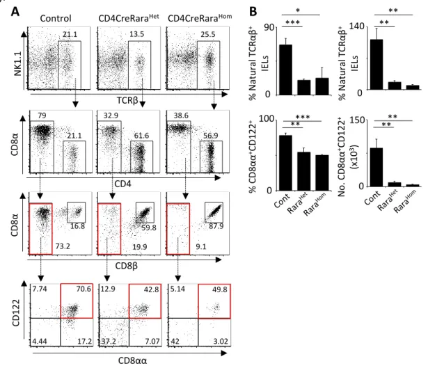
Figure 4: RA signaling controls enteric natural TCRαβ IEL in adulthood. Intraepithelial lymphocytes (IEL) were obtained from intestines of 6 to 8 weeks old CD2CreRara and their WT littermate controls. A. Representative flow cytometry (FACS) plots for DNTCRαβ+ IEL subsets B. Percentage and numbers of
natural DNTCRαβ+ and CD8αα+
CD122+ IEL subsets. Bars represent SEM. Student’s t-test. *p<0.05,
**p<0.01, and ***p<0.001. n=5 for each experimental group
CD8β CD 8α 45.9 48.4 81.5 11.7 91.2 6.98 N o . N at u ra l TCR αβ IE Ls (x10 3) 40 % N at u ra l TCR αβ IE Ls 0 40 *** TCRβ N K1.1 CD4 CD 8α
Control CD2CreRaraHet CD2CreRaraHom
19.2 35.9 46.7 47.2 28.4 10.6 50.8 68.3 88.5 * ** CD8αα CD 122 5.93 35 43.7 15.4 12.1 3.57 17.1 67.1 6.69 3.54 7.48 82.3 0 *** % CD 8α α + C D1 22 + 60 0 *** N o . CD 8α α + CD 122 + (x10 3) 12 0 ****
A
B
2. RA signaling controls intestinal natural IEL compartment
To investigate the role of RA in natural IEL development and function, we explored the impact of RA signal modulation in the generation of these cells.
To this end, we used a mouse line with a truncated form of RARα knocked into the ROSA-26 locus preceded by a STOP signal flanked by two loxP sites (Rara403) [106, 107]. We crossed Rara403 mice to distinct Cre lines, namely CD2Cre and CD4Cre, which induce Rara403 expression at different developmental stages of natural IEL. Thymic precursors and intestinal natural IEL were analyzed by flow cytometry.
In CD2CreRara403 mice, Cre recombinase is expressed under the control of human Cd2 regulatory elements [108]. Analysis of CD2CreR26-eYFP reporter mice revealed that Cre is active after DN1 stage and that virtually all thymic precursors and natural IEL have undergone Cre recombination (Annex 1).
26
A
B
CD8β CD 8α 73.2 16.8 59.8 19.9 87.9 9.1 TCRβ N K1.1 CD4 CD 8αControl CD4CreRaraHet CD4CreRaraHom
21.1 13.5 25.5 79 32.9 38.6 21.1 61.6 56.9 CD 122 7.74 70.6 17.2 4.44 12.9 42.8 7.07 37.2 5.14 49.8 3.02 42 0 140 % N at u ra l T CR αβ + IE Ls 0 90 ** ** * *** % CD 8α α +CD 122 + N o . CD 8α α +CD 122 + (x10 3) 150 0 100 **** 0 ***** % N at u ra l T CR αβ + IE Ls CD8αα
Analysis of CD2CreRara403 mice revealed a profound reduction of natural IEL TCRαβ+
either CD8αα+ or DNTCRαβ+. In addition, TCRαβ+ CD8αα+CD122+ cells were also severely reduced in these mice, indicating a putative impairment of IEL activation in CD2CreRara403 mice (Figure 4).
In order to more specifically define the stage at which RA operates in the IEL lineage we bred Rara403 mice to the CD4Cre line, thus ensuring Cre activity exclusively from the DP stage onwards [109] as confirmed by the analysis of CD4CreR26-eYFP (Annex 2).
Analysis of CD4CreRara403 mice fully confirmed an abrupt reduction of TCRαβ+
natural IEL cell numbers, while conventional IEL cell numbers were unperturbed (Figure 5 and Annex 3). A decrease of CD8αα+CD122+ was also observed in these mice, indicating a minute number of IELs with an activated phenotype. Thus, we conclude that lymphocyte cell-autonomous RA signaling is required for the establishment of a natural IEL compartment.
Figure 5: RA signaling controls enteric natural TCRαβ IEL in adulthood. Intraepithelial lymphocytes (IEL) were obtained from guts of 6 to 8 weeks old CD4CreRara and their WT littermate controls. A.
Representative flow cytometry (FACS) plots for IELs NK1.1-TCRαβ+ IEL subsets B. Percentage and
numbers of natural TCRαβ IEL and CD8αα+
CD122+ subsets. Bars represent SEM. Student’s t-test.
27
3. Regulation of natural IEL by RA signals
In order to gain insight on the mechanisms by which RA controls TCRαβ+ natural IEL
cell numbers we interrogated whether CD2creRara403 IEL maintenance, survival and expansion were altered.
To test proliferative capacity of natural IEL we performed a BrdU assay and measured their incorporation in CD2creRara403Hom IEL. We found similar BrdU incorporation between CD2creRara403 and WT control IEL (Figure 6 A-B) indicating intact proliferation.
To investigate natural IEL survival we determined the expression of the anti-apoptotic genes Bcl2 and Bcl2l1 and determined apoptosis by annexin V staining and flow cytometry. We found that CD2creRara403 IEL had similar apoptosis rates when compared to their WT counterpars. In agreement, anti-apoptotic gene expression was unperturbed or even increases in CD2creRara403 IEL (Figure 6 C-D, G).
The transcription factors AhR and T-bet were shown to regulate peripheral IEL maintenance. AhR is required for the maintenance and proliferation of T lymphocytes at mucosal sites [91]. T-bet is important for the maturation, differentiation and survival of CD8αα+ IEL [87, 88]. To test whether RA could be involved in the regulation of Ahr
and Tbx21 DNTCRαβ+ and CD8αα+ IEL were purified and incubated with RA, BMS493
(an inverse pan-RAR agonist) or vehicle for 12 hours. Quantitative real-time PCR indicated that Ahr and Tbx21 are not main targets of RA signals in IEL (Figure 6 E-F). Collectively, these results indicate that peripheral natural IEL maintenance, survival and expansion are not regulated by RA signals.
B M S 4 9 3 +R A 0 0
28
Figure 6: Expansion and survival of natural IEL is independent of RA signals. Intraepithelial
lymphocytes (IEL) were obtained from guts of 6 to 8 weeks old CD2CreRara and their WT littermate controls. A. BrdU was administered 36 hours before analysis. Representative FACS plots of
TCRαβ+CD88αα+ subset, CD8αβ+
andCD4+ IEL subsets stained for BrdU. B. Percentage of BrdU+ cells in
TCRαβ+CD88αα+
subset, in CD8αβ+ and CD4+ IEL subsets. C. Representative FACS plots of
TCRαβ+CD8αα+ subset, CD8αβ+
andCD4+ IEL subsets stained for annexin V. D. Percentage of annexin V+
cells in TCRαβ+CD88αα+ subset, in CD8αβ+
andCD4+ IEL subsets. E-F. Expression of Ahr and Tbx21 in
DNTCRαβ+ and CD8αα cells accessed by quantitative real time PCR after stimulation with RA or RAR
inhibitors. G. Expression of Bcl2, Bcl2l1 and Ahr was measured by quantitative real time PCR. Results
were normalized to the expression of Gapdh and Hprt. Bars represent SEM. Student’s t-test. *p<0.05,
**p<0.01, and ***p<0.001. n=3 for each experimental group. Data represent 3 independent experiments. CD8αα IELs TCRαβ+ FSC B rd U C D2 C re R ar a H o m C o n tr o l 7.93 5.38 7.14 5.73 7.51 7.43 CD8αβ CD4 % B rd U + IE Ls 0 16 CD8αα CD8αβ CD4 IELs TCRαβ+ FSC A n n e xin V C D2 C re R ar a H e t C o n tr o l 1.37 1.27 1.33 1.23 3.64 3.65 CD8αα CD8αβ CD4 IELs TCRαβ+ 0 16 0 16 % A n n ex in + IE Ls 0 5 CD8αα CD8αβ CD4 IELs TCRαβ+ 0 5 0 5
C
B
A
D
Ahr Bcl2 Bcl2l1 0 1.5 R e la tiv e e xp re ss io n 0 1.5 0 1.5 IELs TCRαβ+CD8αα+G
DN TCRαβ+IELsE
3 0 R el ati ve ex pr es si on 3 0 R el ati ve ex p res si o n Ahr Tbx21 CD8αα IELsF
0,7 0 R el ati ve ex pr es si on 0,7 0 R el ati ve ex p res si o n Ahr Tbx2129
Figure 7: Thymic natural TCRαβ+
IEL precursor population in lymphocyte-autonomous RA signaling-disrupted mice. Thymus from 6 to 8 weeks old CD2CreRara, CD4CreRara and their WT
littermate controls were analysed. A. Representative flow cytometry (FACS) plots of thymic NK1.1
-TCRαβ+
cell subset of CD2creRara mice B. Percentage and numbers of DNTCRαβ+
and of
DNTCRαβ+
CD69+PD-1+ cell subsets. C. Representative flow cytometry (FACS) plots of thymic NK1.1
-TCRαβ+
cell subset of CD4creRara mice. D. Percentage and numbers of DNTCRαβ+ and of
DNTCRαβ+
CD69+PD-1+ cell subsets. Bars represent SEM. Student’s t-test. *p<0.05, **p<0.01, and
***p<0.001.n=3-5 for each experimental group.
% DN TCR αβ + Thymus
TCRαβ+ Control CD2Cre RaraHet CD2Cre RaraHom
CD4 CD 8α 0.6 0 N o . DN TCR αβ + (x10 3) 30 0 40 0 N o . DN TCR β + CD 69 +PD -1 + (x10 3) 10 0 PD-1 CD 69 9.38 34.2 46.7 9.77 37.7 28.8 28.8 4.76 33.8 30.8 31.1 4.33 6.42 32 0.55 47.7 8.88 33.6 0.75 42.1 10.9 53.1 1.07 20.7 * % DN TCR β + CD 69 +PD -1 +
A
B
% DN TCR β + CD 69 +PD -1 +C
D
Thymus TCRαβ+ Control CD4 C D8 α PD-1 C D6 9 9.56 19.6 14.5 29.4 35.9 22.2 32.9 8.96 27.9 28.8 29 14.2 5.96 55.5 2.55 27.8 6.56 54.2 3.09 26.3 7.11 58.9 2.92 21.7 % DN TC Rαβ + 5 0 N o . DN TC Rαβ + (x10 6) 1.2 0 40 0 N o . DN TCR β + CD 69 +PD -1 + (x10 6) 0.3 0 * CD4Cre RaraHet CD4Cre RaraHom4. RA signals are dispensable for natural IEL precursor generation.
Despite significant progress in the understanding of IEL development, the molecular requirements for the generation of natural IEL remain largely unknown [87, 91, 110]. In order to investigate the role of RA in the development of thymic precursors of natural IELs, we employed the CD2CreRara403 and CD4CreRara403 mice to analyze thymocyte progenitor populations by flow cytometry.
30 Analysis of CD2CreRara403 mice revealed that DNTCRαβ+ CD69+PD-1+ IEL precursors were unperturbed (Figure 7). Accordingly, analysis of CD4CreRara403 mice indicated that DNTCRαβ+ CD69+PD-1+ cell numbers were not impared or were even slightly increased (Figure 7).
Altogether, these results demonstrate that lymphocyte cell-autonomous RA signaling is dispensable for the generation of DNTCRαβ+ CD69+ PD-1+ IEL precursors in the
31
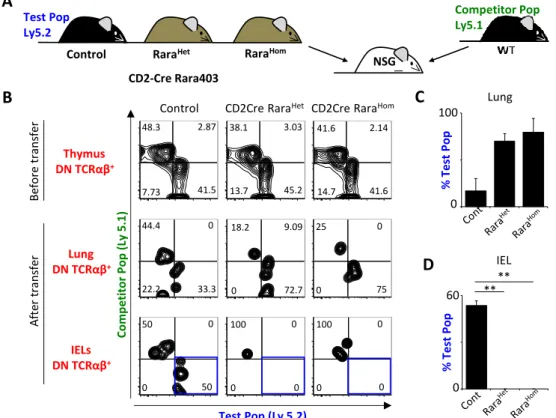
Figure 8: Intraepithelial compartment colonization by thymic natural TCRαβ+ precursors is
controlled by RA signaling. A. Representative scheme of thymic DNTCRαβ+
cell transplantation to perform 48-hour chimeras. B. Representative FACS plots for the cell mixture of test and competitor
thymic DNTCRαβ+
cell subsets before transplantation and colonizing the lung or the intraepithelial
compartment of transplanted animals after 48 hours. C. Percentage of the test population (Ly5.2+ cells) in
the DNTCRαβ+
present in the lung of transplanted animals after 48 hours. D. Percentage of the test
population (Ly5.2+ cells) in the DNTCRαβ+
cell subset colonizing the intraepithelial compartment of transplanted animals after 48 hours. Bars represent SEM. Student’s t-test. *p<0.05, **p<0.01, and ***p<0.001. n=3 for each experimental group.
Test Pop (Ly 5.2) Thymus DN TCRαβ+ IELs DN TCRαβ+ Lung DN TCRαβ+ Co m p e tito r Pop ( Ly 5.1)
Control CD2Cre RaraHet CD2Cre RaraHom
Be fo re tra n sf er After tra n sf er % Test Pop % Test Pop 0 60 100 0 48.3 2.87 41.5 7.73 38.1 3.03 45.2 13.7 41.6 2.14 41.6 14.7 44.4 0 33.3 22.2 18.2 9.09 72.7 0 25 0 75 0 50 0 50 0 100 0 0 100 0 0 0 0
A
B
C
D
** **Control RaraHet RaraHom
NSG CD2-Cre Rara403 Test Pop Ly5.2 Competitor Pop Ly5.1 Lung IEL
5. RA signaling deficient IEL thymic precursors fail to colonize the gut
intraepithelial compartment
Natural IEL precursors were detected in normal numbers or slightly increased in the thymus of Rara403 mice. Thus, our data suggest that their IEL-like genetic identity might be altered, which may compromise their integration in intestinal natural IEL compartment. To test this hypothesis, we interrogated the capacity of these cells to migrate to the intestinal epithelium. To this end, we purified thymic DNTCRαβ+ cells from CD2CreRara403 mice and their WT littermate controls (allotype Ly5.2) as well as thymic IEL precursors for WT mice (Ly5.1). Sequentially we performed competitive chimeras by transferring intravenously the purified cells into NSG mice (Figure 8A).
In contrast to their WT counterparts, transplanted CD2CreRara403 DNTCRαβ+ IEL
progenitors were undetectable in the intestinal epithelium despite being present at other peripheral organs (Figure 8B-D). Thus, our data indicate that RA is essential to confer thymic DNTCRαβ+ precursors with gut homing properties.
32 M FI α 4 β 7 % C C R 9 + % α 4 β 7 + 0 30 *** ** 80 0 Thymus DN TCRαβ+ α4β7 CCR9 M FI C C R9 7200 0 0 500 * * * * * * CD2Cre Rara M FI α 4 β 7 % C C R 9 + % α 4 β 7 + 0 2 * 100 0 * M FI C C R 9 12000 0 0 300 α4β7 CCR9 * * Thymus DN TCRαβ+ CD4Cre Rara
A
B
C
R e la tiv e n u m b e r Re la ti ve n u m b e r Ccr9 Itga4 Itgb7 4 0 10 0 10 0 R el ati ve ex pr es si on R el ati ve ex pr es si on R e la ti ve e xp re ss io n B TSS (Ccr9) A C DExon 1 Exon 2 Exon 3
8 8 2 4 9 4 1 9 -3 4 9 9 5 -329 TSS (Itga4) B A
Exon 1 Exon 2 Exon 3
-65 65 8 -48 30 9 Exon 4 E 1 4 9 5 0 *** *** RaraHet RaraHom Cont RaraHet RaraHom Cont
6. RA controls gut-homing molecules in the thymus
The expression of specific surface gut homing markers, such as CCR9 and α4β7, is required for efficient migration and homing to the intestine[79] [82-85].
We interrogated whether the inability to colonize the gut by natural IEL thymic precursors bearing disrupted RA signaling was caused by gut-homing molecule deregulation. To test this, we analyzed the expression of gut homing markers in thymic precursors DNTCRαβ+ of CD2CreRara403 and CD4CreRara403 mice by flow
cytometry. Our results demonstrate that thymic natural IEL precursors have a large reduction in CCR9 and α4β7 expression (Figure 9A).
Figure 9: RA signaling regulates gut-homing markers expression. A. Thymic DNTCRαβ+ were analysed
in thymus from 6 to 8 weeks old CD2CreRara403 and CD4CreRara403 that were either RaraHet or RaraHom
mice and from their WT littermate controls. Representative histograms for the CCR9 and α4β7 expression
profile in thymic DNTCRαβ+ cells. Percentages of CCR9+ and α4β7+ cells in thymic DNTCRαβ+ cell subset.
Mean fluorescence intensity of CCR9 and α4β7 in thymic DNTCRαβ+ cells. B. Expression of Ccr9, Itga4 and
Itgb7 in thymic DNTCRαβ
+
were quantified by quantitative real time PCR after stimulation with RA or RAR inhibitors. Results were normalized to the expression of Gapdh and Hprt. C. Putative RARE half-sites in Ccr9 and Itga4 loci were previously identified. Bars represent SEM. Student’s t-test. *p<0.05, **p<0.01, and ***p<0.001. n=3-5 for each experimental group. Data represents 3 independent experiments.
33 To test whether RA can regulate the expression of the gut-homing receptors α4β7 and CCR9, WT DNTCRαβ+ thymic precursors were purified and incubated with RA, BMS493 (an inverse pan-RAR agonist) or vehicle for 12 hours. The expression of Ccr9, Itga4 (codifies for α4 integrin) and Itgb7 (codifies for β7 integrin) was quantified by quantitative real-time PCR.
We found that Itga4 and Itgb7 expression was substantially increased upon stimulation with RA relative to BMS493 and control (Figure 9B). Thus, these results suggest that retinoids regulate gut-homing receptors in thymic DNTCRαβ+ natural IEL precursors.
To investigate this hypothesis further, we performed computational analysis of potential retinoic acid response elements (RAREs) in Ccr9, Itga4 and Itgb7 loci (Figure 9C) [111, 112]. Our results identified several potential RAREs in Ccr9 and Itga4, while none were found for Itgb7. Altogether, our data indicate that dietary retinoids imprint thymic IEL precursors with an enteric positioning machinery.
34
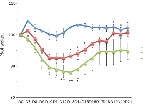
Figure 11: Mice with RA disrupted signaling lose more weight during E.vermiformis infection.
Mice were weighted daily during infection since day 7 until day 21. Blue lines represent control (CD2Cre-)
mice, red lines CD2CreRara403Het mice and green lines represent CD2CreRara403Hom.Bars represent
SEM. Student’s t-test. *p<0.05, **p<0.01, and ***p<0.001. n= 5-8 for each experimental group.
Control CD2CreRaraHet CD2CreRaraHom 100 80 % o f w e ig h t 90 110 D0 D7 D8 D9 D10 D11D12 D13D14D15D16D17D18 D19D20D21 * * * * * * ** ** * * * *
7. Enteric immune barrier depends on lymphocyte cell-autonomous RA
signals
Natural IEL constitute the first layer of immune defense against invading pathogens. We hypothesized that the drastic decrease of natural IEL in mice bearing disrupted RA signaling results in the disruption of the immune barrier.
To test this hypothesis, we investigate the intestinal immunological defense of CD2CreRara403 mice against enteric infectious parasites. To this end, we infected CD2CreRara403 mice with the protozoan Eimeria vermiformis, an organism that causes a natural intestinal infection, and that was shown to be controlled by IEL. [113] [114].
Our data shows that while WT controls lost no more than 2-3% of their weight, CD2CreRara403 mice lost up to 13% of total body mass, in a RA dose-dependent manner, and in a 4-5 days period, a clinically significant dehydration (Figure 11). Reflecting this, infected CD2CreRara403Hom mice appeared sicker than infected control mice, commonly displaying ruffled fur, hunched posture, and listlessness. Accordingly, most CD2CreRara403Hom mice had a delayed recovery, and by day 21 still had significant body weight loss (figure 11). Our data indicate that the intestinal immune defense against intestinal infectious parasite is impaired in mice whose natural IEL lack RA signaling.