This is an Accepted Manuscript, which has been through the Royal Society of Chemistry peer review process and has been accepted for publication.
Accepted Manuscripts are published online shortly after
acceptance, before technical editing, formatting and proof reading. Using this free service, authors can make their results available to the community, in citable form, before we publish the edited article. We will replace this Accepted Manuscript with the edited and formatted Advance Article as soon as it is available.
You can find more information about Accepted Manuscripts in the
author guidelines.
Please note that technical editing may introduce minor changes to the text and/or graphics, which may alter content. The journal’s standard Terms & Conditions and the ethical guidelines, outlined in our author and reviewer resource centre, still apply. In no event shall the Royal Society of Chemistry be held responsible for any errors or omissions in this Accepted Manuscript or any consequences arising from the use of any information it contains.
Accepted Manuscript
Food &
Function
Linking the chemistry and physics of food with health and nutrition
www.rsc.org/foodfunction
ISSN 2042-6496 PAPER T. J. Wooster et al.
Impact of gastric pH profi les on the proteolytic digestion of mixed lg-Xanthan biopolymer gels
Volume 7 Number 1 January 2016 Pages 1–612
Food &
Function
Linking the chemistry and physics of food with health and nutrition
This article can be cited before page numbers have been issued, to do this please use: J. R. Costa, M. Amorim, A. Vilas-Boas, R.V. Tonon, L. M. Cabral, L. Pastrana and M. Pintado, Food Funct., 2019, DOI: 10.1039/C8FO02534G.
Impact of in vitro gastrointestinal digestion on chemical composition,
bioactive properties and cytotoxicity of a Vitis vinifera L. cv. Syrah grape
pomace extract
Authors: Joana R. Costaa, Manuela Amorima, Ana Vilas-Boasa, Renata V. Tononb, Lourdes M. C.
Cabralb, Lorenzo Pastranac, Manuela Pintadoa,*
Affiliation:
a Universidade Católica Portuguesa, CBQF – Centro de Biotecnologia e Química Fina –
Laboratório Associado, Escola Superior de Biotecnologia, Rua Arquiteto Lobão Vital 172, 4200– 374 Porto, Portugal
b Embrapa Agroindústria de Alimentos, Av. das Américas, 29501, 23020-470 Rio de Janeiro, RJ,
Brazil
c INL - International Iberian Nanotechnology Laboratory, Av. Mestre José Veiga s/n, 4715 – 330
Braga, Portugal
*Corresponding author:
mpintado@porto.ucp.pt
CBQF – Centro de Biotecnologia e Química Fina Universidade Católica Portuguesa
Rua Arquiteto Lobão Vital, 172 4200 – 374 Porto, Portugal
Keywords: grape pomace, by-product valorization, digestion, bioactive compounds.
Food
&
Function
Accepted
Manuscript
Published on 14 March 2019. Downloaded by Drexel University on 3/18/2019 1:50:58 PM.
View Article Online
Abstract
Grape pomace is a major by-product worldwide, well-known for its bioactive compounds, such as fibers and phenolic compounds that are popular for their impact upon human health, including gastrointestinal health. The objective of this work was to evaluate the chemical composition and biological activities of an enzymatic grape pomace extract, and to investigate how the gastrointestinal digestion modulates these properties. Grape pomace extract was previously produced using an enzymatic cocktail with xylanase activity and was then exposed to simulated conditions of gastrointestinal digestion, characterised for its chemical composition and screened for antimicrobial, prebiotic and antioxidant activities. The safety of this ingredient after gastrointestinal digestion was also assessed. Grape pomace extract presented high content of dietary fibre and other carbohydrates, including xylooligosaccharides, besides minerals and phenolic compounds. In vitro simulated gastrointestinal digestion allowed to conclude that xylobiose was resistant to gastric conditions, unlike phenolic compounds. The use of 2% (w/v) of this ingredient proved to be a potential carbon source that could be fermented by Lactobacilli and Bifidobacteria spp, even after digestion. The extract also exhibited strong antioxidant and antimicrobial activity against Staphylococcus aureus, Escherichia coli and Pseudomonas
aeruginosa, however, after gastrointestinal digestion, the antioxidant capacity decreased, and
the antimicrobial capacity was strongly reduced or lost. Furthermore, the extract safety was also guaranteed on Caco-2 intestinal cells. This novel and green grape pomace extract proved to be composed by relevant bioactive molecules, including xylooligosaccharides, polyphenols, organic acids and minerals, which provided different biological properties, with potential application in food industry to be used as an ingredient in the development of new functional foods.
Food
&
Function
Accepted
Manuscript
Published on 14 March 2019. Downloaded by Drexel University on 3/18/2019 1:50:58 PM.
View Article Online
1. Introduction
Circular Economy has been receiving increasing attention over the last decade, as it brings together both agroindustrial sustainability and social-economic concerns, throughout the looking for innovative solutions for waste disposal, taking advantage of renewable resources, while improving both anthropological and environmental health.1-3 Agriculture and food
processing industries produce the most significant and promising by-products, which, despite all their biological potential for food, cosmetic or nutraceutical applications, are being used as plant fertilizers or animal feed, or simply incinerated.4
Grapes are one of the most cultivated fruit crops worldwide, with ca. 67 million tons produced annually, from which more than 70% is intended to wine industry, which generates up to 20% of wasted biomass in the form of grape skin, seeds, stems and residual pulp, known as grape pomace (GP).5-7 GP is a lignocellulosic biomass, rich in neutral polysaccharides and bioactive
compounds that has been exhaustively studied for the extraction of its bioactive phenolic compounds, but slight studied concerning the potential of its dietary fiber and polysaccharides. 8-14
In 1998, Saura-Calixto introduced the concept of antioxidant dietary fibre, a natural compound rich in both dietary fibre and polyphenols, which has been reported as beneficial for human health, as it stimulates colonic fermentation and regulates the blood glucose and insulin response, obesity and cardiovascular diseases.16,17 Among other molecules found in GP,
oligosaccharides from the hemicellulose fraction are recognized for their prebiotic activity due to their resistance to gastric acidity and to mammalian enzymatic hydrolysis, and their capacity to selectively stimulate the gut microbiota growth. This property, in combination with other reported bioactivities, makes GP a promising by-product that can be converted into a high added value ingredient with potential food application.17,18
Food
&
Function
Accepted
Manuscript
Published on 14 March 2019. Downloaded by Drexel University on 3/18/2019 1:50:58 PM.
View Article Online
Another bioactivity reported for GP and other alcohol-free winery products is its antimicrobial capacity, especially against the microorganisms related to food spoilage and intestinal infections, including Staphylococcus aureus, Escherichia coli, Salmonella infantis and Candida
albicans.4 The antimicrobial activity of grapes, grape byproducts and wine is usually associated
to the presence of phenolic compounds such as resveratrol and anthocyanins, or even to phytoalexins produced by the grapevine under stress conditions including fungal attack or high UV exposure.4
In order to assure the feasibility of the by-product valorization process, it is necessary an environment friendly method to obtain fibre-rich extracts with potential bioactive properties. In this sense, enzymatic hydrolysis is one of the mildest processes to achieve a final extract with the desired chemical and biological characteristics.19,20
Using such methods, it has previously been developed a novel enzymatic grape pomace extract, which composition rich in fibres and xylooligosaccharides indicates a potential biological activity, although it was not further studied for its chemical composition that could grant these properties. 21
So, the objective of this work was to perform an extensive chemical characterisation of an enzymatic GP extract and evaluate a group of important bioactive properties, including antioxidant, antimicrobial, and prebiotic activities, trying to relate chemical properties to its biological activity. Furthermore, evaluation of these bioactive properties upon in-vitro gastrointestinal digestion was also performed to understand the stability and bioaccessibility of the main bioactive compounds present in this multifunctional extract.
2. Material and Methods
2.1. Raw Material
Food
&
Function
Accepted
Manuscript
Published on 14 March 2019. Downloaded by Drexel University on 3/18/2019 1:50:58 PM.
View Article Online
Syrah grape pomace (Vitis vinifera L. cv. Syrah) from red sparkling production was provided by
Ouro Verde Winery (Miolo Wine Group), located at Vale do São Francisco, Bahia, Brazil. The pomace was oven-dried at 45 ºC for 24 h, milled and sieved to obtain a powder. 21
2.2. GP extract production
GP extract was obtained by enzymatic extraction using the enzymes cocktail produced at our lab by solid state fermentation as previously described.21 Briefly, the enzymatic cocktail, with final
xylanase activity of 100 IU/g, was added to 50 g of grape pomace in sodium acetate buffer 0.2 M at a solid: liquid ratio of 1:18, pH was adjusted to 5.0, and the mixture was incubated at 40 °C with shaking at 200 rpm for 4 hours. The reaction was stopped by heating the test tubes to 100 °C for 5 min, and the supernatant was vacuum-filtered using Whatman No. 1 filter paper. The soluble fraction (GP extract) was then freeze-dried (Telstar LyoQuest, Spain).
GP extract was characterized for moisture content, ashes, total dietary fibre and total protein using AOAC methods22-24, and for its bioactive properties (antioxidant, antimicrobial, and
prebiotic activities), as described later.
2.3. In vitro simulation of gastrointestinal digestion
Simulation of the effect of digestive tract upon GP extract was performed following the method described by Madureira et al.with slight modifications, by dissolving 900 mg of the lyophilized GP extract into 20 mL of ultra-pure water. 25
Mouth Digestion: The pH was adjusted to 6.9, using HCl 1 M. Artificial saliva was simulated by using α-amylase (Sigma-Aldrich Chemistry, St. Louis, Missouri, USA) at 100 U/mL, and added at a rate of 0.6 mL/ min of digestion. Incubation was made during 2 min at 37 ˚C and 200 rpm. Stomach Digestion: The pH was adjusted to 2.0 using HCl 1 M. Gastric juice was simulated by dissolving pepsin (Sigma-Aldrich Chemistry, St. Louis, Missouri, USA) 25 mg/mL, and added at a ratio of 0.05 mL/mL of sample. Incubation lasted 120 min (long digestion), at 37 ˚C and 130 rpm.
Food
&
Function
Accepted
Manuscript
Published on 14 March 2019. Downloaded by Drexel University on 3/18/2019 1:50:58 PM.
View Article Online
Gut digestion: Simulation of gut conditions was performed by initial adjustment of pH to 6.0 using NaHCO3 1 M. The intestinal juice was simulated by dissolving 2 g/L of pancreatin
(Sigma-Aldrich Chemistry, St. Louis, Missouri, USA) and 12 g/L bile salts (Oxoid™, Hampshire, UK). This solution was then added at a concentration of 0.25 mL/mL of sample. All samples were incubated during 1 h, at 37 ˚C and 45 rpm.
After gut digestion, enzymes were removed using 3 kDa cut-off filters and the sample was freeze-dried. This digested GP extract (DGP) was characterised for its bioactive properties (antioxidant, antimicrobial, and prebiotic activities).
All assays were performed in triplicate. After each stage the monosaccharide and oligosaccharide profiles were assessed by HPLC-RI (as described in section 2.3).
2.4. Carbohydrates and organic acids composition
Identification and quantification of carbohydrates and organic acids were performed by High-performance liquid chromatography – HPLC, coupled to refractive index (RI) and diode array (DAD) detectors. Before HPLC analysis, lyophilized samples were re-suspended in ultra-pure water and re-filtered with Millipore Millex syringe filter of 0.22 µm.
2.4.1. Identification and quantification of monosaccharides
Monosaccharides were analyzed by HPLC-RI, using a Beckman Coulter System Gold HPLC with RI detector (Knauer, Berlin, Germany). Identification and quantification were performed with an Aminex HPX-87P (Bio-rad, Berkeley, USA) column, using ultrapure water as mobile phase and an isocratic flow rate of 0.5 mL/ min.26 All compounds were identified and quantified by comparison
to the retention times of pure standards (xylose, fructose, mannose, and glucose) (Sigma Aldrich, St. Louis, USA), and using a calibration curve in the range of concentrations of 0.2 - 2.0 mg/mL.
Food
&
Function
Accepted
Manuscript
Published on 14 March 2019. Downloaded by Drexel University on 3/18/2019 1:50:58 PM.
View Article Online
2.4.2. Quantification of xylooligosaccharides and identification of polysaccharides
Oligosaccharides and polysaccharides were also analyzed by HPLC-RI, using a Waters Ultrahydrogel 120 (7.8 x 300 mm) and a Ultrahydrogel 250 (7.8 x 300 mm) columns, with ultrapure water as mobile phase, and an isocratic flow rate of 0.5 mL/ min.27
Xylooligosaccharides were identified by comparison to the retention times of pure standards: xylobiose (Sigma, St. Louis, USA), xylotriose, xylotetraose, and xylopentose (Megazymes, Wicklow, Ireland), using a calibration curve in the range of concentrations of 0.1 – 2 mg/ mL. High molecular weight polysaccharides were identified by comparison to the retention times of Shodex (Showa Denko K. K., Tokyo, Japan) Pullulan P-82 standards, ranging from P-5 (5.9 kDa) to P-800 (708 kDa).
2.4.3. Identification of organic acids
Organic acids were analyzed in a Beckman Coulter System Gold HPLC equipped with a DAD detector (System Gold 168 Detector, Beckman Coulter) at wavelength of 220 nm, using an Aminex HPX-87H (Bio-rad, Berkeley, USA) column with sulfuric acid 13 mM as mobile phase and an isocratic flow rate was 0.5 mL/ min.26 All organic acids were identified by comparison to the
retention times of pure standards: citric, tartaric, malic, succinic, lactic, formic and acetic acids (Sigma-Aldrich, St. Louis, USA), using a calibration curve in the range of concentrations of 0.1 – 2 mg/ mL.
2.5. Mineral Analysis
The digestion of GP and DGP extracts for mineral analysis was performed following the method described by Roriz et al., with slight modifications. 28 Briefly, 250 mg of samples were mixed in a
Teflon vessel with 6 mL of HNO3 65% and 1 mL of H2O2 30% and heated in a microwave system
(Berghof, Eningen, Germany). Digestion procedure was conducted in five steps: 130 °C for 5 min; 170 °C for 10 min; 200 °C for 15 min; 100 °C for 2 min; and 100 °C for 2 min. After microwave
Food
&
Function
Accepted
Manuscript
Published on 14 March 2019. Downloaded by Drexel University on 3/18/2019 1:50:58 PM.
View Article Online
digestion, samples were cooled down to room temperature and diluted to a final volume of 50 mL.
Microwave-digested samples were then analysed through Inductively Coupled Plasma Atomic Emission Spectrometry (Perkin Elmer, Massachusetts, USA) and quantify through internal standard calibration. All analysis were performed in triplicate.
2.6. Determination of Phenolic Compounds 2.6.1. Total Phenolics
Concentration of total phenolic compounds (TPC) was determined colorimetrically by Folin– Ciocalteu method.29 GP and DGP extracts were dissolved in ultra-pure water, at concentration
of 200 mg/ mL. Quantification was done at 750 nm (UV mini 1240, Shimadzu, Tokyo, Japan) with gallic acid as standard in the range of 0.015–1.00 mg/ mL.
2.6.2. Identification and quantification
Qualitative and quantitative profiles of polyphenols were carried out by HPLC-DAD, according to the method described by Oliveira et al. Analysis was conducted on a Waters Liquid Chromatograph (Waters Alliance, Mildford MA, USA), using a C18 guard column (Symmetry® C18) and an Alltech adsorbosil C18 reversed-phase packing column (250 x 4.6 mm i.d. 5 μm particle size and 125 Å pore size. Mobile phase: Solvent A: acetonitrile 100% with 0.2% TFA; Solvent B: acetonitrile/ water 5:95 (v/) with 0.2% TFA; flow rate = 1 mL/ min. For HPLC analysis, GP and DGP extracts were diluted in ultra-pure water at final concentration of 200 mg/ mL and filtered using 0.45 µm syringe filters. 30
All compounds were identified and quantified by external calibration curve by comparison to pure standards (in the range of concentrations of 1.6 - 105 µg/mL): caffeic acid, (+) cathechin, p-coumaric acid, rutin, syringic acid, vanillic acid, cyanidin-3-galactosidase, cyanidin–3-glucoside,
Food
&
Function
Accepted
Manuscript
Published on 14 March 2019. Downloaded by Drexel University on 3/18/2019 1:50:58 PM.
View Article Online
peonidin-3-o-glucoside, and delphinidin-3-glucoside (Sigma Aldrich, St. Louis, USA); luteolin-7-o-glucoside and neochlorogenic acid (Extrasynthese, France).
2.7. Antioxidant capacity
2.7.1. ABTS Radical Scavenging Activity
The free radical-scavenging activity was measured using the ABTS+ radical cation decolorization
assay.31 Briefly, ABTS+ solution was produced by reacting ABTS (7 mmol/L ) filtered using a 0.45
μm filter (Macherey-Nagel, Düren, Germany) with potassium persulfate (2.45 mmol/L) in the ratio 1: 1 (v/v). The ABTS+ solution was diluted with redistilled water to an absorbance of 0.700 ± 0.02 at 734 nm (Shimadzu 1240 UV–visible spectrophotometer). After addition of 1.0 ml of diluted ABTS+ solution to 10 μL of GP and DGP extracts (dissolved in ultra-pure water at 200 mg/mL), the absorbance reading was done exactly 6 min after initial mixing. Ascorbic acid was used as a standard to prepare a calibration curve in the range of 0.02–0.50 mg/mL.
2.7.2. Oxygen Radical Absorbance Capacity (ORAC)
ORAC assay was performed according to the method described by Contreras et al.32 Briefly, the
reaction was carried out at 40 ºC in 75 mM phosphate buffer saline (PBS, pH 7.4) and the final assay mixture (200 µL) contained 120 µL of fluorescein (70 Nm), 60 µL of AAPH (14 mM) and 20 µL of antioxidant [Trolox (6.25–200 μM) or sample (GP and DGP extracts (200 mg/ mL) also diluted (1:500 – 1:3200) in PBS]. The fluorescence was recorded during 137 min (104 cycles). A FLUOstar OPTIMA plate reader (BMG Labtech, Offnburg, Germany) with 485 nm excitation and 520 nm emission filters was used. The equipment was controlled by the FLUOstar Control software version 1.32 R2 for fluorescence measurement. Black polystyrene 96-well microplates (Nunc, Denmark) were used. AAPH and Trolox solutions were prepared daily and fluorescein was diluted from a stock solution (1.17 mM) in 75 MM PBS (pH 7.4). All reaction mixtures were prepared in duplicate and three independent runs were performed for each sample. The final
Food
&
Function
Accepted
Manuscript
Published on 14 March 2019. Downloaded by Drexel University on 3/18/2019 1:50:58 PM.
View Article Online
ORAC values were expressed as μmol of Trolox equivalent per gram of GP extract (μmol TE/ g of GP extract).
2.8. Antimicrobial activity
Antimicrobial activity was determined against two Gram-positive bacteria - Methicillin-Susceptible Staphylococcus aureus (MSSA) ATCC 25923, Methicillin-Resistant Staphylococcus
aureus (MRSA) CCUG 60578, and two Gram-negative - Escherichia coli ATCC 25922 and Pseudomonas aeruginosa ATCC 10145.
2.8.1. Determination of Minimal Inhibitory Concentrations (MICs)
MICs were determined using a microdilution assay, following the standards for antimicrobial susceptibility testing provided by the Clinical and Laboratory Standards Institute (CLSI).33 Inocula
were prepared by suspending each bacterial colony, previously grown in nutrient agar media for 24 h, in calcium-adjusted Muller-Hinton Broth (MHB) in order to achieve a turbidity equivalent to a 0.5 McFarland standard (1×10⁸ CFU/mL). Inocula were then diluted in the same medium to
reach a bacterial concentration of 106 CFU/mL in each well of a 96-well microplate. The MIC was
determined by observing the lowest concentration of extract that completely inhibited bacterial growth. Sterile MHB was used as negative control. All assays were done in triplicate.
2.8.2. Growth inhibition curves
For determination of growth inhibition curves, inocula were prepared by suspending each bacterial colony into MHB, with a final concentration of ca. 108 CFU/ mL. Twenty microliters of
each inocula were transferred to a 96-well microplate and every well was fulfilled (to final volume of 200 µL) with 2% (w/v) of GP and DGP extracts diluted in MHB. Microplate was incubated in a microplate reader (Multiskan GO, Thermo Scientific) at 37 °C for 24 h, with absorbance measurements at 620 nm registered every hour. Three controls were also
Food
&
Function
Accepted
Manuscript
Published on 14 March 2019. Downloaded by Drexel University on 3/18/2019 1:50:58 PM.
View Article Online
performed: the first one containing inoculum and Mueller-Hinton broth (positive control), the second one containing the solubilized extracts (negative control) and the third one containing only MHB. All assays were done in triplicate.
2.9 Prebiotic potential
The GP extract before and after GIT passage was tested for potential of prebiotic activity using two different in vitro methods using pure probiotic, the first one through the evaluation of growth curves using microplate assay and the other through in vitro fermentation assay to assess the metabolic activity using.
2.9.1. Growth curves via microplate assay
Potential prebiotic effect of GP extract was determined for Bifidobacterium animalis Bo (CSK, Ede, Netherlands), Bifidobacterium longum BG3 (Cell Biotech, Hellerup, Denmark),
Bifidobacterium animalis spp. lactis Bb12, Lactobacillus casei 01 (Chr. Hansen, Hørsholm,
Denmark), and Lactobacillus rhamnosus R11 (Lallemand, Montreal, Canada). Strains were stored at −80 °C in de Man–Rogosa–Sharpe (MRS) broth (Biokar Diagnostics, Beauvais, France) with 30% (v/v) glycerol.
L. casei 01 and L. rhamnosus R11 inocula were prepared by suspending each bacterial colony
into De Man, Rogosa and Sharpe (MRS) broth, achieving a turbidity equivalent to 0.5 McFarland standard, and then diluting to reach the recommended concentration of probiotic bacteria in wells, 5 x 10⁵ CFU/mL. Twenty microliters of each inocula were transferred to a 96-well
microplate and every well was fulfilled (to the final volume of 200 µL) with the GP extract diluted in basal MRS broth without glucose, at concentrations of 1 and 2% (w/v). Microplate was incubated (Multiskan GO, Thermo Scientific) at 37 °C for 24 h with agitation.
B. animalis Bo, B. longum BG3 and B. lactis BB12 inocula were prepared under anaerobic
atmosphere, by suspending each bacterial colony into De Man, Rugosa and Sharpe (MRS) broth
Food
&
Function
Accepted
Manuscript
Published on 14 March 2019. Downloaded by Drexel University on 3/18/2019 1:50:58 PM.
View Article Online
supplemented with 0.05% (v/v) L-cysteine-HCl, achieving a final turbidity equivalent to 0.5 McFarland standard (1x10⁸ CFU/ mL), and then diluted to reach the recommended
concentration of probiotic bacteria in wells, 5 x 10⁵ CFU/ mL. Twenty microliters of each inocula
were transferred to a 96-well microplate and every well was fulfilled (to final volume of 200 µL) with the GP extract, diluted in basal MRS broth without glucose at concentrations of 1 and 2% (w/v). Microplate was sealed with paraffin and incubated at 37 °C for 48 h with agitation, with absorbance measurements at 620 nm registered every hour.
Three controls were also performed: the first one containing inoculum and MRS broth (positive control), the second one containing the solubilized extracts in MRS broth without glucose (negative control) and the third one containing only MRS broth.
2.9.2 In vitro fermentation assay
In vitro fermentation of DGP extract was assessed using only two of the previous strains Bifidobacterium animalis spp. lactis BB12 and Lactobacillus casei 01. Before the assays, all strains
were grown in MRS broth at 37 °C for 16 h. Anaerobic conditions were used for B. animalis Bb12, whereas aerobic atmosphere was used for L. casei 01.
Fermentation assays were carried out with ca. 106 CFU/ mL for each strain, in sterilized media
(121 °C and 15 min) containing MRS-medium without glucose supplemented with 2% (w/v) of DGP extract, 2% (w/v) fructooligosaccharides (FOS, used as reference) and MRS medium without carbon source (negative control) at 37 °C for 48 h. All experiments were carried out in duplicate. Samples of the fermentation broths were withdrawn at 0, 4, 8, 24 and 48 h for metabolites analysis, throughout determination of the pH values and organic acids in the culture media. The pH was evaluated using a digital potentiometer (Crison Instruments, Spain), and organic acids were quantified by HPLC as described in section 2.3.3. Viable cells were quantified by plating on MRS agar (supplemented with cysteine hydrochloride in the case of Bifidobacterium) and
Food
&
Function
Accepted
Manuscript
Published on 14 March 2019. Downloaded by Drexel University on 3/18/2019 1:50:58 PM.
View Article Online
incubation at 37 °C for 48 h (anaerobic and aerobic conditions were used for Bifidobacterium and Lactobacilli strains, respectively).
2.10 Evaluation of cytotoxicity
Cytotoxicity of GP and DGP extracts was assessed in Caco-2 cells through XTT assay. For the assays the 2,3-bis (2-methoxy-4-nitro-5-sulfophenyl)-2H-tetrazolium-5-carboxanilide sodium salt (XTT) solution was prepared as follows. Briefly, a 10 mM of Phenazine Methosulfate (PMS) solution (Sigma-Aldrich, St. Louis, USA) was prepared in PBS (0.01 M; pH 7.4) and a 1 mg/mL XTT solution was prepared in DMEM, previously warmed to 37 °C. Both solutions were sterilized using a 0.22 µm membrane filter (Millipore, Billerica, USA), mixed (2.5 µL of PMS per mL of XTT solution) and aliquots of XTT working solution were frozen until usage.
2.10.1. Cell line growth conditions
Human intestinal cell line (Caco-2) was obtained from Cell Line Services (Appenheim, Denmark). The cells were cultured at 37 °C in a humidified atmosphere of 95% air and 5% CO2, as
monolayers using Dulbecco’s Modified Eagle’s Medium (DMEM) with 4.5 g/L glucose, L-glutamine without pyruvate (Lonza, Verviers, Belgium) containing 10% (v/v) fetal bovine serum (FBS, Biowest, Nuaillé, France) and 1% (v/v) Penicillin-Streptomycin-Fungizone (Lonza, Verviers, Belgium).
2.10.2. XTT assay
GP and DGP extracts biocompatibility was assessed upon the Caco-2 cell line using the XTT colorimetric assay. Briefly Caco-2 cells were seeded at 1 x 10⁵ in the wells of a 96 well microplate
and allowed to adhere. After 24 h, the media was removed and the cells were washed with PBS. Then, this media was added with GP or DGP extracts at two different concentrations (10 and 20 mg/mL). After 24h, 25 µL of XTT working solution were added to each well and the cells were
Food
&
Function
Accepted
Manuscript
Published on 14 March 2019. Downloaded by Drexel University on 3/18/2019 1:50:58 PM.
View Article Online
incubated, in the dark, for 2 h. The optical density (OD) at 485 nm was then measured using a microplate reader (FLUOstar, OPTIMA, BMG Labtech, Ortenberg, Germany). All assays were performed in quintuplicate.
2.11 Statistics
Statistical analysis was performed with IBM SPSS statistic program v 23.0 (Illinois, USA), using t-student test for independent samples and analysis of variance (ANOVA) with Tukey post hoc test. Differences were considered to be significant at a level of p < 0.05.
3. Results and Discussion
3.1 Extract characterization
The nutritional composition of GP enzymatic extract is shown in Table 1. It is possible to observe that GP extract is rich in soluble fibre, other carbohydrates and also in minerals. Glucose and fructose are the simple sugars present in the GP extract, at similar concentrations (p > 0.05), with 14.90 ± 0.73 g/ 100 g of glucose and 11.91 ± 0.43 g/100 g of fructose. The GP extract also presented a high concentration of XOS, especially xylobiose at concentration of 5.57 ± 0.02 g/ 100 g. Xylotriose and xylotetraose were also present but at concentrations that could not be quantified by HPLC –RI (< 0.1 mg/ mL). The amount of extracted xylobiose is in accordance with the results obtained by Kiran and co-workers, who produced XOS from cotton stalk, rice hull, wheat straw, corn cob and sunflower stalks mixture, and obtained yields of 6.94 and 7.22 g/ 100 g xylan.34 The presence of other polysaccharides with molecular weights between 5 - 10 kDa and
near 100 kDa was also identify through HPLC-RI.
Some organic acids were also found in the GP extract, being the acetic acid the most significant one, with a concentration of 2.24 ± 0.13 g/100 g of extract. Citric, tartaric, malic, and succinic acids are also present in the extract at concentrations of 1.95 ± 0.08, 0.34 ± 0.04, 0.30 ± 0.01
Food
&
Function
Accepted
Manuscript
Published on 14 March 2019. Downloaded by Drexel University on 3/18/2019 1:50:58 PM.
View Article Online
and 1.90 ± 0.11 g/100g, respectively. While all these acids are characteristic of grape pomace composition, acetic acid is present at higher quantity due to the enzymatic hydrolysis, as it was used for the acetate buffer preparation. The concentration of ashes present in GP extract was also high and can be explained by the amount of salts used to prepare the buffers (sodium citrate) for the enzymatic extraction, which are present at a concentration of 8.15 ± 0.03 g/ 100 g of dry buffer. Nevertheless, this result indicates that almost all the minerals present in the grape pomace flour (38 g/ kg flour ) were recovered. 21
3.2. Digestion of carbohydrates
In vitro gastrointestinal digestion (GID) models based on human physiology are important tools
in assessment of polysaccharides during gastric and intestinal digestion, particularly because of their simplicity, low cost and bio-accessibility of nutrients.35 The carbohydrate functions that
affect bioavailability and health are determined by the chemical composition and the food matrix where are incorporated, and both of these aspects must be taken into account. As it is difficult to measure directly the biological function associated with different carbohydrate properties, in vitro methods provide rapid and reproducible measures.36
The GID of GP extract was monitored through HPLC-RI quantification of glucose, fructose and xylobiose along the digestion. Results are presented in Figure 1 and indicate the mass (mg) of each sugar present in the initial extract and how they varied along the GI digestion.
Initial amount of glucose was ca. 124 mg and no significant losses were observed during the mouth or stomach digestions (p > 0.05). The major increase was observed after the simulation of intestinal digestion, when the final mass of glucose was 155.01 mg, probably due to the use of pancreatin enzyme, mainly composed by amylases and proteases that could release some glucose monomers from polysaccharides with higher molecular weights.
Food
&
Function
Accepted
Manuscript
Published on 14 March 2019. Downloaded by Drexel University on 3/18/2019 1:50:58 PM.
View Article Online
As expected, xylobiose did not show any losses during upper gastrointestinal digestion and the initial amount of ca. 50 mg maintained until gut digestion. This result confirms its potential to be used as prebiotic, including the microbiota fermentation, as described in sections 3.7 and 3.8. Furthermore, it is possible to observe from HPLC-RI chromatogram that the peak with retention time (RT) of 21.42 min, corresponding to the polysaccharide present in the range of 5-10 kDa, was hydrolyzed during the in vitro GID, especially during gastric digestion (RT=26.39 min), achieving a molecular weight (MW) around 800 Da after gut (retention time 26.47 min). We can also observe that the peak for the substances present in the gut compartment, such as bile salts and pancreatin, appeared very late in HPLC-RI chromatogram, indicating that it did not contain high molecular weight polymers and thus, compounds used to simulate gut digestion did not affect determination of xylobiose nor the MW of GP extract polysaccharide.35 Starch was
reported to be degraded by gastric and intestinal media to oligosaccharides, and could be digested by amylase in small intestine to sugars that can be absorbed by the intestine.37
3.3. Minerals
Owing to the high concentration of ashes in GP extract, ICP-OEP analysis was performed in order to understand what elements were present. As expected, sodium (Na) was the element present in the highest concentration (9.97 ± 0.46 g/100g), followed by potassium (K) (1.59 ± 0.33 g/100 g), as presented in Figure 2. Phosphorous (P), Boron (B), Magnesium (Mg) and Calcium (Ca) are also present at very low concentrations: 5.87 ± 0.42, 0.035 ± 0.02, 0.31 ± 0.27 and 1.58 ± 0.84 mg/ g sample, respectively.
During in vitro GID, part of Na and K present in GP extract were loss and the concentrations that reached gut were 7.72 ± 0.73 (Na) and 0.82 ± 0.072 (K) g/ 100 g sample, significantly lower than the concentrations present in the initial extract (p < 0.05). Part of Mg was also absorbed, as the concentration that reached the gut was only 0.13 mg/ g sample.
Food
&
Function
Accepted
Manuscript
Published on 14 March 2019. Downloaded by Drexel University on 3/18/2019 1:50:58 PM.
View Article Online
The colon contributes to electrolyte homeostasis by conserving sodium, chloride, and potassium. While sodium and chloride can be absorbed against large concentration gradients, potassium absorption requires a favorable concentration gradient.38
3.4. Phenolic Compounds
Total phenolic compounds (TPC) were present in the GP extract at concentration of 239.01 ± 4.44 mg GAE/ g extract. The concentration of phenolic compounds in GP extract is similar to the reported by Ky and Teissedre, who recovered ca. 195.66 to 215.84 mg GAE/ g from grape seed.39
Higher yields of phenolic recovery are usually achieved with the use of organic solvents, such as ethanol or acetone, which were not applied in our extracts.
After GID, the concentration of TPC was significantly reduced to 13.59 ± 0.23 mg GAE/ g sample (p < 0.05). A drastically reduction of phenolic compounds was observed after in vitro GID, with only 5.69% of initial TPC reaching the gut. These results are in accordance with the literature that estimates that 90% of polyphenols are digested before reaching the intestine.40,41 This high
reduction of TPC can be attributed to degradation of anthocyanins, the major phenolic compound present in the grape pomace extract, by the acidic gastric conditions.
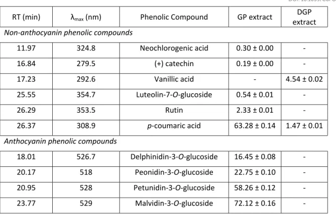
These results were confirmed by HPLC-DAD analysis (Table 2) of both extracts, where it is possible to observe the total anthocyanins degradation, as well as most of polyphenols identified in GP extract, including (+)catechin, rutin, neochlorogenic acid and luteolin. These results are in accordance with other authors that concluded that intestinal digestion induce substancial losses in polyphenol families. Lingua and co-workers observed that only 6% of total phenolics were available in the dialysable fraction of red grapes extract, representing the potential colon available compounds; Sanz- Buenhombre and colleagues recovered part of the vanillic acid after the digestion but verified that (+) catechin was completely degraded. 42,43 It is also possible
to observe that syringic and vanillic acids were not detected in GP extract but were the most abundant compounds in the DGP extract, suggesting that these compounds were initially bound
Food
&
Function
Accepted
Manuscript
Published on 14 March 2019. Downloaded by Drexel University on 3/18/2019 1:50:58 PM.
View Article Online
to other molecules, such as fibres that link with the phenolics through hydrogen bonds, hydrophobic interactions or covalent methyl-esterification, protecting them during the digestion.
The poor biaccessibility of phenols are in accordance with the results obtained by Van de Velde, who simulated the digestion of polyphenols present in blackberries and concluded that less than 98% of total anthocyanins were not available for absorption in the gut.41 These losses are still
not completely understood but are probably due to pH changes along gastrointestinal tract interaction with the digestive enzymes and other chemical reactions that occur during digestion such as interaction with other dietary compounds, or changes in solubility. 42, 44
3.5. Antioxidant capacity
Antioxidant capacity is the most studied bioactivity described for GP extracts, and it is related to the presence of phenolic compounds as well as the high content of antioxidant dietary fiber.14,15
However, the different methods, the different antioxidant standards used and the different references to the basic-unit of sample make it difficult to establish comparisons.13 Hereupon,
antioxidant capacity of GP and DGP extracts was assessed through ABTS and ORAC methods. The ABTS radical scavenging activity of GP extract was 33.98 ± 0.464 mg AA eq/ g extract. Although it is more common to find ABTS activity expressed as Trolox Equivalent (TE), ascorbic acid (AA) is more hydrophilic and therefore, a more suitable reference to compare our water-soluble extract. Our GP extract radical scavenging activity was 7-fold higher than the results obtained by Oliveira et al. for strawberry preparations, and within the range activity reported by Kwak et al. for different Rosa multiflora extracts, which ranged from 9.8 ± 3.2 to 84.8 ± 8.6 µg AA Eq/ mL.46,47 As expected, antioxidant capacity was lower (p < 0.05) for the digested extract,
16.77 ± 0.15 mg AA eq/ g extract, due to the high loss of phenolic compounds during GID, including all the anthocyanins, as discussed above.
Food
&
Function
Accepted
Manuscript
Published on 14 March 2019. Downloaded by Drexel University on 3/18/2019 1:50:58 PM.
View Article Online
Antioxidant activity determined by ORAC assay was 3500 ± 0.058 µmol TE/ g extract and it is in agreement with the results obtained by Dudonné et al. for different aqueous plant extracts, such as Cistus ladaniferus (leaf) – 1410 ± 53 µmol TE/ g, Eucalyptus globulus (leaf) – 2846 ± 134 µmol TE/ g, Jasminium grandiflorum (flower) – 2330 ± 64 µmol TE/ g or Vanilla planifolia – 1593 ± 12 µmol TE/ g.48 After GID, the oxygen radical absorbance capacity of GP extract was reduced to
385.80 ± 0.150 µmol TE/ g (p < 0.05), confirming the results obtained by ABTS method that GID reduces the antioxidant capacity.
Overall, this loss of antioxidant activity confirms the poor bioaccessibility of phenolic compounds described before, as these compounds largely contribute to the antioxidant bioactivity.
3.6.Antimicrobial activity
The Minimal Inhibitory Concentrations (MIC) of the GP extract were 14 mg/ mL for Gram-negative bacteria (E. coli and P. aeruginosa) and 16 mg/ mL for Gram-positive (MSSA and MRSA), enlightening that all microorganisms were susceptible to GP extract.
Growth inhibition curves were performed upon MSSA, MRSA, Escherichia coli and Pseudomonas
aeruginosa. These microorganisms were selected as representative of Gram positive and
Gram-negative bacteria and using a marker of multiresistant bacteria such as MRSA. Growth inhibition curves for selected microorganisms, in the presence of GP and DGP extracts at concentration of 2% (w/v), as measured by turbidity at 660 nm, are presented in Figure 3. Corroborating the MIC determined (1.4-1.6 %) before, from Figure 3, the 2% GP inhibited completely the growth of all strains tested.
It is also possible to observe that the antimicrobial potential of the GP extract was affected by the digestion process. DGP extract did not affect the growth of Gram-negative bacteria (losing the antimicrobial activity) and although it still exhibits some reduction of the Gram-positive bacteria growth, is the reduction observed is much lower than with the GP extract before digestion.
Food
&
Function
Accepted
Manuscript
Published on 14 March 2019. Downloaded by Drexel University on 3/18/2019 1:50:58 PM.
View Article Online
The antimicrobial activity of the GP extract may be attributed to the presence of several components, including minerals such as sodium and potassium, anthocyanins and some phenolic acids, organic acids such as acetic, citric and tartaric acids; besides the presence of XOS with a well-known antimicrobial activity.49-51 In fact, it is well-known that natural extracts display
higher antimicrobial activity than selected compounds. Bassole et al. attributed antibacterial activity of fruit extracts to an overall synergy between bioactive compounds with antimicrobial properties and other minor components.52 In fact, the loss of antimicrobial activity after GID of
GP extract could be explained by this hypothesis, as the reduction on the antimicrobial potential could be related to the loss of part of these components, including the total degradation of anthocyanins (Table 2) and other phenolic compounds, the loss of ca. 30% of sodium and 50% of potassium, and the absorption of part of the organic acids during the digestion.
The results observed in the present work are in agreement with the literature: Xu et al. evaluated the antimicrobial activity of four different GP extracts on S. aureus and E. coli, and verified an inhibition of S. aureus (MICs ranging 40.6 to 250 mg/ ml); Jayaprakasha and co-workers obtained a grape seed extract with antimicrobial activity against MSSA, E. coli and P. aeruginosa and also found to be the most effective antibacterial fraction against Gram-positive bacteria when compared to Gram-negative bacteria; Oliveira and co-workers obtained Merlot CO2 supercritical
extracts with high activity on S. aureus, Bacillus cereus, E. coli and P. aeruginosa.4,53,54
Once again, it was confirmed the high susceptibility of the bioactive compounds present in GP extract to GIT and their loss of bioaccessibility, suggesting the need of an encapsulation system able to protect these compounds along the gastrointestinal tract.
3.7. Prebiotic effect
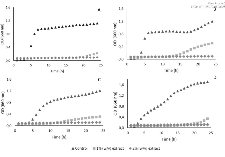
The prebiotic activity of GP extract was studied on 5 strains in basal MRS medium without glucose, at concentrations of 1 and 2% (w/v). Figure 4 present the growth of evaluated
Food
&
Function
Accepted
Manuscript
Published on 14 March 2019. Downloaded by Drexel University on 3/18/2019 1:50:58 PM.
View Article Online
Bifidobacteria and Lactobacillus strains, respectively, as measured by turbidity at 660 nm.
Frutooligosaccharides (FOS) at the same concentrations were also used as control.
All the probiotic microorganisms grew in the presence of GP extract, increasing their growth (OD at 660 nm) along the first 10 h of fermentation for Bifidobacteria and 18 h for Lactobacilli. The use of 2% (w/v) of GP extract promoted higher growth up of assayed microorganisms than 1% (w/v) of GP extract, as expected. FOS proved to be the best carbon source, at 2% (w/v) for Bifidobacteria and at 1% for Lactobacilli (Figure 4). There were not significant differences on the maximum growth of all probiotic strains used when the MRS was supplemented with 1% (w/v) of FOS or with 2% (w/v) of GP extract, except for L. rhamnosus that presented a lower growth. On the other hand, GP extract allowed Bifidobacterium spp. to achieve the maximum OD faster, between 9 and 11 hours compared to the 16 – 20 hours in the presence of FOS.
The results obtained for the L. casei 01 are somehow different from the results obtained by Gullón et al., who achieved a faster growth of this microorganism in the presence of FOS than with XOS derived from Eucalyptus globulus wood, rice husks, wheat bran and from barley wastes.55
Results indicate that GP extract can stimulate a fast growth of different probiotic strains, possibly due to the presence of glucose and XOS. These results are in accordance with the well-established prebiotic activity described for XOS including the selective stimulation of gut microbiota growth as the GP extract also presented antimicrobial activity against some pathogens, as described above.56,57 These beneficial properties of XOS are related to the
improvement of gastrointestinal functions as well as increase or change in the composition of short chain fatty acids, increased faecal weight and mineral absorption, immune stimulation, and decreased colonic pH values.56
3.8 In vitro fermentation of GP extract
Food
&
Function
Accepted
Manuscript
Published on 14 March 2019. Downloaded by Drexel University on 3/18/2019 1:50:58 PM.
View Article Online
In order to evaluate the effectiveness of DGP extract as a fermentable carbohydrate source for probiotic bacteria, in vitro fermentation assays were carried out using a Lactobacilli (L. casei 01) and a Bifidobacteria strain (B. animalis Bb12). Table 3 shows the number of viable cells (expressed as log CFU/mL) for both strains, as well as the pH value evolution in the culture media. FOS were used as positive control once they are already a well-known prebiotic; a negative control (without carbon source) was also used.
The number of viable cells of Bifidobacterium Bb12 reached 7.93 log CFU/ mL with FOS, and 8.14 log CFU/ mL with the GP extract, after 48 hours of fermentation, allowing to conclude that both carbon sources stimulated the growth of this probiotic strain. As expected, without carbon source there was not any cell growth, confirming that the Bb12 growth was stimulated by the fermentable carbon sources, and significantly improved by the use of DGP (p < 0.05).
Regarding Lactobacillus casei 01, the number of viable cells grew until 24 hours (7.54 and 7.95 log CFU/ mL for FOS and GP extract, respectively) and stabilized between 24 and 48 hours (7.54 and 7.93 log CFU/ mL for FOS and GP extract, respectively). These results for La01 are somehow expected, as it is generally known that Lactobacilli spp. usually reach the maximum growth after ca. 24 hours.
With the pH values evolution along the fermentation process, a similar behavior was observed i.e., media with carbon source in its composition suffered a decrease of pH values, while the negative control was slightly affected by changes in pH values. For the media fermented by Bb12, pH reduction was more pronounced in the presence of FOS than with GP extract (4.93 and 5.21, respectively), while for the media fermented by La01, a reverse effect was observed: the pH value after 48 hours was lower with GP extract (5.30) than with FOS (5.85).
HPLC-DAD analysis of short-chain fatty acids (SCFA) in cell-free supernatants confirmed the production of acetic, butyric, formic, and propionic acids during fermentation. Table 4 shows the concentrations of these organic acids along the 48 hours of fermentation. An overview of this table allows to observe that acetic acid was the SCFA produced in the highest quantity,
Food
&
Function
Accepted
Manuscript
Published on 14 March 2019. Downloaded by Drexel University on 3/18/2019 1:50:58 PM.
View Article Online
followed by propionic acid, while butyric acid presented the lowest concentrations in all the assays. It is known that the production of acetic acid by Bifidobacteria spp. is higher than the production by Lactobacilli spp., which is observed in the fermentation with FOS; when using GP extract, this difference was only observed after 24 hours.55 Furthermore, the presence of acetic
acid in higher quantity could be due to the fact that this organic acid was used for the production of the extract.21
Regarding the Bifidobacterium strain fermentation, the production of acetic and formic acids was significantly higher (p < 0.05) when the GP extract was used as carbon source, while the propionic acid was higher when using FOS, after 24 hours. The production of butyric acid during fermentation with Bb12 was not affected by the carbon sources, as it was not found significant differences in the concentration of this SCFA.
Regarding the fermentation with the Lactobacillus strain, the same behavior in the production of acetic and propionic acids was found, i.e, the use of GP extract improved the production of these SCFA comparing to FOS. On the other hand, the production of butyric acid was improved by the use of GP extract (p < 0.05) and the production of formic acid did not present significant differences (p > 0.05) after 4 hours of incubation.
Selective fermentations of prebiotic carbohydrates by Bifidobacteria and Lactobacilli depend on different factors, including the strain and substrate.59 GP and its extract contain significant
amounts of carbohydrates (approximately 50 g/ 100g) and showed that it can act as a fermentable source of polysaccharides, as they can promote the growth of both Bb12 and La01 and produce organic acids.
3.9 Cytotoxicity
Toxicity of GP extract before and after digestion was evaluated through XTT cell proliferation assay upon Caco-2 intestinal cells, using extracts concentrations of 1 and 2% (w/v), as these were
Food
&
Function
Accepted
Manuscript
Published on 14 March 2019. Downloaded by Drexel University on 3/18/2019 1:50:58 PM.
View Article Online
the concentrations that showed potential bioactivities. Figure 5 presents the metabolism inhibition of Caco-2 cells in the presence of GP and DGP extracts.
The XTT cell proliferation assay is an effective method to measure cell growth and drug sensitivity in tumor cell lines.60 GP extract at concentration of 2% promoted Caco-2 cells
metabolism by 37.05 ± 2.56% and by 20.91 ± 2.27%, respectively. After the digestion of GP extract, it is possible to observe a slightly, but not significant (p > 0.05), inhibition of metabolism: 5.75 ± 2.81% and 9.43 ± 1.90% for DGP extract at concentrations of 2 and 1%, respectively. It is possible to conclude that GP extract is safe to be used as food ingredient, at concentrations up to 2% (w/v). It is also important to correlate the Caco-2 cells viability with the antimicrobial and prebiotic activities, once 2% (w/v) of GP extract was enough to proliferate probiotic strains and inhibit some pathogenic bacteria growth.
4. Conclusions
This paper described the potential bioactive properties of a grape pomace extract produced through a novel eco-friendly method. Freeze-dried GP extract presented high content of total dietary fibre, other carbohydrates such as xylobiose, minerals and phenolic compounds. In vitro digestion simulation of GP extract allowed to conclude that xylobiose was resistant to gastrointestinal conditions, and that this extract has a prebiotic potential, at concentration of 2% (w/v), establishing a potential carbon source that can be fermented by Lactobacillus and
Bifidbacterium spp. Grape pomace extract proved to have antimicrobial activity against
pathogenic bacteria, with large spectrum being active against Gram-negative and Gram-positive strains, although this effect was lost after the gastrointestinal digestion. Similar impact of gastrointestinal digestion occurred to polyphenols and consequently to the antioxidant capacity, which strongly decreased after digestion.
The reduction on the antimicrobial and antioxidant capacities of the GP extract after the GID process suggests the need for an alternative system to protect it from the harsh gastrointestinal
Food
&
Function
Accepted
Manuscript
Published on 14 March 2019. Downloaded by Drexel University on 3/18/2019 1:50:58 PM.
View Article Online
conditions, such as an encapsulation system, to guarantee its ability to act on the intestinal environment. These results allow to conclude that a multifunctional extract with different bioactive properties could be obtained from GP presenting relevant potential for food applications as ingredient in the development of new functional foods.
Conflict of Interest
There are no conflict of interests.
Acknowledgments
This project was supported by National Funds from FCT through projects Multibiorefinery (POCI-01-0145-FEDER-016403) and UID/Multi/50016/2013.
Food
&
Function
Accepted
Manuscript
Published on 14 March 2019. Downloaded by Drexel University on 3/18/2019 1:50:58 PM.
View Article Online
References
1. P. Ghisellini, C. Cialani and S. Ulgiati, A review on circular economy: the expected transition to a balanced interplay of environmental and economic systems. Journal of Cleaner Production, Journal of Cleaner Production, 2016, 114, 11 – 32.
2. R. Liguori and V. Faraco, Biological processes for advancing lignocellulosic waste biorefinery by advocating circular economy, Bioresource Technology, 2016, 215, 13 – 20.
3. M. Geissdoerfer, P. Savaget, N. M. P. Bocken and E. J. Hultink, The circular economy – A new sustainable paradigm, Journal of Cleaner Production, 2017, 143, 757 – 768.
4. D. A. Oliveira, A. A. Salvador, A. Smânia Jr., E. F.A. Smânia, M. Maraschin and S. R. S. Ferreira, Antimicrobial activity and composition profile of grape (Vitis vinifera) pomace extracts obtained by supercritical fluids, Journal of Biotechnology, 2013, 164, 423 – 432.
5. FAO - Food and Agricultural Organization of the United Nations Statistics Division, http://faostat3.fao.org/home/E, (accessed July 2016).
6. M. Spanghero, A. Z. M. Salem and P. Robinson, Chemical composition, including secondary metabolites, and rumen fermentability of seeds and pulp of Californian (USA) and Italian grape pomaces, Animal Feed Science and Technology, 2009, 152 (3-4), 243 – 255.
7. K. R. Corbin, Y. S. Y. Hsieh, N. S. Betts, C. S. Byrt, M. Henderson, J. Stork, S. DeBolt, G. B. Fincher and R. A. Burton, Grape marc as a source of carbohydrates for bioethanol: Chemical composition, pre-treatment and saccharification, Bioresource Technology, 2015, 193, 76 – 83.
8. F. Bonilla, M. Mayen, J. Merida and M. Medina, Extraction of phenolic compounds from red grape marc for use as food lipid antioxidants, Food Chemistry, 1999, 66 (2), 209 – 215. 9. C. Negro, L. Tommasi and A. Miceli, Phenolic compounds and antioxidante activity from red
grape marc extracts, Bioresource Technology, 2003, 87 (1), 41 – 44.
10.M. Pinelo M. Rubilar, M. Jerez, J. Sineiro and M. J. Núñez, Effect of solvent, temperature, and solvent-to-solid Ratio on the total phenolic content and antiradical activity of extracts from different components of grape pomace, Journal of Agricultural and Food Chemistry, 2005, 53
(6), 2111 – 2117.
11. G. Spigno, L. Tramelli and D. M. De Faveri, Effects of extraction time, temperature and solvent on concentration and antioxidant activity of grape marc phenolics, Journal of Food
Engineering, 2007, 81 (1), 200 – 208.
12. T. Vatai, M. Škerget and Ž Knez, Extraction of phenolic compounds from elder berry and different grape marc varieties using organic solvents and/or supercritical carbon dioxide,
Journal of Food Engineering, 2009, 90 (2), 246 – 254.
Food
&
Function
Accepted
Manuscript
Published on 14 March 2019. Downloaded by Drexel University on 3/18/2019 1:50:58 PM.
View Article Online
13. A. Llobera and J. Cañellas, Dietary Fibre content and antioxidant activity of Manto Negro red grape (Vitis vinifera): pomace and stem, Food Chemistry, 2007, 101 (2), 659 – 666.
14. C. Beres, F. F. Simas-Tosin, I. Cabezudo, S. P. Freitas, M. Iacomini, C. Mellinger-Silva and L. M. C. Cabral, Antioxidant dietary fibre recovery from Brazilian Pinot noir grape pomace, Food Chemistry, 2016, 201, 145 – 152.
15. F. Saura-Calixto, Antioxidant dietary fibre product: a new concept and a potential food ingredient, Journal of Agricultural and Food Chemistry, 1998, 46 (10), 4303 – 4306.
16. M. Elleuch, D. Bedigian, O. Roiseux, S. Besbes, C. Blecker and H. Attia, Dietary fibre and fibre-rich by-products of food processing: characterization, technological functionality and commercial applications: a review, Food Chemistry, 2011, 124, 411 – 421.
17. M. B. Roberfroid, G. R. Gibson, L. Hoyles, A. L. McCartney, R. Rastall, I. Rowland, D. Wolvers, B. Watzl, H. Szaiewska, B. Stahl, F. Guarner, F. Respondek, K. Whelan, V. Coxam, M. J. Davicco, L. Léotoing, Y. Wittrant, N. M. Delzenne, P. D. Cani, A. M. Neyrinck and A. Meheust, Prebiotics effects: metabolic and health benefits, British Journal of Nutrition, 2010, 104 (2), 61–63. 18. W. F. Broekaert, C. M. Courtin, K. Verbeke, T. Van De Wiele, W. Verstraete and J. A. Delcour,
Prebiotic and other health related effects of cereal derived arabinoxylans, arabinoxylan- oligosaccharides and xylooligosaccharides, Critical Reviews in Food Science and Nutrition, 2011, 51, 178 – 194.
19. P. Binod and A. Pandey, in Pretreatment of biomass: processes and technologies, ed. A. Pandey, S. Negi, P. Binod and C. Larroche, Elsevier, Amesterdam, Netherlands, 1st edition,
2015, Chapter 1, 3-6.
20. S. Chamorro, A. Viveros, I. Alvarez, E. veja and A. Brenes, Changes in polyphenol and polysaccharide content of grape seed extract and grape pomace after enzymatic treatment,
Food Chemistry, 2012, 133 (2), 308 – 314.
21. J. R. Costa, R. V. Tonon, L. M. F. Gottschalk, M. C. P. A. Santiago, C. Mellinger-Silva, L. Pastrana, M. Pintado and L. M. C. Cabral, Enzymatic production of xylooligosaccharides from Brazilian Syrah grape pomace flour: a green alternative for adding value to agricultural by-products, Journal of the Science of Food and Agriculture, 2019, 99 (3), 1250-1257.
22. AOAC, 2010, AOAC Test method 931.04. 23. AOAC, 2010, AOAC Test method 2001.11. 24. AOAC, 2007, AOAC Test method 994.13.
25. R. Madureira, M. Amorim, A. M. Gomes, M. Pintado and F. X. Malacata, Protective effect of whey cheese matrix on probiotic strains exposed to simulated gastrointestinal conditions,
Food Research International, 2011, 44, 465 – 470.
Food
&
Function
Accepted
Manuscript
Published on 14 March 2019. Downloaded by Drexel University on 3/18/2019 1:50:58 PM.
View Article Online
26. G. Zappa, L. Conterno and V. Gerbi, Determination of organic acids, sugars, diacetyl, and acetoin in cheese by high performance liquid chromatography, Journal of Agricultural and
Food Chemistry, 2001, 49, 2722 – 2726.
27. B. Gullón, P. Gullón, F. Tavaria, M. Pintado, A. M. Gomes, J. L. Alonso and J. C. Parajó,
Structural features and assessment of prebiotic activity of refined
arabinoxylooligosaccharides from wheat bran, Journal of Functional Foods, 2014, 6, 438 – 449.
28. M. Roriz, S. M. P. Carvalho and M. W. Vasconcelos, High relative air humidity influences minerals accumulation and growth in iron deficient soybean plants, Frontiers in Plant Science, 2014, 5, 726.
29. V. L. Singleton and J. A. Rossi, Colorimetry of total phenolics with Phosphomolybdic-Phosphotungstic Acid Reagents, American Journal of Enology and Viticulture, 1965, 16, 144 - 158.
30. C. M. Oliveira, A. S. Barros, A. C. S. Ferreira, A. M. S. Silva, Influence of the temperature and oxygen exposure in red Port wine: A kinetic approach, Food Research International, 2015 75, 337 – 347.
31. M. S. Gião, M. L. González-Sanjosé, M. D. Rivero-Pérez, C. I. Pereira, M.E. Pintado and F. X. Malcata, Infusions of Portuguese medicinal plants: dependence of final antioxidant capacity and phenol content on extraction features, Journal of the Science of Food and Agriculture, 2007, 14, 2638 – 47.
32. M. d. M. Contreras, B. Hernández-Ledesma, L. Amigo, P. J Martín-Álvarez and I. Recio, Production of Antioxidant Hydrolyzates from a Whey Protein Concentrate with Thermolysin: Optimization by Response Surface Methodology, LWT-Food Science and Technology, 2011,
44, 9-15.
33. Clinical and Laboratory Standards Institute, 2012, Document M07-A9, 32 (2), 9th ed.
34. E. U. Kiran, O. Akpinar and U. Bakir, Improvement of enzymatic xylooligosaccharides production by the co-utilization of xylans from different origins, Food and Bioproducts
Processing, 2013, 91, 565 – 574.
35. J. Hu, S. Nie, F. Min and M. Xie, Artificial simulated saliva, gastric and intestinal digestion of polysaccharide from the seeds of Plantago asiatica L., Carbohydrate Polymers, 2013, 92, 1143 – 1150.
36. K. N. Englyst and H. N. Englyst, Horizons in Nutritional Science – Carbohydrate bioavailability, British Journal of Nutrition, 2005, 94, 1 – 11.
Food
&
Function
Accepted
Manuscript
Published on 14 March 2019. Downloaded by Drexel University on 3/18/2019 1:50:58 PM.
View Article Online
37. K. W. Heaton, S. N. Marcus, P. M. Emmett and C. H. Bolton, Particle size of wheat, maize, and oat test meals: effect on plasma glucose and insulin responses and on the rate of starch digestion in vitro, American Journal of Clinical Nutrition, 1988, 47, 675 – 682.
38. S. F. Phillips and J. Giller, The contribution of the colon to electrolyte and water conservation in man, Journal of Laboratory and Clinical Medicine, 1973, 81, 733-746.
39. I. Ky and P. Teissedre, Characterization of Mediterranean grape pomace seed and skin extracts: polyphenolic content and antioxidant activity, Molecules, 2015, 20, 2190 – 2207. 40. T. Tarko, A. Duda-Chodak and N. Zajac, Digestion and absorption of phenolic compounds
assessed by in vitro simulation methods, Roczniki Państwowego Zakładu Higieny, 2013, 64
(2), 79-84.
41. F. Van de Velde, M. E. Pirovani and S. Drago, Bioaccessibility analysis of anthocyanins and ellagitannins from blackberry at simulated gastrointestinal and colonic levels, Journal of Food
Composition and Analysis, 2018, 72, 22- 31.
42. M. S. Lingua, D. A. Wunderlin, M. V. Baroni, Effect of simulated digestion on the phenolic components of red grapes and their corresponding wines, Journal of Functional Foods, 2018,
44, 86-94.
43. M. Sanz-Buenhombre, S. Villanueva, C. Moro, L. Tomás-Cobos, B. Viadel, A. Guadarrama, Bioavailability and the mechanism of action of a grape extract rich in polyphenols in cholesterol homeostasis, Journal of Functional Foods, 2016, 21, 178 - 185.
44.F. Giusti, E. Capuano, G. Sagratini, N. Pellegrini, A comprehensive investigation of the behaviour of phenolic compounds in legumes during domestic cooking and in vitro digestion,
Food Chemistry, 2019, 285, 458-467.
45.B. Gullón, M. E. Pintado, X. Barber, J. Férnandez-López, J. A. Pérez-Álvarez and M. Viuda-Martos, Bioaccessibility, changes in the antioxidant potential and colonic fermentation of date pits and apple bagasse flours obtained from co-products during simulated in vitro gastrointestinal digestion, Food Research International, 2015, 78, 169-176.
46. A. Oliveira, E. Alexandre, M. Coelho, C. Lopes, D. Almeida and M. Pintado, Effects of grape variety, harvest date, fermentation vessel and wine ageing on flavonoid concentration in red wines Food Chemistry, 2015, 171, 370-378, 2015.
47. C. S. Kwak, H. I. Choi and J. Yang, Antioxidant activity of Rosa multiflora Thunb. flower extract
and suppressive activity on proinflammatory mediator production in
lipopolysaccharidestimulated RAW 264.7 macrophages, Functional Foods in Health and
Disease, 2016, 6 (5), 265 – 278.
48.S. Dudonné, X. Vitrac, P. Coutière, M. Woillez and J. Mérillon, Comparative study of
Food
&
Function
Accepted
Manuscript
Published on 14 March 2019. Downloaded by Drexel University on 3/18/2019 1:50:58 PM.
View Article Online