Faculdade de Engenharia da Universidade do Porto
Development of Multifunctional Solid Lipid
Nanoparticles for Theranostic Applications in
Rheumatoid Arthritis
João Pedro Albuquerque e Costa
Dissertation for the Master Thesis in Bioengineering
Major in Biomedical Engineering
Supervisor: Bruno Sarmento
Co-Supervisor: Salette Reis
ii
© João Albuquerque, 2014 - This Page was intentionally left blank -
iii
Summary
This work was submitted as a Master Thesis in partial fulfillment of the requirements for the degree of Master of Science in Bioengineering at the Faculty of Engineering, University of Porto. It was conducted under the guidance of Professor Bruno Filipe Carmelino Cardoso Sarmento, PhD, Affiliated Researcher at INEB – Instituto de Engenharia Biomédica and Assistant Professor at Instituto Superior de Ciências da Saúde-Norte (ISCS-N), CESPU, and under the co-supervision of Professor Maria de La Salette de Freitas Fernandes Hipólito Reis Dias Rodrigues, Associate Professor at Faculty of Pharmacy, University of Porto and Affiliated Researcher at GABAI – REQUIMTE.
The research experimental work was performed in the Faculty of Pharmacy, University of Porto, at the department of Chemical Sciences – Applied Chemistry in collaboration with INEB and ISCS-N.
Thesis defense took place at the Faculty of Engineering, University of Porto, on the 15th of
iv
v
Abstract
Rheumatoid Arthritis (RA) is the most common autoimmune disease related to the joints and one of the most severe. Despite the intensive investigation, RA inflammatory process remains unknown and finding effective and long lasting therapies that specifically target RA is a challenging task. In RA the pro-inflammatory macrophages persist in the inflammation site and frequently overexpress cytokines and other biomolecule factors that amplify even more the inflammatory process. However, during RA, the macrophages also overexpress the CD64 surface marker that drives the search for new specific RA therapies.
This work proposed an innovative approach for RA therapy, taking advantage of the new emerging field of nanomedicine and the tools that it offers for targeted therapies. This study aimed to develop a targeted theranostic system for intravenous administration, using Solid Lipid Nanoparticles (SLN), a biocompatible and biodegradable colloidal delivery system, widely researched for medical applications, to function as a drug delivery system. The SLNs were encapsulated with methotrexate (MTX) and superparamagnetic iron oxide nanoparticles (SPIONs), to be used as therapeutic and imaging agents, respectively. The SLNs were also surface-functionalized with an anti-CD64 antibody that specifically targets RA-infected macrophages.
A total of eight different cetyl palmitate and stearic acid SLN formulations were produced using in an organic solvent-free emulsification-sonication method that combined high shear homogenization and ultra-sonication in order to compare the influence of each component present (MTX, SPIONs and anti-CD64) on NP characteristics. Particle size was assess by dynamic light scattering and analyzed by transmission electron microscopy and surface charge (zeta potential) mas measured by phase analysis light scattering. The placebo formulations showed sizes around 160 nm and zeta potentials of – 40 mV. Results also showed that MTX did not influence significantly NP properties, whereas SPIONs encapsulation caused an increase in both size and zeta values. The antibody conjugation caused an increased in zeta potential as expected but an unexpected decrease in NP size was observed. However, all the formulations presented sizes below 200 nm and zeta values lower than -12 mV, indicating suitable characteristics as nanosystems for intravenous administration. The stability of these formulations was also proven up to one month for the non-conjugated formulations. Nanoparticle morphology was analyzed by transmission electron microscopy (TEM).
TEM photographs indicated that the SPIONs were encapsulated inside the SLN matrix. Also, it was possible to observe small deformity and aggregation of NPs, while formulations without SPIONs presented a spherical shape with little aggregation. FT-IR was used to confirm the presence of MTX in the SLNs as well as the successful conjugation of the antibody to the SLN.
vi
MTX association efficiency was determined by UV/Vis spectrophotometry, rendering values non-lower than 98% for both MTX-loaded SLNs and MTX- and SPIONs-loaded SLNs.
In vitro studies were performed with THP-1 cells and enabled to assess the cytotoxicity of the developed formulations. MTT and LDH assays demonstrated that the formulations were biocompatible and presented low cytotoxicity a concentrations lower than 500 µg/mL, but there were no significant changes when comparing the different formulations at the same concentrations unexpectedly.
This study could provide an effective and viable approach for future theranostic strategies. It was proven that the proposed NP were not cytotoxic, that both a therapeutic and imaging agent could be co-encapsulated and the SLN functionalized for a potential future application such as anti-body specific targeting.. The proposed formulations are, therefore, promising candidates for future theranostic applications.
vii
“A single dream is more powerful than a thousand realities”
viii
ix
Acknowledgements
I would like to express my gratitude to both Professor Bruno Sarmento and Professor Salette Reis, for giving me the opportunity to work with their respective research groups and for their availability, support and guidance during the thesis research and experimental work. And also to all the members of both the already mentioned research groups for their assistant and welcoming attitude.
I would also like to thank Catarina Moura, for all of her assistance, prompt availability, support and, above all, patience over these last few months.
I would also like to acknowledge Engineering and Metal&Bio, for all the moments and lessons that they have provided, and I hope that they will continue to provide, over the last five years of my life. In particular to all the wonderful people that begun their studies in the Faculty of Engineering in the year 2009 and to my three precious goddaughters, Tweety, Teló and Mourinho, as well as the newest member of the family, Leppy, my granddaughter for always being there whenever I need, not matter the reason or time.
I would also like to thank, from the bottom of my heart, my family but specially my parents for always, always being there of basically making me the man I am today. To Leonor for all of her love and patience, for always being there over the last two years of my life.
Last I also like to thank a man who is no longer among, but that it had a great influence in getting me here, to my high school Math teacher José Carlos Carvalho, may his soul rest in peace.
x
xi
Table of Contents
Summary ... iii Abstract ... v Acknowledgements ... ix Table of Contents ... xiList of Figures ... xiii
List of tables ... xv Glossary ... xvii 1. Introduction ... 1 1.1. The Joint ... 1 1.2. Rheumatoid Arthritis ... 2 1.2.1. The Disease ... 2
1.2.2. Diagnosis, Management and Treatment ... 3
1.3. Nanomedicine ... 4
2. State of the Art ... 7
2.1. Lipid Nanotechnology ... 7
2.1.1. Liposomes and Micelles ... 8
2.1.2. Solid Lipid Nanoparticles ... 10
2.1.3. Nanostructured Lipid Carriers ... 11
2.2. Nanoparticle Functionalization ... 13
2.2.1. Functionalization Methods ... 13
2.2.1.1. Lipid NPs Functionalization ... 14
2.2.2. Functionalization Purposes ... 14
3. Rationale and Strategy of the Thesis ... 17
4. Materials and Methods ... 19
4.1. Materials ... 19
4.2. Methods ... 19
4.2.1. Preparation of Multifunctional Nanoparticles ... 19
4.2.1.1. Optimization of Nanoparticle production method ... 19
4.2.1.2. Definitive Nanoparticle production method ... 22
4.2.1.3. Antibody conjugation to the Solid Lipid Nanoparticles ... 23
xii
4.2.2.1. Dynamic light scattering and phase analysis light scattering ... 24
4.2.2.2. Transmission Electron Microscopy ... 25
4.2.2.3. MTX Quantification ... 26
4.2.2.4. Fourier Transform Infrared spectroscopy ... 27
4.2.2.5. Antibody Quantification ... 27
4.2.3. In Vitro Studies ... 28
4.2.3.1. MTT Assay ... 28
4.2.3.2. LDH Assay ... 30
4.2.4. Statistical analysis ... 31
5. Results and discussion ... 33
5.1. Nanoparticle characterization ... 33
5.1.1. Nanoparticle size, Polydispersion Index and Zeta Potential ... 34
5.1.1.1. Nanoparticle formulations stability ... 35
5.1.2. Transmission electron microscopy ... 37
5.1.3. MTX association efficiency ... 40
5.1.4. Antibody conjugation ... 41
5.2. In vitro Assays ... 42
5.2.1. Cellular viability and cytotoxicity ... 42
Conclusions ... 45
Future work ... 47
xiii
List of Figures
Figure 1-1 – Structure of a synovial joint. Adapted from [1]. ... 1 Figure 1-2 – Pathogenesis of RA: synovial and systemic inflammation. Adapted from [7]. ... 3 Figure 1-3 – Schematic representation of a multifunctional NP. Adapted from [17]. ... 5 Figure 2-1 - Three drug incorporation models (solid solution model (left), core-shell models
with drug-enriched shell (middle) and drug-enriched core (right)). Adapted from [40]. ... 8 Figure 2-2 – Schematic representation of a liposome (left) and a micelle (right), both
transversely sectioned. Adapted from [50]. ... 8 Figure 2-3 – Geometry of phosphatidylcholine (pink), lysophosphatidylcholine (blue),
arachidonic acid (green) and their tendency to self-assemble. Adapted from [52]. ... 9 Figure 2-4 – Schematic representation of the structure of a solid lipid nanoparticle (SLN).
Adapted from [41]. ... 10 Figure 2-5 – Schematic representation of the three types of NLCs: imperfect type (top),
amorphous type (middle) and multiple type (bottom). Adapted from [56]. ... 12 Figure 2-6 – Schematic representation of the four main approaches for NP functionalization:
(a) electrostatic attachment, (b) covalent attachment to ligand, (c) attachment to a co-factor and (d) covalent attachment to the NP surface. Adapted from [58]. ... 13 Figure 3-1 – Schematic representation of the proposed strategy for theranostic application
in RA. ... 17 Figure 4-1 – Schematic representation of the NP preparation procedure. ... 23 Figure 4-2 – Schematic representation of the NP functionalization procedure’s several
reactions. Adapted from [35]. ... 24 Figure 4-3 – Standard curve for MTX quantification using spectrophotometry. ... 26 Figure 4-4 – Standard curve for total protein quantification using spectrophotometry. ... 28 Figure 4-5 – Standard calibration curve for cell seeding density on the MTX assay. Mean
Values ± Standard deviation values shown, with n=3. ... 29 Figure 5-1 – Multifunctional NP formulations prepared by an organic solvent-free
emulsification-sonication method that combined high shear homogenization and ultra-sonication. (A) cetyl palmitate SLNs, (B) MTX-loaded cetyl palmitate SLNs, (C) SPIONs-loaded cetyl palmitate SLNs and (D) MTX- and SPIONs-SPIONs-loaded cetyl palmitate SLNs. .... 33 Figure 5-2 – Transmission electron photographs of (A1, A2) cetyl palmitate NPs, (B1, B2)
MTX-loaded cetyl palmitate NPs, (C1, C2) SPIONs-loaded cetyl palmitate and (D1, D2) MTX- and SPIONs-loaded cetyl palmitate. (A1, B1, C1, D1) total magnification of 15000x with a scale bar corresponding to 1 µm. (A2, B2, C2, D2) total magnification of 40000x with a scale bar corresponding to 0.2 µm. ... 38 Figure 5-3 – Transmission electron photographs of (E1, E2) anti-CD64 conjugated cetyl
palmitate NPs, (F1, F2) anti-CD64 conjugated MTX-loaded cetyl palmitate NPs, (G1, G2) anti-CD64 conjugated SPIONs-loaded cetyl palmitate and (H1, H2) anti-CD64 conjugated MTX- and SPIONs-loaded cetyl palmitate. (E1, F1, G1, H1) total
xiv
magnification of 15000x with a scale bar corresponding to 1 µm. (E2, F2, G2, H2) total magnification of 40000x with a scale bar corresponding to 0.2 µm... 39 Figure 5-4 – FT-IR spectrum for MTX-loaded SLNs, placebo SLNs and free MTX. ... 40 Figure 5-5 FT-IR spectrum for Anti-CD64 conjugated SLNs, non-conjugated SLNs and free
anti-CD64. ... 41 Figure 5-6 – Effect of the developed NP formulations on differentiated THP-1 cells viability
as a function of the different NP concentrations (500, 50, 5, 0.5 and 0.05 µg/mL) tested. Values represented as mean ± standard deviation (n≥3; * statistical significant differences assessed with p≤0.05). ... 43 Figure 5-7 – Cytotoxicity of the developed NP formulations on differentiated THP-1 cells as
a function of the different NP concentrations (500, 50, 5, 0.5 and 0.05 µg/mL) tested. Values represented as mean ± standard deviation (n≥3; * statistical significant differences assessed with p≤0.05). ... 43
xv
List of tables
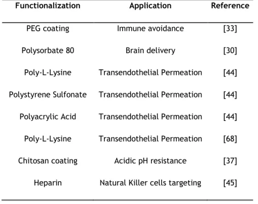
Table 2-1 – Applications of some specific functionalized SLNs. ... 15 Table 4-1 - Physicochemical properties of the NP formulations. Size, PdI and Zeta potential
for all the formulations considering both Witepsol E85 and cetyl palmitate SLNs for different homogenization and sonication times. ... 20 Table 4-2 - Physicochemical properties of the NP formulations. Size, PdI and Zeta potential
for all the formulations considering both Witepsol E85 and cetyl palmitate SLNs with oleic and stearic acid. ... 21 Table 4-3 - Physicochemical properties of the NP formulations. Size, PdI and Zeta potential
for all the formulations considering cetyl palmitate SLNs with stearic acid at different %. ... 21 Table 4-4 - Physicochemical properties of the NP formulations. Size, PdI and Zeta potential
for all the formulations for cetyl palmitate SLNs with surfactant at different %. ... 22 Table 5-1 – Physicochemical properties of the NP formulations. Size, PdI and Zeta potential
for all the formulations and MTX association for the MTX-loaded and MTX- and SPIONs-loaded SLN prior to NP functionalization and post-functionalization. ... 34
xvi
xvii
Glossary
List of Abbreviations
ACPA Anti-Citrullinated Peptides Antibody
AU Arbitrary Units
BBB Brain-Blood-Barrier BSA Bovine Serum Albumin
CMC Critical Micelar Concentration DLS Dynamic Light Scattering
DMARD Disease-Modifying Anti-Rheumatic Drug DMEM Dulbecco’s Modified Eagle’s Medium DMSO Dimethyl Sulfoxide
EDC 1-Ethyl-3-(3-dimethylaminopropyl) Carbodiimide Hydrochloride FEUP Faculdade de Engenharia da Universidade do Porto
FFUP Faculdade de Farmácia da Universidade do Porto FLS Fibroblast-Like Synoviocytes
FT-IR Fourier Transform InfraRed
GABAI Grupo de Análises Bioquímicas Ambientais e Industriais HPH High Pressure Homogenization
HPLC High Performance Liquid Chromatography
Ig Immunoglobulin
IL Interleukin
INEB Instituto Nacional de Engenharia Biomédica ISCS-N Instituto Superior de Ciências da Saúde – Norte LDH Lactate Dehydrogenase
mAb Monoclonal Antibody
MES 2-Morpholinoethanesulfonic Acid MLS Macrophage-Like Synoviocytes
xviii
MMP Matrix MetalloProteinase MRI Magnetic Resonance Imaging
MTT 3-(4,5-Dimethylthiazol-2-yl)-2,5-diphenyltetrazolium bromide
MTX Methotrexate
NHS N-Hydroxysulfosuccinimide NLC Nanostructured Lipid Carrier
NP Nanoparticle
NSAID Non-Steroidal Anti-Inflammatory Drug PALS Phase Analysis Light Scattering PBS Phosphate Buffer Saline PdI Polydispersity Index PEG Polyethylene Glycol
RA Rheumatoid Arthritis
REQUIMTE Rede de Química e Tecnologia SEM Scanning Electron Microscopy SLN Solid Lipid Nanoparticle
SPION Superparamagnetic Iron Oxide Nanoparticle TEM Transmission Electron Microscopy
TNF-α Tumor Necrosis Factor – alfa
1
1.
Introduction
1.1. The Joint
Muscles pull on bone to make them move, but movement would be impossible without joints. Joints allow the skeleton to be flexible, enabling it to bend in certain locations that in turn allows movement at different degrees around those locations while, simultaneously, providing protection for the same [1]. A joint, or articulation, is an anatomical location where two or more bones come together and can be classified into three groups depending on the type of structures that binds these bones: fibrous, cartilaginous and synovial [1, 2].
In a fibrous joint bones are united by a fibrous, collagen rich, connective tissue and have no joint cavity. This type of joint can be found between the bones of the skull, for instance. Cartilaginous joints are similar to fibrous joints but, in their turn, they are connected by cartilage, being it hyaline or fibrocartilage. An example of a cartilaginous joint is the manubriumsternal joint (between the manubrium and sternum). Synovial joints are more complex than fibrous and cartilaginous joints. In a synovial joint bones are not directly joined, the articular extremities of the bone are encapsulated in a synovial cavity by dense connective tissue and often strengthened by complementary ligaments as shown in Figure 1-1. An example of a synovial joint is the acromioclavicular joint (between the acromium and the clavicle) [1, 2].
Figure 1-1 – Structure of a synovial joint. Adapted from [1].
The different constituents of a synovial joint are described in detail below:
1. Joint capsule – composed by an outer fibrous capsule that may be attached to auxiliary ligaments, and an inner synovial membrane, also known as synovium, that secretes the
2
synovial fluid and contains two cell types, macrophage-like synoviocytes (MLS) and fibroblast-like synoviocytes (FLS). The joint capsule stabilizes and provides protection to the articulation;
2. Joint cavity – space comprised by the joint capsule and filled with synovial liquid. This liquid, containing hyaluronic acid and lubricin, provides lubrication, shock absorption and nourishment to the joint and bones;
3. Articular cartilage – hyaline cartilage covers the bone inside the synovial cavity increasing shock absorption and reducing friction between bones during movements [1-3].
Joints can also be classified according to the type and degree of movement allowed: i) synarthrosis, when they allow little to no mobility, ii) amphiarthrosis, when they allow slight mobility, and iii) diarthrosis, when they allow free mobility. This functional characterization is associated with their structural classification, thus most synarthrosis joints are fibrous joints, most amphiarthrosis joints are cartilaginous joints and all diarthrosis are synovial joints [1, 2].
1.2. Rheumatoid Arthritis
1.2.1. The Disease
Rheumatoid arthritis (RA) is a chronic systemic inflammatory disease of uncertain cause that affects 1% of the population of the developed world, being more common among women than men. RA usually develops in the fourth and fifth decade of life, with 80% of the total cases occurring between the ages of 35 and 50 [4-7]. It is also theorized that some individuals are more genetically susceptible to contract this disease than others [6, 8]. RA is often induced by an external agent (like cigarette smoking, infection or trauma) that triggers an autoimmune reaction, leading to synovial hypertrophy and chronic joint inflammation along with potential for extra-articular manifestations [4-8].
RA is usually a clinical syndrome spanning several disease subsets, which all lead towards a final common pathway in which persistent inflammation and associated damage to articular cartilage and underlying bone are present (figure 1-2). The primary symptoms of RA are pain, stiffness and swelling of the joints resulting in impaired physical function and movement. These symptoms are often accompanied by inherent symptoms such as fever and malaise. Although there are several associated mechanisms that trigger RA inflammation, the exact pathway that leads to it was not yet been determined [4-8].
The primary inflammation of RA starts in the synovium. An external factor induces the overproduction of TNF-α by MLS and FLS. In response, the tissue of the synovial lining enters hyperplasia, as a result of MLS and FLS proliferation and accumulation. This increased number of synoviocytes leads to an even higher production of TNF-α and an overproduction of many pro-inflammatory cytokines such as interleukin-6 (IL-6) and interleukin-1 (IL-1) that drive and maintain
3 the persistent inflammation and induce joint destruction, by increasing the production of matrix metalloproteinases (MMPs) and activating osteoclasts [4, 6, 7, 9]. It has also been observed that, in RA, FLS show abnormal behavior and may spread to other joints, supporting hypothesis that RA starts in joints [6].
Figure 1-2 – Pathogenesis of RA: synovial and systemic inflammation. Adapted from [7].
Autoantibodies also play an important role in RA by wrongly targeting the body’s own molecules as foreign or pathogenic. The main autoantibodies usually found in RA are rheumatoid factors (primarily IgM and IgA rheumatoid factors) and antibodies directed against citrullinated peptides (ACPA) located in the outer cellular membrane. In fact, 50-80% of all patients present at least one of these antibodies [6, 8]. It has also been registered that the presence of ACPA leads to histologically different types of RA, patients with ACPA (ACPA-positive) tend to have more lymphocytes in the synovial tissue; on the other hand, patients without ACPA (ACPA-negative) have increased fibrosis. Additionally, ACPA-positive RA is associated with increased joint damage and low remission rates [6].
1.2.2. Diagnosis, Management and Treatment
Early diagnosis and treatment have been recognized as essential for improving clinical outcomes in patients with rheumatoid arthritis. RA diagnosis is usually achieved by bloods tests or imaging techniques such as X-rays or magnetic resonance imaging (MRI) [4-6, 10]. However, diagnosis is somewhat difficult in the early stages of the disease since diagnostic criteria were developed from data obtained in patients with established rheumatoid arthritis, and therefore, are not readily applicable [11, 12].
4
In particular, MRI as demonstrated a greater sensitivity to early RA signs when compared to clinical examination or traditional radiology imaging. MRI allows for a direct visualization of the joint and enables the identification and analysis of the earliest histological and physiological alterations, making it a promising tool for an early RA diagnosis [5, 10-12].
Currently, there is no cure for RA, but some treatments are able to improve symptoms and hinder disease progression. These therapies aim to minimize symptoms such as articular pain and swelling, prevent bone erosion and deformity, and maintain joint functionality for daily activity. The best results with these treatments are obtained when they are initiated early and aggressively [4-6, 11, 13, 14].
Disease management is achieved primarily by using two main classes of medication: i) analgesics and non-steroidal anti-inflammatory drugs (NSAIDs) that lessen joint pain and stiffness, and disease-modifying anti-rheumatic drugs (DMARDs) that reduce joint swelling and pain, decrease acute-phase markers, limit progressive joint damage and improve function, by facilitating movement [5-7, 9, 13, 14].
The dominant DMARD in RA management is methotrexate (MTX), due to its proven efficacy, safety profile and low cost. MTX is an analogue of folic acid that ultimately disrupts cellular folate metabolism by inhibiting its target enzyme, the di-hydrofolate reductase. This drug seems to exert its anti-inflammatory effects by acting at different levels of the pathophysiological signaling cascade, directly inhibiting proliferation, pro-inflammatory cytokine production and inducing apoptosis in cells involved in immune and inflammatory reaction. These combined effects explain the overall anti-inflammatory action of MTX in the sustained management of RA [5, 6, 9].
New therapies are also been studied that consider the use of biological agents that target TNF-α and kinase receptors, in an attempt to disrupt RA signaling pathways, as well as the use of glucocorticoids to diminish synovial inflammation [6, 7, 13-16]. Despite recent developments and intensive research in RA, there is no definitive cure for the disease. Additionally, most of these treatments are also not targeted, leading to drug accumulation in undesired locations that may cause complications [6, 7, 13, 14].
1.3. Nanomedicine
Nanotechnology is the understanding and manipulation of materials whose dimensions range between 1-100 nm and has been in the forefront of much research in the past decades. Although nanotechnology comprises dimensions between 1-100 nm other systems consider materials with more than 100 nm to be applications of nanotechnology [17]. Traditionally, nanoparticles (NPs) are made of metals, ceramics and inorganic semiconductors. However, NPs can also be made of organic polymers, colloids or biomolecules, including DNA, proteins and lipids. This alternative approach is known as soft nanotechnology. This research led to innumerous applications, in particular the
5 application of this technology to medicine gave rise to nanomedicine [18-23]. As a consequence of the intensive work done in the area of nanotechnology, biocompatible colloidal systems, such as matrix NPs, micellar systems and conjugates have been developed. These systems enable nanomedicine applications for drug delivery and tissue regeneration strategies [17, 20, 22, 24].
Nanomedicine allowed the development of many drug delivery systems. Due to the side-effects of many drugs, nanoparticulate drug delivery systems were designed in order to achieve fewer complications. The main objective of using colloidal drug delivery systems is the reduction of systemic side-effects and maintenance of appropriate drug concentration in the required place. In these systems NPs are more interesting when compared with microparticles, since they possess a greater cell uptake, which is desirable when the drug is intended to penetrate the cellular membrane [20, 22, 24, 25].
The possibility of NP multi-functionalization opens the path for a novel and widespread medical applications based on nanomedicine strategies. (Figure 1-3). NPs have the potential to carry therapeutic and/or imaging agents, for theranostic applications. They can be conjugated to a specific ligand, such as antibodies, to target a specific tissue or organ or facilitate cellular penetration. They can also be used to avoid/bypass bio-barriers and macrophage uptake, by coating the NP with polyethylene glycol (PEG), for example [26-36].
Figure 1-3 – Schematic representation of a multifunctional NP. Adapted from [17].
Although nanomedicine has successfully achieved targeted drug delivery, this would not be possible without the prior identification of novel and specific cell surface markers. These markers are overly expressed in specific tissues or in specific disease-infected tissues and allow an accurate targeting to the tissues in question [28, 30-34, 37].
6
The multi-functionalization of NPs also offers new opportunities for nanomedicine applications, as it allows a simultaneous conjugation of therapeutic and diagnostic agents in a single approach. In the light of the new nanomedicine advancements, new theranostic approaches are being studied in order to provide more efficient diagnostic, treatment and monitoring of many diseases [21, 22, 32].
Nanomedicine approaches have been studied using several different NPs, like polymeric NPs [26, 29], magnetic NPs [27] and lipid NPs [28, 31, 38]. For the purposes of this work, lipid NPs, in particular solid lipid nanoparticles (SLNs), will be used. They will be more thoroughly discussed in the following sections.
7
2. State of the Art
In biology, naturally occurring nanostructures have been a source of inspiration for new nanotechnological designs and hybrid-nanostructures made of biological and non-biological, organic and inorganic building blocks. Lipids, with their amphiphilicity, diversity of head and tail chemistry, and antifouling properties that block nonspecific binding to lipid-coated surfaces, provide a powerful toolbox for nanotechnology [18, 19, 21-23].
2.1. Lipid Nanotechnology
The first attempt to incorporate lipophilic drugs in lipid droplets was the parenteral fat emulsion method developed in the 1960s, which is still widely used in the clinic for the parenteral application of poorly water-soluble drugs [23, 39]. But it was only in 1991 that the first generation of lipid NPs, the SLNs, were developed [23, 39-41].
Lipids possess desirable properties for applications in nanomedicine. They are biocompatible, biodegradable and well tolerated all over the body making them very versatile when considering various administration routes. Since the lipids used in the production of lipid nanoparticles naturally exist in the body, their degradation products are easily metabolized by the organism. Therefore lipid nanoparticles for drug delivery applications are associated to little or none systemic cytotoxicity [30, 31, 37, 42-45].
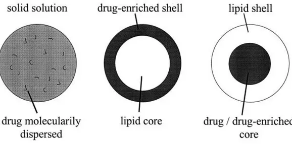
When designing a lipid nanoparticle for drug delivery applications three main types of particle models can be considered: i) solid solution with the drug molecularly dispersed, ii) lipid rich core with drug-enriched shell, and iii) lipid shell with drug-enriched core, as illustrated in figure 2-1. The first model allows for a constant release of drug, as the second and third models enable an immediate and a controlled release, respectively [25, 39-41].
Lipids also have the ability to self-assemble when in aqueous solution into different structures, depending on the lipids used. This enables fast and simple production methods that may also help to lower the cost of NP production and facilitate scale-up of the method used [23, 39, 46, 47].
As previously stated, different types of lipids are used to produce different types of lipid nanoparticles. The three main types of lipid nanoparticles are: i) liposomes and micelles, ii) SLNs, and iii) nanostructured lipid carriers (NLCs) [23, 25, 39, 41, 48, 49]. These different types of lipid nanoparticles are described in detail bellow.
8
Figure 2-1 - Three drug incorporation models (solid solution model (left), core-shell models with drug-enriched shell (middle) and drug-drug-enriched core (right)). Adapted from [40].
2.1.1. Liposomes and Micelles
Liposomes and micelles are small NPs formed by the self-assembly of amphipilic lipids in aqueous environment. Liposomes are bilayer vesicles, that may be uni- or multi-lamellar, while micelles are monolayer aggregates (figure 2-2) [23, 49-51]. In liposomes the polar heads of the lipids (hydrophilic) are located outwards in the bilayer, faced towards the inner aqueous core and the outer environment. This aqueous core represents an advantage in the use of liposomes for drug delivery as both hydrophilic and lipophilic drugs can be loaded inside the NP, in the core and inside the bilayer membrane of the NP, respectively. In micelles, on the other hand, the polar heads surround the entire lipid aggregate and the apolar tails (hydrophobic) are turned inwards, resulting in a more dense solely hydrophobic structure [49-51].
Figure 2-2 – Schematic representation of a liposome (left) and a micelle (right), both transversely sectioned. Adapted from [50].
9 Both liposomes and micelles may be derived from the hydration of lipid films or from disrupting cellular membranes. However they only form if a specific lipid concentration is achieved in the medium, i.e. if the critical micelar concentration (CMC) is reached [23, 49-51].
A key parameter in determining whether liposomes or micelles will be formed is the lipid geometry, since it has great influence in the packing and arranging of the lipids (figure 2-3). Cylindrical lipids, such as phosphatidylcholine, tend to arrange themselves to form lipid bi-layers, favoring liposomal structures, whereas conical lipids, such as lysophosphatidylcholine, tend to form micelles [49-51]. Some lipids, such as arachidonic acid, are known to have an inverted cone geometry and are capable of forming inverse micelles, relying on surfactants to stabilize the hydrophobic tails of the lipid [51, 52].
Figure 2-3 – Geometry of phosphatidylcholine (pink), lysophosphatidylcholine (blue), arachidonic acid (green) and their tendency to self-assemble. Adapted from [52].
As previously described, liposomes and micelles are usually produced by self-assembly in aqueous environment and can be easily functionalized recurring to the polar groups existing on their surface. This functionalization can be performed either before or after particle production. It is also possible to incorporate lipids that “react” to specific environments into the lipid layer, making the liposome or micelle responsive to certain conditions, such as low pH [23, 49-51].
Liposomes have found several different applications as NP-based drug delivery systems such as increase skin penetration after topical administration for meloxicam delivery [53], multiple modification for integrin targeting in triple-negative breast cancer [54] and coating with thermosensitive polymers for ultrasound-mediated drug delivery [55].
10
2.1.2. Solid Lipid Nanoparticles
SLNs are submicron sized nanoparticles, in the range of 50-1000 nm, composed of physiologically tolerated lipid components which are in a solid state at room temperature. These NPs consist of a solid lipid matrix that allows the encapsulation and protection of drugs against degradation and modification of the drug release profile [23, 39-42]. Several different lipids can be used to compose the matrix that has to be stabilized by a surfactant (emulsifier) or combination of surfactants that can highly influence the properties of the resulting particles [30, 43, 46-48, 56].
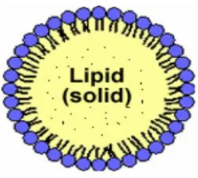
SLNs are seen as an alternative carrier system to polymeric NPs which is identical to oil-in-water (o/w) emulsions, but where the polymer of the emulsion is replaced by a solid lipid, shown in figure 2-4. Thanks to their structure and composition SLNs combine many of the advantages of polymeric nanoparticles, fat emulsions and liposomes [23, 25, 39, 41, 57, 58]. SLNs allow a controlled release of the encapsulated drug, with excellent biocompatibility and high drug content, increased stability and bioavailability of the bioactive compounds. These NPs are also capable of incorporating hydrophilic and hydrophobic drugs and can be easily subjected to commercial sterilization procedures. However there are a few disadvantages associated with SLNs such as high water content in the dispersions and the possible expulsion of drug during storage due to a shift in crystallization forms [23, 25, 39-41, 57, 59].
Figure 2-4 – Schematic representation of the structure of a solid lipid nanoparticle (SLN). Adapted from [41].
SLNs can be produced by several different methods from solid lipids, emulsifying agents and water/other solvent. The two most commonly used methods are High Pressure Homogenization (HPH), hot or cold, and w/o/w double emulsion [23, 41, 46, 47, 56]. Both methods are relatively simple and fast, at a relatively low cost, require no special solvents and easily allow scale-up production. HPH may cause drug degradation (high temperatures in hot homogenization) or coalescence (by high pressure) but require no organic solvents of any kind. Whereas double emulsion requires organic solvents but allows the encapsulation of thermo-sensible drugs [46, 47, 56].
11 SLNs have been used for several different applications by different groups such as to encapsulate SPIONs for controlled drug delivery [60], delivery systems of camptothecin to the brain by intravenous administration [31], oral administration of insulin after modification with stearic acid-actaarginine [61] and coated with chitosan in order to increase drug bioavailability after oral administration [37]. The functionalization of SLNs may be complicated because solid lipids that constitute the NPs possess few exposed polar groups, which are the targets of most functionalization methods [23, 40, 41, 48]. However some success has achieved by using electrostatic layer-by-layer assembly [44, 45], by functionalizing to surfactant instead of the lipids themselves [33, 62], by coating the surface of positively-charged SLNs with polymeric material like chitosan and then functionalizing the polymer [37] and by embedding phospholipids in the SLN and functionalizing them [33, 38].
2.1.3. Nanostructured Lipid Carriers
NLCs are similar to SLNs in several aspects and due to this share many of the same properties and advantages as them (described in the previous section). The main difference between NLCs and SLNs is that the first are produced from a blend of liquid and solid lipids, whereas the latter are produced solely of solid lipids [23, 25, 39, 59]. This compositional variation alters the structure of NP, SLNs tend to form perfect crystals while NLCs inhibit the crystallization process and produce imperfections in the lattices. This mixture of lipids in NLCs can be tailored in order to achieve a pretended nanostructure with the goal to better accommodate the drug or to alter its release profile. NLCs were developed in order to surpass the SLNs potential drug expulsion caused by crystalline form shift, this was achieved by producing a NP without a perfect crystallization of the lipid matrix, as previously described [23, 25, 39].
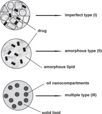
Three main types of NLCs have been described regarding their structure: i) imperfect type, ii) amorphous type and iii) multiple type, [25, 39, 41, 59] as shown in figure 2-5.
12
Figure 2-5 – Schematic representation of the three types of NLCs: imperfect type (top), amorphous type (middle) and multiple type (bottom). Adapted from [59].
Imperfect type NLCs have an imperfect structured solid matrix, this is achieved by mixing different lipids (liquid and solid) that cause imperfections in the crystallization of the lipid matrix. These imperfections are what accommodate the drug, additionally mixing small of chemically different liquid lipids increases incompatibility, consequently increasing the number of imperfections and the drug payload. Amorphous type NLCs possess a solid amorphous matrix without any specific structure that prevents drug expulsion caused by crystallization form shift since the matrix is amorphous and not crystalline. This is achieved by mixing solid lipids with special lipids such as isopropylmyristate. Multiple type NLCs consist of multiple oil in fat in water (O/F/W) double emulsions, where the drug is solubilized in the lipophilic phase and its solubility decreases during cooling and storage. Many drugs have higher solubility in liquid lipids than in solid lipids and therefore a higher payload can be obtained using this type of NLC [25, 39, 59].
NLCs have been proposed for applications of different natures such as an attempt to increase cellular uptake by modifying the NLCs with chitosan hydrochlorides [63], optimization of intranasal delivery of artemether [64] and to try and enhance in vitro activity by surpassing drug resistance of Doxorubicin and Ducosahexaenoic acid in cancer cells [65].
13
2.2. Nanoparticle Functionalization
Nanomedicine has brought a new tool for medical applications laid on the ability to multi-functionalize the drug delivery systems for innumerous purposes. These functionalization can be obtained by different methods, depending on the type of NP and its composition, and designed for different ends, both of which are described in the following sections.
2.2.1. Functionalization Methods
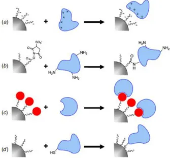
NP functionalization can be achieved by four main approaches (shown in figure 2-6): i) electrostatic attachment, ii) covalent linkage to a NP ligand, iii) attachment to a co-factor bound to the ligand or the NP itself, and iv) direct covalent linkage to the NP surface [27, 33, 34, 66, 67].
Figure 2-6 – Schematic representation of the four main approaches for NP functionalization: (a) electrostatic attachment, (b) covalent attachment to ligand, (c) attachment to a co-factor and (d) covalent attachment to the NP surface. Adapted from [66].
Electrostatic attachment is the simplest approach that requires no type of chemical reaction consisting of an electrostatic adsorption between differently charged NP and biomolecule. In order to perform this method the surface of the NP has to be electrically charged (negative or positive) so that a biomolecule with the opposite charge can be adsorbed. This method is already routinely used as an electron dense marker in histology [27, 33, 66, 67].
Another general method for NP functionalization is covalent linking to a NP ligand, occurring when a biomolecule (usually a protein or peptide) is able to react with a ligand to form a covalent bound. This linkage is very stable and resistant and is also widely used for surface modification in several NPs applications. The most commonly used functional groups for this reaction are amine and carboxylic acid groups. The method of direct covalent linkage is very similar to this method but instead of the
14
biomolecule reacting with a functional group on the end of the ligand it reacts with one directly on NP surface [33, 66, 67].
Alternatively, NP functionalization can be achieved attachment to a co-factor. This is usually performed by using specific labeling strategies of bioconjugation: firstly a labeling molecule is attached to the NP surface or NP ligand and after this a second molecule (linked to the desired biomolecule) with great affinity for the label is added creating a stable linking complex. The most commonly used linkage is biotin-streptavidin that allows for up to 4 biotin molecules to be linked to 1 streptavidin and also has a nearly covalent dissociation constant. Note that the antibody conjugation falls into this category of linkage, since the antibody will be linked to the NP and recognize specific epitopes binding only to them [33, 66, 67].
2.2.1.1. Lipid NPs Functionalization
The functionalization of lipid NPs is generally obtained by covalent linkage, directly to the surface or an attached ligand, or by bioconjugation methods. These methods usually imply the use of a lipid “anchor” with an amine, phosphate or carboxylic acid in the head portion, since not all lipids possess functional groups that are capable of functionalization reactions. This lipid anchor is usually pre-functionalized and can be incorporated during NP synthesis or post-synthesis.
Several groups have been able to successfully functionalize SLNs for innumerous nanomedicine applications using the previously described methods, or similar methods with slight variations, as shown in table 2-1.
2.2.2. Functionalization Purposes
A NP functionalization can be performed to achieve a variety of purposes, Table 2-1 shows some examples of SLNs with functionalization for specific applications. Note that the Table 2-1 only shows SLN applications because they are the focus of this work, but the applications described in this section are transversal to all NPs. Most NP surface functionalization aim to: i) decrease identification and elimination by the immune system in the blood stream, ii) enable specific targeting of the NP to the pretended tissue or organ, or iii) facilitate the entry of the NP into the cells [26-29, 32-34, 67].
The coating of the NP with long chain hydrophilic polymers allows the creation of stealth NPs that are not easily recognized by the immune system and therefore have increased circulation time [26, 29, 32, 33]. The use of polyethylene glycol (PEG) chains has been highly described for this type of application and has demonstrated excellent efficacy [26, 29].
Some molecules have the ability to permeate a cell by using specific selective channels in the cellular membrane. These channels could be used as an advantage to increase NP uptake by placing the same molecules in the surface of the NP [33, 34, 66, 67]. This technique has already been
15 successfully applied, for instance, in the coating NP with polysorbate 80 to help penetrate the Brain-Blood-Barrier (BBB) [30].
NP surface coating with targeting molecules is the most common application of NP surface functionalization, as the molecules are capable of recognizing specific cell membrane markers and attaching only to them, enabling highly specific cell targeting [33, 34, 66, 67]. The use of antibodies as targeting agents is widely described in the literature for targeted drug delivery for example [33, 34].
Table 2-1 – Applications of some specific functionalized SLNs.
Functionalization Application Reference
PEG coating Immune avoidance [33]
Polysorbate 80 Brain delivery [30]
Poly-L-Lysine Transendothelial Permeation [44]
Polystyrene Sulfonate Transendothelial Permeation [44]
Polyacrylic Acid Transendothelial Permeation [44]
Poly-L-Lysine Transendothelial Permeation [68]
Chitosan coating Acidic pH resistance [37]
16
17
3. Rationale and Strategy of the Thesis
In this work, an approach to attempt targeted therapy and simultaneous imaging is proposed. This study aims to develop a targeted theranostic system for intravenous administration, consisting of the encapsulation of MTX (RA therapeutic drug) and SPIONs (MRI contrast agent) within SLNs. These SLNs are functionalized with a monoclonal antibody (mAb) against the MLS specific cell surface receptor, CD64. A representation of the strategy is shown in figure 3-1.
This work was divided in to two separate phases. Firstly a research on the fundamental basics and the current state of the art was performed as well as a planning of the work to be done in the experimental phase of the work. This phase took place during the first semester of the 2013/2014 school year, from October to January. The second place was accomplished during the second semester of the same school year, from February to June, and consisted mainly of experimental research work, conducted in the department of Chemical Sciences of the Faculty of Pharmacy, University of Porto.
Considering the experimental research work, the first step was NP production and characterization. After the process of optimization, four different formulations were prepared: i) cetyl palmitate SLNs, ii) MTX-loaded cetyl palmitate SLNs, iii) SPIONs-loaded cetyl palmitate SLNs and iv) MTX- and SPIONs-loaded cetyl palmitate SLNs. All of these formulations were then extensively characterized in terms of NP diameter, polydispersion, zeta potential and morphology. The NP content of MTX and SPIONs were also determined. The four formulations were also conjugated with an antibody and its presence was verified and quantified. This extensive characterization enabled comparison of NPs properties and the assessment of each of the formulation’s components effect on those properties. Finally in vitro assays were performed, using the monocytic cell line THP-1, in order to assess cellular viability and NP’s cytotoxicity.
18
19
4. Materials and Methods
4.1. Materials
The lipids cetyl palmitate and Witepsol E85 were gently provided by Gattefossé (Saint Priest Cedex, France) and Sasol (Joanesburgo, Gauteng, South Africa), respectively. Stearic acid was purchased from Merck (© Merck KGaA, Darmstadt, Germany). MTX was obtained by courtesy of Excella (Excella GmbH, Feucht, Germany). Iron Oxide Nanocrystals (10 nm) coated with oleic acid and dispersed in chloroform (25 mg/mL) were kindly provided by Ocean Nanotech (Ocean Nanotech Inc., Springdale, AR, USA). Anti-Human CD64 (Fc gamma Receptor 1) antibody solution (1 mg/mL) was purchased from eBioscience (eBioscience Inc., San Diego, CA, USA). LDH Cytotoxicity Detection Kit was from Takara Bio (Takara Bio Inc., Shiga, Japan). Twee 60, dimethyl sulfoxide ACS reagent ≥99.9% (DMSO), dichloromethane (ACS reagent, ≥99.5% contains 50 ppm amylene as stabilize), ethyl acetate (ACS reagent, ≥99.5%), 2-morpholinoethanesulfonic acid low moisture content, ≥99% (MES), 1-ethyl-3-(3-dimethylaminopropyl) carbodiimide hydrochloride purum, ≥98.0% (EDC), N-hydroxysulfosuccinimide 98% (NHS), Thiazolyl Blue Tetrazolium Bromide 98% (MTT), Triton™ X-100 for molecular biology, Trypan Blue powder and phorbol 12-myristate 13-acetate (PMA) were purchased from Sigma-Aldrich (St. Luis, MO, USA). Dulbecco’s Modified Eagle’s Medium (DMEM), Dulbecco’s phosphate buffer saline 10x pH 7.4 (PBS), fetal bovine serum (FBS), Penicillin-Streptomycinm and Fungizone ® Antimycotic were purchased from Gibco® (Invitrogen Corporation, Paisley, UK). Aqueous solutions were prepared with double-deionized water (Arium Pro, Sartorius AG, Göttingen, Germany). THP-1 (ATCC® TIB-202TM) cells were generously provided by Professor Susana
Santos and Doctor Marta Freitas, INEB
4.2. Methods
4.2.1. Preparation of Multifunctional Nanoparticles
4.2.1.1. Optimization of Nanoparticle production method
In the first attempt of NP production both cetyl palmitate and Witepsol E85 were used in an organic solvent-free emulsification-sonication method that combined high shear homogenization and ultra-sonication. As standard procedure, 500 mg of solid lipid (cetyl palmitate and Witepsol E85) and 100 mg of surfactant (Tween 60) were heated to 70ºC, in order to fuse the lipids. After the lipids were totally fused, 4.400 mL of water at 70ºC were added to the fused lipid-surfactant mixture, and the resulting solution was immediately homogenized with a high-shear homogenizer (YSTRAL GMBH X10/20-E3, Ballrechten-Dottingen, Germany) for 30 seconds at 12000 rpms to produce a lipid-in-water micro-emulsion. This micro-emulsion was afterwards re-homogenized using a probe-type sonicator (model VCX-130 with a VC 18 probe, Sonics&Materials Inc., Newtown, CT, USA) for 5 minutes at 80%
20
amplitude to produce the NPs. The resulting NP suspension was stored in glass flasks at room temperature until further use.
Table 4-1 - Physicochemical properties of the NP formulations. Size, PdI and Zeta potential for all the formulations considering both Witepsol E85 and cetyl palmitate SLNs for different homogenization and sonication times. Lipid used in SLN Homogenization time (sec) Sonication
time (min) Size (nm) PdI
Zeta Potential (mV) Witepsol E85 30 5 798.5 ± 16.8 0.372 ± 0.004 -34.27 ± 0.66 Witepsol E85 30 10 954.5 ± 3.40 0.372 ± 0.016 -27.14 ± 0.96 Witepsol E85 30 15 762.9 ± 37.5 0.350 ± 0.022 -28,04 ± 1,05 Witepsol E85 60 5 781.1 ± 125.8 0.334 ± 0.019 -22.39 ± 0.87 Witepsol E85 60 10 639.3 ± 68.1 0.346 ± 0.003 -26.03 ± 0.64 Witepsol E85 60 15 338.3 ± 9.6 0.334 ± 0.026 -29.02 ± 1.14 Witepsol E85 120 5 729.2 ± 68.3 0.388 ± 0.006 -28.31 ± 0.76 Witepsol E85 120 10 661.9 ± 30.6 0.333 ± 0.030 -28.16 ± 0.95 Witepsol E85 120 15 536.0 ± 21.6 0.379 ± 0.004 -21.99 ± 2.69 Cetyl Palmitate 30 5 466.4 ± 16.7 0.248 ± 0.019 -36.67 ± 0.78 Cetyl Palmitate 30 10 333.0 ± 8.2 0.280 ± 0.010 -36.18 ± 0.75 Cetyl Palmitate 30 15 471.2 ± 7.8 0.286 ± 0.010 -36.35 ± 0.92 Cetyl Palmitate 60 5 455.7 ± 3.6 0.277 ± 0.008 -36.52 ± 0.83 Cetyl Palmitate 60 10 426.2 ± 2.2 0.277 ± 0.004 -34.02 ± 0.69 Cetyl Palmitate 60 15 430.0 ± 5.9 0.247 ± 0.011 -33.57 ± 1.86 Cetyl Palmitate 120 5 397.3 ± 6.3 0.234 ± 0.015 -31.71 ± 1.04 Cetyl Palmitate 120 10 437.3 ± 7.1 0.252 ± 0.010 -29.20 ± 1.01 Cetyl Palmitate 120 15 415.3 ± 7.1 0.246 ± 0.017 -37.13 ± 0.49
In an attempt to better understand the effects of the homogenization process in NPs properties new NPs were prepared, using the same method that was previously described but increasing both homogenization and sonication times. It was found that increasing the homogenization times (30 seconds, 1 minute and 2 minutes) or the sonication times (5 minutes, 10 minutes and 15 minutes)
21 resulted in little changes to the NP properties and so no alterations were done to the original method (table 4-1).
To later facilitate NP functionalization the number of available functional groups, in particular carboxylic terminal groups, on the NP surface should be increased as much as possible. In order to achieve, this the addition of a fatty acid was proposed. Fatty acids have long hydrophobic chains that enable an easy integration into the SLN matrix and possess a terminal carboxylic acid group that could be used for antibody conjugation. Two fatty acids were chosen and assessed (Table 4-2) for this: i) stearic acid (an 18-C long saturated fatty acid) and ii) oleic acid (an 18-C long unsaturated acid fatty acid). The results showed that the NP formulation with more promising characteristics were composed of cetyl palmitate and stearic acid and, therefore, the other formulations were discarded at this point. To further optimize the cetyl palmitate/stearic acid formulation several proportions of acid were used (2.5%, 5%, 7.5% and 10%) and best properties were obtained using 7.5% (regarding total formulation weight) of stearic acid (Table 4-3).
Table 4-2 - Physicochemical properties of the NP formulations. Size, PdI and Zeta potential for all the formulations considering both Witepsol E85 and cetyl palmitate SLNs with oleic and stearic acid.
Lipid used in SLN Fatty acid Size (nm) PdI Zeta Potential (mV)
Witepsol E85 Oleic acid 607.0 ± 18.1 0.358 ± 0.008 -41.10 ± 3.10 Witepsol E85 Stearic acid 660.9 ± 56.0 0.309 ± 0.020 -36.93 ± 1.42 Cetyl palmitate Oleic acid 426.6 ± 150.3 0.241 ± 0.025 -39.08 ± 1,21 Cetyl palmitate Stearic acid 408.6 ± 16.4 0.229 ± 0.031 -41.11 ± 0.35
Table 4-3 - Physicochemical properties of the NP formulations. Size, PdI and Zeta potential for all the formulations considering cetyl palmitate SLNs with stearic acid at different %.
Lipid used in SLN Stearic acid % Size (nm) PdI Zeta Potential (mV)
Cetyl palmitate 2.5% 607.0 ± 18.1 0.358 ± 0.008 -28.84 ± 2.33 Cetyl palmitate 5.0% 660.9 ± 56.0 0.309 ± 0.020 -34.58 ± 1.50 Cetyl palmitate 7.5% 426.6 ± 150.3 0.241 ± 0.025 -40.04 ± 0.96 Cetyl palmitate 10.0% 408.6 ± 16.4 0.229 ± 0.031 -39.62 ± 1.45
The selected formulation (cetyl palmitate with 7.5% stearic acid) was still presenting dimensions that were too large for the pretended application. On a first attempt to reduce NP diameter the NP suspension were filtrated (using disposable syringes and cellulose filters with several pore sizes). Filtration greatly improved NP diameter but it was shown to retain a large amount of NPs in the filters, causing a large amount of mass loss that would be translated in the future to large drug loss. Because of this issue filtration was discarded from the process. In another attempt to reduce NP size a decrease in total lipid quantity (600 mg to 400 mg of total formulation) and an increase in the
22
lipid/surfactant proportion (16%, 20%, 25%, 30% and 35% of the formulation total) was considered (the latter results can be observed in Table 4-4). The formulation that presented the best NP characteristics was composed of 400 mg of total mass with theoretical 35% of surfactant.
Table 4-4 - Physicochemical properties of the NP formulations. Size, PdI and Zeta potential for all the formulations for cetyl palmitate SLNs with surfactant at different %.
Lipid used in SLN Surfactant (%) Size (nm) PdI Zeta Potential (mV)
Cetyl palmitate 16% 434.2 ± 12.8 0.284 ± 0.015 -37.65 ± 1.31 Cetyl palmitate 20% 340.3 ± 5.3 0.231 ± 0.007 -49.78 ± 0.70 Cetyl palmitate 25% 255.6 ± 4.0 0.206 ± 0.008 -47.27 ± 1.19 Cetyl palmitate 30% 214.4 ± 3.6 0.200 ± 0.008 -46.78 ± 0.61 Cetyl palmitate 35% 164.6 ± 1.9 0.189 ± 0.008 -44.33 ± 1.30
4.2.1.2. Definitive Nanoparticle production method
SLNs composed of cetyl palmitate and stearic acid were prepared by an organic solvent-free emulsification-sonication method that combined high shear homogenization and ultra-sonication (figure 4-1). As standard procedure, 230 mg of cetyl palmitate, 30 mg of stearic acid and 140 mg of Tween 60 were heated to 70ºC (temperature above the fusion point of all the lipids in the mixture), in order to fuse the lipids. 4.400 mL of water at 70ºC was added to fused lipid-surfactant mixture and the resulting solution was immediately homogenized with a High-shear Homogenizer (YSTRAL GMBH X10/20-E3, Ballrechten-Dottingen, Germany) for 30 seconds at 12000 rpms to produce a lipid-in-water micro-emulsion. This micro-emulsion was afterwards re-homogenized using a probe-type sonicator (model VCX-130 with a VC 18 probe, Sonics&Materials Inc., Newtown, CT, USA) for 5 minutes at 80% amplitude to produce the NPs. The resulting NP suspension was stored in glass flasks at room temperature until further use.
The encapsulation of both MTX and SPIONs was achieved during the production method [35]. 4 mg of MTX were added prior to the lipid fusing and dissolved, as much as possible, in fused solid lipid + surfactant mixture. 40 µl of SPION dispersion were added after the lipid fusion and before the addition of the water.
23
Figure 4-1 – Schematic representation of the NP preparation procedure.
Four different formulations were prepared: i) SLN-Placebo (cetyl palmitate NPs not containing MTX or SPIONs), ii) SLN-MTX (cetyl palmitate NPs containing only MTX), iii) SLN-SPIONs (cetyl palmitate containing only SPIONs) and iv) SLN-MTX/SPIONs (cetyl palmitate containing both MTX and SPIONs).
4.2.1.3. Antibody conjugation to the Solid Lipid Nanoparticles
As previously stated, the SLN were further functionalized with an anti-CD64 antibody that is able to recognize macrophage’s CD64 (present in the macrophage cell membrane and over expressed in RA). This recognition is performed by the antibody’s Fc region and because of this any attempt of antibody conjugation must be done through the antibody’s Fab fragment in order to maintain its functionality [69]. Considering this, the conjugation reaction performed in two steps: i) the NP’s carboxylic groups (present in the stearic acid that was integrated into the SLN matrix) were activated in the presence of EDC and NHS, and ii) the antibody’s primary amine (Fab fragment) reacted with the activated carboxylic group to form a stable amide bound between the SLN surface and the antibody [35]. 230 mg of Cetyl Palmitate 30 mg of Stearic Acid 140 mg of Tween 60 Heat up till 70ºC Dissolve 4 mg of MTX Add 40 µL of SPIONs add 4.400 mL of water High Shear Homogenization for 30 secs at 12000 rpms Sonication for 5 minutes at 80% amplitude Store in glass flasks at
room temperature - Characterization
- Functionalization - Etc
24
Figure 4-2 –Schematic representation of the NP functionalization procedure’s several reactions. Adapted from [35].
The NP formulations were diluted in 10 mL of MES buffer, pH 5.0 (this value of pH ensures maximum EDC attachment to carboxylic groups), so that the final concentration is 10 mg of NPs per mL of buffer. The NPs were then activated by adding 1 mL of 0.1 M EDC and 1 mL of 0.7 M NHS (both dissolved in MES buffer, pH 5.0) to the previously prepared suspension. The NP suspension was then kept at room temperature under moderate stirring for 1h. 1 mL of the activated-NP suspension was transferred to an Eppendorf® tube and 10 µL of the anti-CD64 antibody solution were added to the
latter. The resulting suspensions were then homogenized with a vortex and incubated at 4ºC for 24h. The functionalized NPs were then stored at 4ºC until further usage [35].
4.2.2. Nanoparticle characterization
4.2.2.1. Dynamic light scattering and phase analysis light
scattering
Dynamic light scattering (DLS), also known as photon correlation spectroscopy or quasi-elastic light scattering, is a commonly used technique for the determination of size distribution profiles in the sub-micron range. The technique is based on the Brownian motion, that is, particles and molecules are in constant random motion and the speed at which they move is related to their size (smaller particles move faster that larger ones). The movement speed can be determined by measuring the changes of the scattered intensity at a specific angle through time and, taking into account the temperature of the suspension, the hydrodynamic radius of the NPs can be determined was well as its size distribution [70].
25 Zeta potential is a physical property related to the charge of a particle and can be used to infer on the stability of a suspension or system. When the modulus of the zeta potential is high the particles tend to repel each other, due to strong electric charges, and hence do not tend to agglomerate. However if the modulus is low the electric charges will not have enough force to repel each other and the particles will have a tendency to form agglomerates. The zeta potential can be measured by phase analysis light scattering (PALS), that relies on applying an electric field to the suspension that will force the particles to move the electrodes depending on their charge. The movement speed of the particles is dependent of the field applied and of the particles zeta potential, since the field is known the zeta potential can be easily determined [71].
The NP formulations were characterized in terms of particle diameter, diameter distribuition profile (polydispersity index - PdI) and zeta potential by upper mentioned techniques. Mean diameter and PdI were determined by DLS using a 90Plus Particle Size Analyzer (Brookhaven Instruments Corporation, Holtsville, NY, USA) and the zeta potential was assessed by PALS using a ZetaPALS Zeta Potential Analyzer (Brookhaven Instruments Corporation, Holtsville, NY, USA), at 660 nm, at 20ºC with a detection angle of 90º. The formulations were previously diluted in water, the non-conjugated NPs 400x and the conjugated NPs 4x, to achieve a final concentration of 250 µg of NP per mL for all formulations. All measurements were performed with three independent batches of NPs, with six runs each.
4.2.2.2. Transmission Electron Microscopy
Transmission electron microscopy (TEM) uses an electron beam to illuminate ultra-thin samples (under 0.5 µm thick). The electrons interact with the sample and those that pass through it are collected by a system of electromagnetic lenses that greatly magnifies and focus the image. The image formed is collected and displayed in an imaging device, such as a florescent screen, on a layer of photographic film or, more commonly, its detected by a sensor such as a CDD camera.
Prior to the TEM analysis, the non-conjugated NP suspensions were diluted 100X in water to achieve a 1 mg/mL concentration, since the conjugated NPs were already at this concentration (result of the functionalization procedure). Samples were prepared by placing 10 µL of NP dispersion on a copper-mesh grid (one grid for each different sample) and after 2 minutes the excess of suspension was removed with filter paper. As a contrasting agent, 10 µL of 0.75% uranyl acetate solution were placed on the grid and after 30 seconds the excess was removed using filter paper. The grids were after observed in a JEM-1400 Transmission Electron Microscope (JEOL Ltd.), with an accelerating voltage of 80 kV.
26
4.2.2.3. MTX Quantification
Spectrophotometry is a technique that allows for a quantitative measurement of the amount of radiation that a samples absorbs. A beam of light (visible or not) passes through a sample and the amount of light that reach the detector is measured. The amount of light that a molecule absorbs, at a specific wavelength, can then be used to determine the amount of that molecule that is present in the solution. UV/Vis spectrophotometry was used to determine the amount of MTX present in the formulations using an indirect method, i.e. the amount of drug that was present outside the NPs was dosed, and the association efficiency was determined by subtracting the amount of MTX that remained in the aqueous phase to total amount of MTX (equation 1).
(1)
Association efficiency (%) =
Total amount of MTX−Amount of MTX in the supernatantTotal amount of MTX
× 100
Firstly the NP suspensions were diluted 200x and filtrated with Amicon ® Ultra Centrifugal Filters Ultracell-50 kDa (EMD Millipore, Darmstadt, Germany) at 3500 G for 30 minutes using a HeraeusTM
MultifugeTM X1R centrifuge (Thermo Scientific, Waltham, MA, USA). The resulting supernatant was
collected for MTX quantification by spectrophotometry. The absorbance of the supernatant was then read using a V-660 UV/Vis Spectrophotometer (Jasco Inc., Easton, MD, USA), and the values of the absorbance were registered at 299 nm wavelength (MTX absorbance peak). The readings of each sample, containing MTX, were corrected using the supernatant resulting from filtration of the respective formulation without MTX (SLN-placebo was used as baseline for SLN-MTX, and SLN-SPIONs was used as baseline for SLN-MTX/SPIONs) in order to minimize formulation interferences. Each sample reading was performed in triplicate (each from an independent batch of NP formulations).
A calibration curve was constructed using standard solutions of MTX with the final concentrations of 0.000625, 0.00125, 0.0025, 0.005, 0.010, 0.020, 0.030 and 0.040 mg/mL. In order to minimize reading interferences these standard solutions were prepared in the supernatant resulting from the previously described filtration of SLN-placebo NP suspension.
Figure 4-3 – Standard curve for MTX quantification using spectrophotometry.
y = 24,562x + 0,1614 R² = 0,9794 0 0,2 0,4 0,6 0,8 1 1,2 1,4 0 0,005 0,01 0,015 0,02 0,025 0,03 0,035 0,04 0,045 Abso rbance ( AU) MTX concentration (mg/mL)
27
4.2.2.4. Fourier Transform Infrared spectroscopy
Fourier transform infrared (FT-IR) spectroscopy is a technique that allows the identification of samples specific molecular bonds that can used to identify components. It uses an infrared beam that passes through the sample and collects an absorbance/transmittance spectrum whose peaks correspond to the frequencies of vibrations between the bonds of the chemical elements present in the sample. Because each different material is a unique combination of atoms, the infrared spectrum works as molecule fingerprint making it possible to identify, with great certainty, the components of the sample[72]. FT-IR analysis proved extremely useful in determining the presence of both MTX and antibody within the NP formulations.
Prior to FT-IR analysis the NP formulations were lyophilized, firstly 1 mL of NP suspension was frozen at -80ºC for 30 minutes and after place in a Bench Top Pro with Omnitronics (SP Scientific, Warminster, PA, USA) at -80ºC at 150 mTorr for 18h. The resulting powders were then characterized by FT-IR analysis, using a Frontier FT-IR Spectrometer with Universal ATR Sampling Accessory (PerkinElmer, Waltham, MA, USA). Each of the infrared spectrum was collect with 32-scans with 4 cm-1 resolution in the mid-infrared region (3700 to 600 cm-1).
4.2.2.5. Antibody Quantification
The amount of antibody conjugated to the NPs was indirectly determined by quantifying the total amount of protein of the formulation that was not linked to the NPs. In order to separate the free antibody from the NP-conjugated antibody the conjugated NP suspension was centrifuged at 33000 G for 5h, and the supernatant was carefully collected.
The antibody was after quantified using a colorimetric method with the Thermo ScientificTM
Coomassie (Bradford) Protein Assay Kit. This kit allows for a quick and viable quantification of the total amount of protein present – when Coomassie dye binds to a protein, in acidic medium, it produces an immediate color change from brown to blue, resulting in a shift of the maximum absorbance peak from 465 nm to 595 nm [73].
Firstly a calibration curve for the amount of protein was constructed (Figure 4-3) using standard bovine serum albumin (BSA) solutions with concentrations of 25, 20, 15, 10, 5, 2.5 and 0 µg/mL of BSA in PBS. In order to measure the absorbance of both standard solutions and samples, the micro microplate protocol was used. 150 µL of the standard solutions/samples were transferred to a 96-well plate, in triplicate, and 150 µL of the coomassie reagent was added to those plates. The 96-96-well plate was stirred for 30 seconds and left to incubate at room temperature protected from the light. The plate was then read at 595 nm using a SynergyTM HT Multi-mode Microplate Reader (Biotek
![Figure 1-1 – Structure of a synovial joint. Adapted from [1].](https://thumb-eu.123doks.com/thumbv2/123dok_br/15721703.1070543/19.892.224.697.690.973/figure-structure-synovial-joint-adapted.webp)
![Figure 1-2 – Pathogenesis of RA: synovial and systemic inflammation. Adapted from [7]](https://thumb-eu.123doks.com/thumbv2/123dok_br/15721703.1070543/21.892.137.780.230.603/figure-pathogenesis-ra-synovial-systemic-inflammation-adapted.webp)
![Figure 1-3 – Schematic representation of a multifunctional NP. Adapted from [17].](https://thumb-eu.123doks.com/thumbv2/123dok_br/15721703.1070543/23.892.258.652.595.959/figure-schematic-representation-multifunctional-np-adapted.webp)