Volume 2013, Article ID 523761,15pages http://dx.doi.org/10.1155/2013/523761
Research Article
Antileukotriene Reverts the Early Effects of
Inflammatory Response of Distal Parenchyma in
Experimental Chronic Allergic Inflammation
Nathália Brandão Gobbato,
1Flávia Castro Ribas de Souza,
1Stella Bruna Napolitano Fumagalli,
1Fernanda Degobbi Tenório Quirino dos
Santos Lopes,
1Carla Máximo Prado,
2Milton Arruda Martins,
1Iolanda de Fátima Lopes Calvo Tibério,
1and Edna Aparecida Leick
11Medicine Department, School of Medicine, University of S˜ao Paulo(USP), 01246-903 S˜ao Paulo, SP, Brazil 2Biological Science Department, Universidade Federal de S˜ao Paulo(UNIFESP), 09972-270 Diadema, SP, Brazil
Correspondence should be addressed to Edna Aparecida Leick; leick51@yahoo.com.br
Received 2 May 2013; Revised 23 July 2013; Accepted 24 July 2013
Academic Editor: Alexandre Paula Rogerio
Copyright © 2013 Nath´alia Brand˜ao Gobbato et al. his is an open access article distributed under the Creative Commons Attribution License, which permits unrestricted use, distribution, and reproduction in any medium, provided the original work is properly cited.
Aims. Compare the efects of montelukast or dexamethasone in distal lung parenchyma and airway walls of guinea pigs (GP) with chronic allergic inlammation.Methods. GP have inhaled ovalbumin (OVA group-2x/week/4weeks). Ater the 4th inhalation, GP were treated with montelukast or dexamethasone. Ater 72 hours of the 7th inhalation, GP were anesthetised, and lungs were removed and submitted to histopathological evaluation.Results. Montelukast and dexamethasone treatments reduced the number of eosinophils in airway wall and distal lung parenchyma compared to OVA group (� < 0.05). On distal parenchyma, both treatments were efective in reducing RANTES, NF-�B, and ibronectin positive cells compared to OVA group (� < 0.001). Montelukast was more efective in reducing eotaxin positive cells on distal parenchyma compared to dexamethasone treatment (� < 0.001), while there was a more expressive reduction of IGF-I positive cells in OVA-D group (� < 0.001). On airway walls, montelukast and dexamethasone were efective in reducing IGF-I, RANTES, and ibronectin positive cells compared to OVA group (� < 0.05). Dexamethasone was more efective in reducing the number of eotaxin and NF-�B positive cells than Montelukast (� < 0.05).Conclusions. In this animal model, both treatments were efective in modulating allergic inlammation and remodeling distal lung parenchyma and airway wall, contributing to a better control of the inlammatory response.
1. Introduction
Asthma plays an important role as a major cause of mortality and morbidity all over the world. Data from the World Health Organization estimates that 100 millions to 150 millions
people worldwide have a diagnosis of asthma [1], and around
180.000 people around the world die, every year, from this disease. Moreover, asthma afects people in school age and economically active people, having a high social and economic impact with lost productivity and reduced partici-pation in family life. herefore, it is considered a public health problem with high cost medications and hospitalizations of
patients, enhancing the importance of continuing studying asthma.
Bronchial asthma is a chronic inlammatory disorder involving chronic airway inlammation, tissue remodeling, and declined airway function. he asthma physiopathology involves mediators and many cells, mainly eosinophils which plays important role through the release of speciic mediators
[2], and the degree of eosinophilia has been related with the
disease severity in some asthmatic patients [3].
factors can be involved in airway inlammation, including insulin-like growth factor-1 (IGF-I) which is upregulated
dur-ing this event [4], contributing with the remodeling process
[5]. Furthermore, we may also consider the extracellular
matrix (ECM) proteins, as ibronectin for example. Increased deposition of ibronectin and collagen into the subepithelial space of the airways is observed in all forms of asthma and
appears very early in the disease [6].
Much of the subjacent inlammation of asthma can be mediated by the transcription nuclear factor-kappa B
(NF-�B), which is involved in the regulation of many of
the inlammatory proteins that are expressed in asthmatic
airways [7].
he importance of distal lung responses to the global pulmonary alterations enhancing asthma symptoms,
partic-ularly on severe asthma, has been recently addressed [8–10].
he inlammation process in the peripheral airways has a
highainity to cysteinyl leukotriene-1 receptors (CysLT1Rs),
and it is known that inhaled therapies are insuicient to
achieve the small airways [11]. It is also important to highlight
that important features of remodeling seem to occur mainly
in the small airways [12], emphasizing the importance of
targeting peripheral inlammation to achieve the disease control.
Our group had previously shown that in the ovalbumin-induced guinea pig asthma model, repeated allergen exposure leads to chronic allergic airway and distal lung parenchyma inlammation, characterized by an inlux of eosinophils, mast cells, and allergen-speciic T cells, a h2-type cytokine
pattern and airway and tissue remodeling [8,13,14].
Corticosteroids are the irst-line therapy to threat asthma patients and were considered the most important drugs to improve hyperreactivity and bronchial hyperresponsiveness. However, antileukotrienes may also be considered to control asthma patients since cysteinyl leukotrienes (CysLTs) have been known to play an important role in bronchoconstric-tion and airway inlammabronchoconstric-tion. CysLTs are synthesized from arachidonic acid and most of their actions are mediated by the
CysLT1Rs, a G protein-coupled receptor. CysLTs have many
pulmonary actions, including human airway smooth muscle contraction, chemotaxis, mucous secretion, smooth muscle
proliferation, and increase in vascular permeability [15–18].
Clinical trials with anti-leukotriene drugs in mild and moderate persistent asthmatics have shown improvement in pulmonary function, symptoms, and need for rescue
medications and a reduction in asthma exacerbations [19,
20]. It has also been shown that antileukotrienes reduce
sputum and mucosal eosinophils in subjects with asthma
[21]. However, recent long duration trials have evaluated the
impact of antileukotrienes in comparison to glucocorticoids
and showed that symptoms, spirometry,�2-agonist use, and
also quality of life were improved to a greater extent with
glucocorticoids [22,23].
Using the same experimental model described in the present study, we observed that both montelukast or dexam-ethasone treatments diminished collagen iber content, met-allopeptidase inhibitor-1 (TIMP-1), matrix metmet-allopeptidase-
metallopeptidase-9 (MMP-metallopeptidase-9) and transforming growth factor (TGF-�) in distal
lung parenchyma. Nonetheless, concerning elastic iber con-tent, dexamethasone’s treatment did not reduce this response
[24]. hese foundings are in agreement with the results
observed by Goleva et al. [25], in the evaluation of asthmatic
patients resistant or not to corticosteroids.
Concerning these aspects, we considered pertinent to evaluate the efects of corticosteroids (dexamethasone) and antileukotrienes (montelukast sodium) in the eosinophilic inlammation and remodeling process modulated by ibronectin and IGF-I in an animal model of chronic allergic inlammation, studying both compartments, airway walls, and distal lung parenchyma.
2. Material and Methods
Guinea pigs (GP) were maintained in an animal facility with
a 12-hour light/dark cycle and fed water and chowad libitum.
GP received humane care in compliance with the “Guide for
care and use of laboratory animals” [26], and all experiments
described in this study were previously approved by the Institutional Review Board of the University of S˜ao Paulo (S˜ao Paulo, Brazil/CAPpesq number 0276/09).
2.1. Experimental Groups. Four groups of GP were studied:
(a) saline group (SAL,� = 8); (b) ovalbumin-sensitized GP
(OVA, � = 8); (c) ovalbumin-sensitized GP treated with
montelukast (OVA-M,� = 8); (d) ovalbumin-sensitized GP
treated with dexamethasone (OVA-D,� = 8).
2.2. Induction of Chronic Pulmonary Allergic Inlammation. Male Hartley GP, weighing 300–400 g, were placed in a
plexi-glass box (30×15×20 cm) coupled to an ultrasonic nebulizer
(Soniclear, S˜ao Paulo, Brazil). A solution of ovalbumin (OVA, Grade V, Sigma Chemical Co., Saint Louis, MO, USA) diluted in 0.9% NaCl (sterile saline solution) was prepared. he animals received seven inhalations during four weeks with
increasing concentrations of OVA (1∼5 mg/mL) in order to
avoid tolerance. Control animals received aerosolized normal saline. he solution was continuously aerosolized into the environment until respiratory distress occurred, or until 15
minutes had elapsed, as previously described [13,14,27]. he
observer who made the decision to withdraw the guinea pig from the inhalation box was blinded to the treatment status of the animal.
2.3. Montelukast and Dexamethasone Treatments. In order to avoid interference with sensitization, only ater twenty-four hours of the fourth inhalation guinea pigs started to receive
either daily oral montelukast sodium 10 mg⋅kg−1(four hours
before inhalations of ovalbumin and at the same hour on the
other days) or dexamethasone (5 mg⋅kg−1/day i.p.) [13,28].
2.4. Morphometric Studies. Ater 4 weeks, guinea pigs were anesthetized with sodium pentobarbital (50 mg/kg intraperi-toneally), tracheostomized, and ventilated at 60 breaths/min (Harvard Apparatus South Natick, MA). Ten minutes ater OVA challenge, a positive end-expiratory pressure of 5 cm
were occluded at the end of expiration. GP were exsan-guinated via the abdominal aorta, and lungs were removed en bloc.
Lungs were ixed with bufered 4% formaldehyde. Sec-tions representing peripheral areas were processed for
paraf-in embeddparaf-ing. Histological sections of 3–5�m in thickness
were stained and evaluated by persons blinded to the protocol design. Morphometric analysis was performed with a light microscope (Nikon, E200, City, Japan), with an integrating eyepiece (composed of 100 points and 50 lines) using a
point-counting technique [13,14,29].
hree to ive airways randomly selected from each lung
slide were focused at a magniication of×1,000. he number
of eosinophils and positive cells for IGF-I, eotaxin, RANTES,
ibronectin, and NF-�B was determined as numbers of points
of the integrating eyepiece falling on areas of cells or positive cells in three randomly selected areas of each airway wall divided by the number pointing in the airway wall area
(104�m2) [13,14,29].
To measure the number of positive cells in distal lung parenchyma, we counted in 10 ields per slide the number of eosinophils and positive cells for IGF-I, eotaxin, RANTES,
ibronectin, and NF-�B in each ield divided by alveolar
tissue area. Measurements were expressed as cells/104�m2,
at×1,000 magniication (104�m2), divided by the number
pointing in the parenchyma area (104�m2) [29,30].
2.5. Measurement of Eosinophil Density. Five�m thick slides
of lung were stained with Luna [13, 31]. We analyzed the
density of eosinophils within the alveolar septa and in airway walls as described above. Measurements were expressed as
cells/104�m2.
2.6. Immunohistochemistry. Immunohistochemistry was performed with IGF-I (1:75; Santa Cruz—Sc 9013),
anti-NF-�B (1:50; Santa Cruz—Sc 109), anti-ibronectin (1:400;
DAKO 0245), anti-eotaxin (1:100; Santa Cruz—Sc 6181), and anti-RANTES (1:400; Santa Cruz—Sc 1410) antibodies, by
peroxidase method [24,29].
Using a 100-point grid (area: 104�m2at×1,000
magnii-cation), the number of the positive cells for IGF-I, eotaxin,
RANTES, ibronectin, and NF-�B was counted as mentioned
above inSection 2.4.
2.7. Data Analysis. Values are expressed as mean±standard
error (SEM). Statistical analysis was performed using
Sig-maStat sotware (SPSS Inc, Chicago, IL, USA). Data were evaluated by one-way analysis of variance (ANOVA) and
multiple comparisons were made usingHolm-Sidakmethod.
hePvalue<0.05 was considered signiicant [32].
3. Results
3.1. Measurements of Eosinophils Density. Figure 1(a)presents the mean and SEM values of eosinophilic recruitment in distal lung parenchyma. We observed a signiicant increase
in the number of eosinophils (cells/104�m2) in OVA group
(9.91 ± 0.70) compared to control (SAL group:2.62 ± 0.26).
here was a decrease of eosinophils in OVA-M (3.03 ± 0.28)
and OVA-D (4.17 ± 0.36) compared to OVA(� < 0.05). here
were no signiicant diferences between OVA-M group and OVA-D group.
Figure 1(b) presents the mean and SEM values of
eosinophilic recruitment in airway walls. We observed a
sig-niicant increase in the number of eosinophils (cells/104�m2)
in OVA group (17.72±3.79) compared to control (SAL group:
4.89 ± 0.97). here was a decrease of eosinophils in OVA-M (11.55±0.87) and OVA-D (8.05±0.67) compared to OVA(� <
0.05). here were no signiicant diferences between OVA-M
group and OVA-D group.
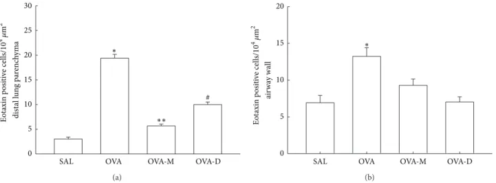
3.2. Measurements of Eotaxin Expression. Considering
eotaxin expression in inlammatory cells (Figure 2(a)), we
observed a signiicant increase in the number of eotaxin
positive cells (104�m2) in distal lung parenchyma in OVA
group (19.38 ± 0.79) compared to control (SAL group:
3.00 ± 0.39, � < 0.001). here was a decrease of eotaxin
positive cells in OVA-M (5.65±0.38) and OVA-D (9.9±0.54)
compared to OVA(� < 0.001). here was a signiicant
reduction of eotaxin positive cells in OVA-M compared to
OVA-D (� < 0.001).
Figure 2(b) shows eotaxin expression in inlammatory
cells in airway walls. We observed a signiicant increase in the
number of eotaxin positive cells (104�m2) on airway walls in
OVA group (13.23 ± 1.18) compared to control (SAL group:
6.92 ± 0.99,� < 0.001). here was a signiicant decrease of
eotaxin positive cells in OVA-M (9.29 ± 0.86) and in OVA-D
(7.03 ± 0.70) compared to OVA (� < 0.001). here were no diferences between OVA-D and OVA-M groups.
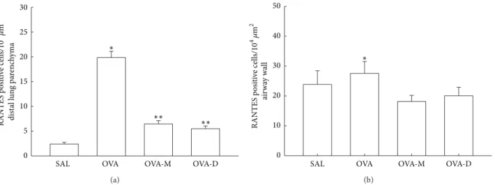
3.3. Measurements of RANTES Expression. he RANTES
expression in distal lung parenchyma is shown inFigure 3(a).
here was a signiicant increase in RANTES expression
(pos-itive cells/104�m2) in OVA group (19.84 ± 1.25) compared
to control (SAL group:2.40 ± 0.35,� < 0.001). here was
a signiicant decrease of RANTES positive cells in OVA-M (6.44 ± 0.66) and in OVA-D (5.47 ± 0.54) compared to OVA (� < 0.001). here were no diferences between OVA-D and OVA-M groups.
Figure 3(b) shows RANTES expression in airway walls
(positive cells/104�m2). here was a signiicant increase in
RANTES expression in OVA group (27.56 ± 1.83) compared
to control (SAL group:18.88 ± 1.31,� < 0.05). here was
a signiicant decrease of RANTES positive cells in OVA-M (18.14±1.30) and in OVA-D (20.05±1.24) compared to OVA (� < 0.05). here were no diferences between OVA-D and OVA-M groups.
3.4. Measurements of IGF-I. Considering IGF-I expression in
inlammatory cells (Figure 4(a)), we observed a signiicant
increase in the number of IGF-I positive cells (104�m2)
in distal lung parenchyma in OVA group (22.89 ± 1.16)
compared to control (SAL group:4.87 ± 0.93,� < 0.001).
SAL OVA OVA-M OVA-D
Eos
in
o
p
h
il
s
dist
al l
u
n
g pa
re
nc
h
yma (
10
4 �
20
15
10
5
0
∗
∗∗
m
2 )
(a)
ai
rw
ay
wall (
10
4�
m
2)
SAL OVA OVA-M OVA-D
Eos
in
o
p
h
il
s
30
25
20
15
10
5
0
∗∗ ∗
(b)
Figure 1: (a) Mean and SEM values of eosinophil in distal lung of GP that previously inhaled normal saline or ovalbumin, and ater the 4th inhalation, GP were treated with montelukast (OVA-M group) and dexamethasone (OVA-D group).∗� < 0.05compared to the other groups.
∗∗� < 0.05compared to SAL group. (b) Mean and SEM values of eosinophil in airway wall of GP that previously inhaled normal saline or
ovalbumin, and ater the 4th inhalation, GP were treated with montelukast (OVA-M group) and dexamethasone (OVA-D group).∗� < 0.05 compared to the other groups.∗∗� < 0.05compared to SAL group.
30
25
20
15
10
5
0
SAL OVA OVA-M OVA-D
∗∗ ∗
Eo
taxin p
osi
ti
ve
cells/
10
4 �
m
2
dist
al l
u
n
g pa
re
nc
h
yma
#
(a)
SAL OVA OVA-M OVA-D
20
15
10
5
0
∗
Eo
taxin p
osi
ti
ve
cells/
10
4�
m
2
ai
rw
ay
wall
(b)
Figure 2: (a) Mean and SEM values of eotaxin positive cells in distal lung of GP that previously inhaled normal saline or ovalbumin, and ater the 4th inhalation, GP were treated with montelukast (OVA-M group) and dexamethasone (OVA-D group). ∗� < 0.001compared to the other groups. ∗∗� < 0.001compared to SAL and OVA-D groups. #� < 0.001compared to SAL group. (b) Mean and SEM values of Eotaxin positive cells in airway wall of GP that previously inhaled normal saline or ovalbumin, and ater the 4th inhalation, GP were treated with montelukast (OVA-M group) and dexamethasone (OVA-D group).∗� < 0.001compared to SAL, OVA-M and OVA-D groups.
(� < 0.001). here was a signiicant reduction of IGF-I
positive cells in OVA-D compared to OVA-M (� < 0.001).
here were no diferences between OVA-D compared to SAL group.
Figure 4(b) presents the mean and SEM values of
IGF-I positive cells (104�m2) in airway walls. We observed a
signiicant increase in expression of IGF-I positive cells in
OVA group (50.70 ± 1.43) compared to control (SAL group:
25.70 ± 1.79). here was a decrease of IGF-I positive cells in
OVA-M (25.53±1.36) and OVA-D (20.90±4.35) compared to
OVA (� < 0.001). here were no diferences between OVA-M
and OVA-D groups.
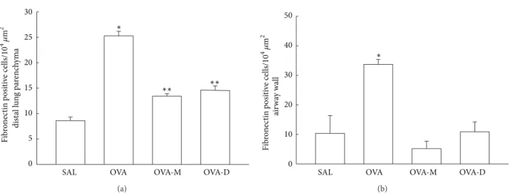
3.5. Measurements of Fibronectin Positive Cells. Figure 5(a)
shows the number of ibronectin positive cells (104�m2) in
distal lung parenchyma. We observed a signiicant increase in the number of ibronectin positive cells in OVA group (25.25 ± 0.90) compared to control (SAL group:8.61 ± 0.70,
� < 0.001). here was a signiicant decrease in the number of
ibronectin positive cells in OVA-M (13.41 ± 0.47) and
OVA-D (14.61 ± 0.82), compared to OVA group (� < 0.001). here were no signiicant diferences between OVA-D and OVA-M groups.
Figure 5(b)shows the number of ibronectin positive cells
30
25
20
15
10
5
0
SAL OVA OVA-M OVA-D
∗∗
∗∗ ∗
RANTES p
osi
ti
ve
cells/
10
4 �
m
2
dist
al l
u
n
g pa
re
nc
h
yma
(a)
SAL OVA OVA-M OVA-D
30
20
10
0
∗
RANTES p
osi
ti
ve
cells/
10
4�
m
2
ai
rw
ay
wall
50
40
(b)
Figure 3: (a) Mean and SEM values of RANTES positive cells in distal lung of GP that previously inhaled normal saline or ovalbumin and ater the 4th inhalation GP were treated with montelukast (OVA-M group) and dexamethasone (OVA-D group).∗� < 0.001compared to the other groups.∗∗� < 0.001compared to SAL group. (b) Mean and SEM values of RANTES positive cells in airway wall of GP that previously inhaled normal saline or ovalbumin and ater the 4th inhalation GP were treated with montelukast (OVA-M group) and dexamethasone (OVA-D group). ∗� < 0.05compared to the other groups.
30
25
20
15
10
5
0
SAL OVA OVA-M OVA-D
∗∗ ∗
IG
F
-I p
osi
ti
ve
cells/
10
4�
m
2
dist
al l
u
n
g pa
re
nc
h
yma
(a)
30
20
10
0
SAL OVA OVA-M OVA-D
∗
IG
F
-I p
osi
ti
ve
cells/
10
4�
m
2
ai
rw
ay
wall
60
50
40
(b)
Figure 4: (a) Mean and SEM values of IGF-I positive cells in distal lung of GP that previously inhaled normal saline or ovalbumin, and ater the 4th inhalation GP were treated with montelukast (OVA-M group) and dexamethasone (OVA-D group). ∗� < 0.001compared to the other groups. ∗∗� < 0.001compared to SAL and OVA-D groups. (b) Mean and SEM values of IGF-I positive cells in airway wall of GP that previously inhaled normal saline or ovalbumin, and ater the 4th inhalation, GP were treated with montelukast (OVA-M group) and dexamethasone (OVA-D group).∗� < 0.001compared to the other groups.
in the number of ibronectin positive cells in OVA group (33.69 ± 1.66) compared to control (SAL group:10.35 ± 5.98,
� < 0.001). here was a signiicant decrease in the number of
ibronectin positive cells in OVA-M (5.21±2.47) and OVA-D
(10.88 ± 3.33) animals compared to OVA group (� < 0.001). here were no signiicant diferences between OVA-D and OVA-M groups.
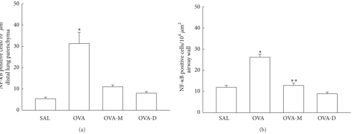
3.6. Measurements of NF-�B. Figure 6(a)shows the NF-�B expression in distal parenchyma. We observed a signiicant
increase in the number of NF-�B (positive cells/104�m2) in
OVA group (31.38 ± 5.21) compared to control (SAL group:
5.38 ± 0.79,� < 0.001). here was a signiicant decrease in
the number of NF-�B positive cells in OVA-M (11.04 ± 0.86)
and OVA-D (8.06 ± 0.70) compared to OVA group (� <
0.001). here were no diferences between M and OVA-D groups.
Figure 6(b)shows the NF-�B expression in airway walls.
We observed a signiicant increase in the number of
NF-�B (positive cells/104�m2) in OVA group (26.20 ± 1.34)
compared to control (SAL group:11.95 ± 0.96, � < 0.05).
here was a signiicant decrease in the number of NF-�B
positive cells in OVA-M (12.84 ± 1.09) and OVA-D (8.95 ±
0.78) compared to OVA group (� < 0.05). Nevertheless,
30
25
20
15
10
5
0
SAL OVA OVA-M OVA-D
dist
al l
u
n
g pa
re
nc
h
yma
∗∗ ∗∗
∗
Fib
ro
n
ec
tin p
o
si
ti
ve
cells/
10
4 �
m
2
(a)
ai
rw
ay
wall
50
40
SAL OVA OVA-M OVA-D
30
20
10
0
∗
Fib
ro
n
ec
tin p
o
si
ti
ve
cells/
10
4 �
m
2
(b)
Figure 5: (a) Mean and SEM values of ibronectin positive cells in distal lung of GP that previously inhaled with normal saline or ovalbumin, and ater the 4th inhalation, GP were treated with montelukast (OVA-M group) and dexamethasone (OVA-D group). ∗� < 0.001compared to the other groups. ∗∗� < 0.001compared to SAL group. (b) Mean and SEM values of Fibronectin positive cells in airway wall of GP that previously inhaled normal saline or ovalbumin, and ater the 4th inhalation, GP were treated with montelukast (OVA-M group) and dexamethasone (OVA-D group).∗� < 0.001compared to the other groups.
no diferences between OVA-M and OVA-D groups when compared to SAL group.
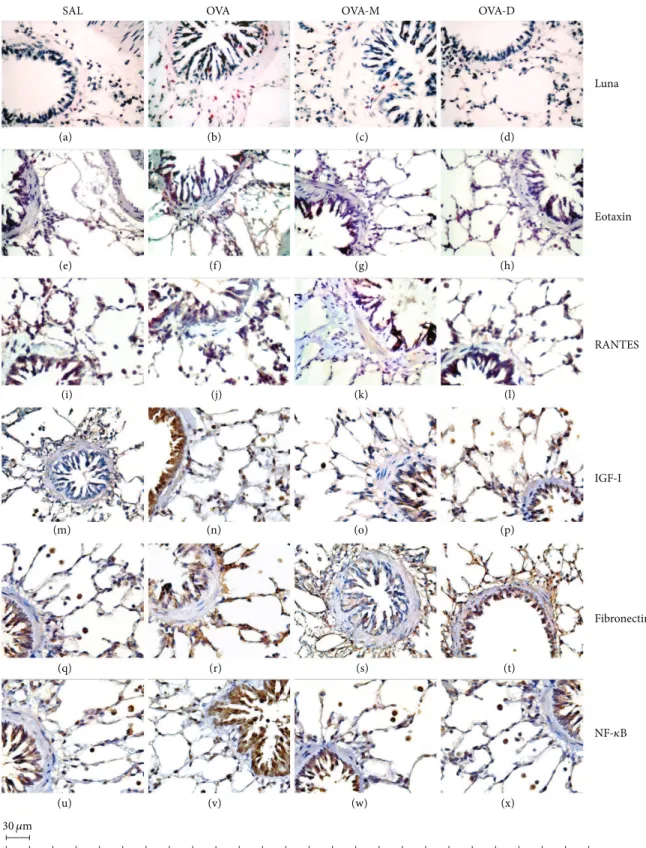
3.7. Representative Photomicrographs of Airway Walls.
Figure 7 shows representative photomicrographs of guinea
pigs airway wall samples. We observed a signiicant increase in the number of eosinophils, eotaxin positive cells, RANTES positive cells, IGF-I positive cells, ibronectin positive cells,
and NF-�B positive cells in OVA group compared to control
(SAL group). here was a signiicant decrease in eosinophils, eotaxin positive cells, RANTES positive cells, IGF-I positive
cells, ibronectin positive cells, and NK-�B positive cells in
OVA-M and OVA-D compared to OVA group.
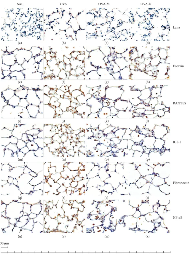
3.8. Representative Photomicrographs of Distal Lung Parenchyma. Figure 8 presents representative photo-micrographs of guinea pigs distal parenchyma samples. We observed a signiicant increase in the number of eosinophils, eotaxin positive cells, RANTES positive cells, IGF-I positive
cells, ibronectin positive cells, and NF-�B positive cells in
OVA group compared to control (SAL group). here was a signiicant decrease in eosinophils, eotaxin positive cells, RANTES positive cells, IGF-I positive cells, ibronectin
positive cells, and NK-�B positive cells in OVA-M and
OVA-D compared to OVA group.
It is important to mention that two more control groups were performed: saline groups treated either with mon-telukast (SAL-M) or dexamethasone (SAL-D). here were no statistical signiicant diferences among the three control groups, so we showed only one of them.
4. Discussion
Although corticosteroids are highly recommended as a irst line therapy for asthma, it is important to highlight that
montelukast can have a consistent beneit in controlling asthma symptoms and can be used as an alternative for those patients who have diiculties using inhaled corticosteroids (especially young children) or have some kind of intolerance
with dexamethasone therapy [33]. Furthermore, a reduced
adherence with inhalants for asthma compared to those therapies which are orally administrated has been addressed
[34,35].
To elucidate the role of antileukotrienes and corticos-teroids in the control of changes in asthma remodeling, we evaluated the efects of montelukast or dexamethasone treat-ments on eosinophilic recruitment, including its activation by detecting eotaxin and RANTES positive cells in airways and lung parenchyma in guinea pigs (GP), with chronic allergic lung inlammation. Furthermore, to better comprehend the mechanisms involved in the remodeling process, we evalu-ated IGF-I and ibronectin in distal lung parenchyma and
airway walls. Considering the importance of NF-�B, one
of the most important transcriptional factors involved in asthmatic responses, we also analyzed the expression of this protein in both pulmonary compartments.
We initiated these treatments only one day ater the fourth inhalation, because in a previous study, using passive
cutaneous anaphylaxis technique (PCA) [10], we observed
an increase in speciic immunoglobulin G1 (IgG1) homo-cytotropic anaphylactic antibodies in GP that received four inhalations with ovalbumin (at the end of the second week of this protocol).
Dexamethasone is considered a very potent anti-in-lammatory therapy available for asthma. Its efects are prob-ably related to a broad anti-inlammatory actions, includ-ing a decrease in airway iniltration of lymphocytes and
eosinophils [36]. However, it is important to emphasize that
one of the major challenges in using corticosteroids is related
50
40
30
20
10
0
SAL OVA OVA-M OVA-D
∗
dist
al l
u
n
g pa
re
nc
h
yma
NF
-𝜅
B p
osi
ti
ve
cells/
10
4�
m
2
(a)
50
40
SAL OVA OVA-M OVA-D
30
20
10
0
NF
-𝜅
B p
osi
ti
ve
cells/
10
4 �
m
2
ai
rw
ay
wall ∗
∗∗
(b)
Figure 6: (a) Mean and SEM values of NF-�B positive cells in distal lung of GP that previously inhaled normal saline or ovalbumin, and ater the 4th inhalation, GP were treated with montelukast (OVA-M group) and dexamethasone (OVA-D group). ∗� < 0.001compared to the other groups. (b) Mean and SEM values of NF-�B positive cells in airway wall of GP that previously inhaled normal saline or ovalbumin, and ater the 4th inhalation, GP were treated with montelukast (OVA-M group) and dexamethasone (OVA-D group).∗� < 0.05compared to the other groups.∗∗� < 0.05compared to OVA-D group.
treatments that can improve the quality of life of asthmatic patients.
CysLTs play a major role in the pathophysiology of asthma, activating and recruiting some inlammatory cells, promoting airway remodeling, and increasing bronchial
hyperreactivity and vascular permeability [38,39].
Antileukotrienes are a new class of anti-inlammatory drugs, which includes, Zairlukast, Montelukast, Pranlukast, and Zileuton. Montelukast is known to be a highly selective, pharmacological antagonist of type 1 cysteinyl leukotriene
receptors (CysLT1Rs) which can recognize the cysteinyl
leukotrienes (CysLTs) LTD4 and LTC4/LTE4 expressed on
some inlammatory cells such as basophils, neutrophils, and lymphocytes and also on some structural cells such as ibrob-lasts, myoibrobibrob-lasts, smooth muscle cells, and epithelial cells
[38].
Since montelukast acts as a potent antagonist of the proinlammatory aspects of the CysLTs, it reduces asth-matic inlammation and airway resistance and also prevents bronchoconstriction, controlling asthma inlammation and
improving inlammatory and pulmonary responses [40,41].
Many studies in patients with asthma demonstrated a decrease in the number of eosinophils in the airways
ater treatment with corticosteroids [42]. Corticosteroids
can cause the induction of eosinophils apoptosis by the mechanism known as programmed cell death, acting as a
target therapy in asthmatic patients [43]. McMillan et al.
[21] showed in mice chronically exposed to ovalbumin that
chronic administration of budesonide was able to decrease airway hyperreactivity, as well as leukocyte iniltration, and decreased production of h2 mediators such as interleukin 4 (IL-4), interleukin 12 (IL-12), and eotaxin-1.
Concerning eosinophil recruitment, we observed that both treatments tested were eicient in reducing this iniltra-tion in distal lung and in airway walls.
Not only cysteinyl leukotrienes can act as a chemotactic for eosinophils, but also RANTES, eotaxin and eotaxin 2 are chemotactic substances that are involved during eosinophilic
recruitment [44–46].
Eotaxin and RANTES are known as members of the C-C family of chemokines. he most important character-istic of the C-C chemokines is their facility to chemoat-tract and activate inlammatory leucocytes, particularly lym-phocytes, monocytes, eosinophils, and basophils, and also some stromal cells such as endothelial and smooth muscle
cells [47]. Another efect of eotaxins is their ability to
cause immunoglobulin E(IgE)-independent degranulation of
basophils [48].
A family of receptors mediates the activities of C-C chemokines; in general, any C-C chemokine can bind to sev-eral C-C receptors. Eotaxin is exceptional and binds only to the chemokine receptor type 3 (CCR3). Furthermore, eotaxin can chemoattract and activate eosinophils speciically, while RANTES are less speciic to eosinophils, chemoattracting and activating other inlammatory cells as monocytes and
lymphocytes [49].
Montelukast has mainly antieosinophil properties [50,
51]. his could be one of the possible explanations in
our study suggesting why montelukast showed a better performance when compared to dexamethasone treatment while reducing eotaxin positive cells in distal parenchyma. Another possibility is that eotaxins are considered to be more speciic than RANTES when activating eosinophils during the inlammation process.
A huge number of cells expressing eotaxin mRNA and protein in the bronchial mucosa area of atopic asthmatics compared with controls have been demonstrated by Ying et
al. [52] and Mattoli et al. [53]. Furthermore, Ying et al. [52]
SAL OVA OVA-M OVA-D
Luna
Eotaxin
RANTES
Fibronectin IGF-I
(a) (b) (c) (d)
(e) (f) (g) (h)
(i) (j) (k) (l)
(m) (n) (o) (p)
(q) (r) (s) (t)
(u) (v) (w) (x)
NF-𝜅B
30�m
SAL OVA OVA-M OVA-D
Luna
Eotaxin
RANTES
Fibronectin IGF-I
(a) (b) (c) (d)
(e) (f) (g) (h)
(i) (j) (k) (l)
(m) (n) (o) (p)
(q) (r) (s) (t)
(u) (v) (w) (x)
NF-𝜅B
30�m
of binding only to CCR3, plays a role in the speciic recruit-ment of eosinophils to the asthmatic bronchial mucosa, a phenomenon which in turn regulates asthma severity.
In many asthmatic patients, the addition of anti-leukotriene can be a valuable approach for uncontrollable asthma regardless of treatment with inhaled corticosteroid despite the fact that little is known about its molecular mechanism.
Wu et al. [54] studying to the efect of a 3-day course of
high-dose montelukast on mediators of airway inlammation induced by a single allergen challenge in sensitized mice showed that eotaxin protein and mRNA expression in the lung remained unchanged.
Uguccioni et al. [55] using pranlukast (another
anti-leukotriene) in vitro, evaluated eosinophil transmigration across human umbilical vein endothelial cells in response to LTD4, eotaxin, RANTES, and platelet-activating fac-tor (PAF). hey showed that pranlukast did not modify eosinophil transmigration in response to eotaxin, RANTES, or PAF.
Nevertheless, in the present study, both treatments tested were eicient in reducing eotaxin positive cells in distal lung and in airway walls. Although montelukast treatment showed more eiciency in reducing eotaxin positive cells in distal lung parenchyma compared to dexamethasone treatment, both of them reduced these parameters in airway walls.
Some studies have shown the chemoattractant power activity of RANTES for eosinophils in airway allergic
inlammation. Gonzalo et al. [56] showed that treatment
with methylated RANTES (a CCL5 antagonist) decreased eosinophilia ater antigen challenge by blocking the activa-tion of RANTES and its binding to the receptor.
Regarding the evaluation of RANTES positive cells, we noted that both treatments have shown efectiveness either in distal lung parenchyma or in the airway wall contributing to a better control of asthma.
Lung remodeling and chronic inlammation are impor-tant characteristics of asthma. It has been shown that mod-ulation of ibroblasts into myoibroblastic phenotype, with addition of particular contractile aspects, indispensable for connective tissue remodeling during the wound healing
pro-cess in asthmatics airways [57]. Nonetheless, the connection
between ibroblasts, myoibroblasts, and smooth muscle cells
needs to be better investigated [58].
Fibroblasts are responsible for producing a wide array of extracellular matrix components like collagen, reticular and elastic ibers, laminin, ibronectin, hyaluronic acid, glycopro-teins, and proteoglycans amorphous extracellular substance
[59, 60]. Nam et al. [61] showed that primary bronchial
ibroblasts were susceptible to mechanical stimuli during diferentiation into myoibroblasts. Furthermore, they have addressed that ibroblasts from asthmatics showed higher potential for tissue ibrosis when compared to control group. Myoibroblasts are known to be contractile cells with biochemical and morphologic aspects that stay somewhere
between ibroblasts and smooth muscle cells [62,63].
TGF-� is the major regulator that promotes myoibroblast
dif-ferentiation through its capacity to accumulate intracellular
contractile proteins, ibronectin containing extra type III
domain A (EDAcFN), and collagen density [57].
hrough inlammatory mesenchymal and epithelial cells, a multimeric form of cellular ibronectin is produced, and it is deposited in the ibrils of the extracellular matrix, containing variable portions of the extra type III domain A and B (EDA
and EDB) sequences [64]. hese domains are known to play
an important role in the contribution of EDAcFN to ibroblast
activation [65] and wound healing process [66].
Fibronectin matrix formation is a dynamic, cell-dependent process that is tightly regulated. he loss of this regulation promotes an increase in the deposition of ibronectin and other extracellular matrix (ECM) molecules in the subepithelial matrix in patients with asthma. A speciic site of ibronectin modulates cell contractility, collagen gel contraction, and cell migration. Furthermore, the increased or inappropriate exposure of this site in matrix ibronectin may lead to abnormal tissue remodeling by enhancing and/or prolonging cell contractility and by altering the rate of reepithelialization. he development of new strategies to control the extent and duration of the exposure of cells to matrix-speciic epitopes can supply a functional method to
structure normal tissue remodeling in asthma [6].
he cysteinyl leukotriene LTD4 causes a potentiation of ibronectin-induced migration of human lung ibroblasts
[67], contributing to the airway remodeling process.
Further-more, Tokuriki et al. [68] observed that the leukotriene D4
acts as a precipitating factor during the remodeling mediated by endothelin-1, and antileukotrienes have a role in prevent-ing aberrant extracellular matrix degradation. An experimen-tal mice model of asthma showed that the antileukotrienes inhibit the process of airway remodeling, including the inlux of eosinophils into the lungs, eosinophil degranulation, the release of h2 cytokines, hyperplasia of mucous glands, hypersecretion mucus, hyperplasia of smooth muscle cells,
collagen deposition, and pulmonary ibrosis [69].
In this study, we observed an increase in ibronectin positive cells in chronic allergic lung inlammation model. Both treatments tested were able to decrease the number of ibronectin positive cells in distal lung parenchyma and in distal airway walls.
Fibrosis and asthma are highly linked by the increased deposition of extracellular matrix at speciic places in the airway wall. Growth factors may be considered during
asthma remodeling. Increased expression of TGF-� [70]
has been shown in lung ibrosis and plays an important role in the excessive matrix deposition and also in cell proliferation. IGF-I is known to be a highly potent mitogen for ibroblasts and smooth muscle cells, acting on ibroblasts as a progression factor pushing the cells from a G1 phase to mitosis in the cell cycle, activating them to make the
collagen synthesis [71]. It is important to emphasize that
collagen deposition at the lamina reticularis contributes to the remodeling process, an asthma characteristic, enhancing the role that IGF-I can play as an important mediator of inlammation and remodeling in asthmatic airways.
Muz et al. [72] showed that airway collagen
in collagen deposition/lung ibrosis. In addition, Henderson
Jr. et al. [73] showed that montelukast could signiicantly
reduce airway remodeling in a mouse model of chronic asthma, addressing montelukast efect in some aspects of the remodeling process.
Veraldi et al. [5] showed that insulin-like growth factor
binding protein-3 IGFBP-3 levels are increased in bron-choalveolar lavage (BAL) luid of asthmatic patients 48 hours ater allergen challenge, suggesting that this is the most important protein that IGF binds, developing airway remodeling in asthma through this modulation. Considering this, the creation of an IGFBP-3 antagonist could be a promise therapeutic target for asthma treatment.
Rajah et al. [74] using cultured airway smooth muscle
cells addressed the synergism between IGF and inlammatory
agents such as leukotriene D4and interleukin 1-beta, showing
that these agents induce the secretion of an IGFBP protease which cleaves the IGFBPs secreted by airway smooth cells. his event allows IGF to stimulate proliferation, emphasizing that IGFBP proteases act as a critical element in asthma
and other diseases. Rajah et al. [75] also reported that
antimatrix metalloproteinase (MMP-1) acts as an IGFBP protease induced by leukotrienes that plays an important role in modulating IGF action in airway smooth muscle cells. Our indings showed that the anti-leukotriene montelukast reduced IGF levels in distal lung parenchyma and airway walls in experimental models of chronic allergic inlamma-tion.
Hoshino et al. [76] have investigated the efects of inhaled
corticosteroids in asthmatic patients. hey observed a signif-icant decrease in thickness of the subepithelial collagen and also an important reduction in the number of ibroblasts and the expression of IGF-I, suggesting that the decrease in colla-gen thickness ater the treatment with inhaled corticosteroid could be a result of blocking the transcription of the IGF-I gene. In our study, we observed a decrease in IGF with both treatments. However, dexamethasone treatment was more eicient in reducing IGF-I positive cells in distal parenchyma when compared to montelukast treatment.
Corticosteroids are known to play an important role in the therapy of a large number of diseases, especially diseases
of the gastrointestinal tract and liver [77]. Gayan-Ramirez
et al. [78] showed that corticosteroid treatment in rats was
highly linked with a reduction in IGF-I serum levels mainly ater triamcinolone treatment that resulted from a decrease in IGF-I manifestation in the liver.
Our indings showed that dexamethasone was more ei-cient when compared to montelukast treatment in reducing the expression of IGF-I in distal lung parenchyma. his could be explained since this growth factor is synthesized and released by the liver and can be mediated by the action of
corticosteroids [79].
Further studies are needed to ascertain the molecular mechanisms involving IGF-I and the current therapies.
NF-�B is a transcription factor, and it plays an important
role in expression of lots of proinlammatory genes, which leads to the synthesis of growth factors, adhesion molecules,
and chemokines [80].
Lin et al. [81] showed that total lung extracts from
brownNorway rats exhibit enhanced NF-�B activity ater the
ovalbumin (OVA) sensitization and challenge model. here
is also a relation between NF-�B and nitric oxide (NO).
When inducible nitric oxide synthase (iNOS) is activated, it generates a high concentration of NO through the activation
of some inducible nuclear factors, including the NF-�B [82].
Our results showed a higher eicacy of corticosteroids
compared to montelukast treatment in reducing NF-�B in
airway walls. his can be explained by the broad mecha-nism that corticosteroids play, by switching of the expres-sion of multiple genes, including adheexpres-sion molecules and
chemokines [83].
NF-�B activity is highly regulated by its interaction with
I�B proteins. One possible explanation to corticosteroids
higher eicacy in reducing NF-�B is that they can be
involved in regulating the expression of I�B proteins. Another
possibility is that NF-�B is regulated during the interaction
with other coactivators, such as transcriptional co-activator proteins (CBP/p300), which are highly known to interact
with NF-�B promoting transcription [83].
Arachidonate 5-lipoxygenase (ALOX5) is an enzyme that
synthetizes the cysteinyl leukotrienes LTC4, LTD4, LTE4,
and LTB4. ALOX5 inhibition can be associated with an
improvement in asthma outcome, and it is known that the ALOX5 gene promoter has binding-sites for some
transcrip-tion factors, including NF-�B. However, not all asthmatic
patients will respond to antileukotrienes since those who have mutant alleles at the ALOX-5 should not have clinical beneits
[84]. his could be a possible explanation of the relative lack
of response by montelukast treatment in reducing NF-�B in
airway walls.
Considerable attention has been given to the signaling
pathways related with the NF-�B activation. hese signaling
protein molecules can be, in the near future, a potential
therapeutic target to block NF-�B activation, interrupting
the asthma process. Further studies are needed to better understand how these molecules interact with each other
[85].
Despite the fact that there is current evidence of the importance of these treatments on airways regarding the control of main morphofunctional alterations present on asthma, the present study enlightens that isolated treatment with these drugs was chiely capable of controlling also distal lung parenchyma alterations.
It is worthy to mention that, in the present study, all of the drugs were systemically administered, not through inhalation that probably contributed to the eicacy of the treatment on distal lung parenchyma.
better understand the mechanisms involved in the patho-physiology of asthma. We observed important results on the relation of the available treatments for allergic diseases and the immune response on the asthmatic patient. Our goal is a continuous feedback loop to accelerate the translation of data into knowledge from the basic sciences into the development of new treatments, translating the indings from clinical trials into everyday practice.
5. Conclusion
In this animal model, both corticosteroid and montelukast treatments were efective in the control of the inlamma-tory response in distal lung parenchyma and distal airway walls. But it is noteworthy to emphasize the contribution of montelukast in the resolution of some inlammatory and remodeling aspects in this experimental model, mainly in the control of distal lung parenchyma functional and histopathological alterations.
In conclusion, asthma is a chronic disease and corti-costeroids are considered to be a gold standard medical therapy. However, since corticosteroids can have a wide range of side efects, it is essential that studies of therapeutic eicacy consider alternative treatments. Antileukotrienes are an orally, safe administered, nonsteroidal therapy for treating mild persistent asthma, especially in young children. hey are an option for the treatment of asthma, based on many of its biological efects described above.
herefore, our data contributes to a better understanding of the inlammatory and remodeling processes in both treatments in this experimental model of asthma and may assist researchers to further studies based on their expected outcomes.
Conflict of Interests
he authors declare that they have no conlict of interests.
Authors’ Contribution
Nath´alia Brand˜ao Gobbato designed and performed the major part of the experiments and morphometric analysis, performed the statistical analysis and drated the manuscript. Fl´avia Castro Ribas de Souza and Stella Bruna Napolitano Fumagalli assisted in performing the experiments. Carla M´aximo Prado helped with the statistical analysis and in the preparation of the manuscript. Fernanda Degobbi Ten´orio Quirino dos Santos Lopes assisted in the inter-pretation of the manuscripst. Iolanda de F´atima Lopes Calvo Tib´erio and Milton Arruda Martins participated in the design and discussion of the study. Edna Aparecida Leick supervised the study, participated in its design and in elucidation of results as well as in the elaboration of the manuscript. All authors read and approved the inal manuscript.
Acknowledgments
his study was supported by the following Brazilian Scientiic Agencies: Coordenac¸˜ao de Aperfeic¸oamento de Pessoal de Nivel Superior (CAPES), Fundac¸˜ao de Amparo `a Pesquisa do Estado de S˜ao Paulo (FAPESP), and Conselho Nacional de Desenvolvimento Cient´ıico and Tecnol´ogico (CNPq), Brazil (LIM-20-HC-FMUSP).
References
[1] World Health Organization,World Health Report 2000 Making a Diference, WHO, Geneve, Switzerland, 2000.
[2] A. B. Kay, “he role of eosinophils in the pathogenesis of asthma,”Trends in Molecular Medicine, vol. 11, no. 4, pp. 148– 152, 2005.
[3] J. Bousquet, “Workshop summary: relating inlammatory changes in asthma to clinical status,”Respiratory Medicine, vol. 94, pp. S32–S33, 2000.
[4] R. M. Pascual and S. P. Peters, “Airway remodeling contributes to the progressive loss of lung function in asthma: an overview,” Journal of Allergy and Clinical Immunology, vol. 116, no. 3, pp. 477–486, 2005.
[5] K. L. Veraldi, B. T. Gibson, H. Yasuoka et al., “Role of insulin-like growth factor binding protein-3 in allergic airway remodeling,” American Journal of Respiratory and Critical Care Medicine, vol. 180, no. 7, pp. 611–617, 2009.
[6] D. C. Hocking, “Fibronectin matrix deposition and cell contrac-tility: implications for airway remodeling in asthma,”Chest, vol. 122, no. 6, pp. 275S–278S, 2002.
[7] M. R. Edwards, N. W. Bartlett, D. Clarke, M. Birrell, M. Belvisi, and S. L. Johnston, “Targeting the NF-�B pathway in asthma and chronic obstructive pulmonary disease,”Pharmacology and herapeutics, vol. 121, no. 1, pp. 1–13, 2009.
[8] P. Angeli, C. M. Prado, D. G. Xisto et al., “Efects of chronic L-NAME treatment lung tissue mechanics, eosinophilic and extracellular matrix responses induced by chronic pulmonary inlammation,”American Journal of Physiology—Lung Cellular and Molecular Physiology, vol. 294, no. 6, pp. L1197–L1205, 2008. [9] T. Lanc¸as, D. I. Kasahara, C. M. Prado, I. F. L. C. Tib´erio, M. A. Martins, and M. Dolhnikof, “Comparison of early and late responses to antigen of sensitized guinea pig parenchymal lung strips,”Journal of Applied Physiology, vol. 100, no. 5, pp. 1610– 1616, 2006.
[10] A. S. Nakashima, C. M. Prado, T. Lanc¸as et al., “Oral tolerance attenuates changes in in vitro lung tissue mechanics and extracellular matrix remodeling induced by chronic allergic inlammation in guinea pigs,”Journal of Applied Physiology, vol. 104, no. 6, pp. 1778–1785, 2008.
[11] L. Bjermer, “History and future perspectives of treating asthma as a systemic and small airways disease,”Respiratory Medicine, vol. 95, no. 9, pp. 703–719, 2001.
[12] M. Dolhnikof, L. F. F. da Silva, B. B. de Araujo et al., “he outer wall of small airways is a major site of remodeling in fatal asthma,”Journal of Allergy and Clinical Immunology, vol. 123, no. 5, pp. 1090–1097, 2009.
[14] I. F. L. C. Tib´erio, G. M. G. Turco, E. A. Leick-Maldonado et al., “Efects of neurokinin depletion on airway inlammation induced by chronic antigen exposure,” American Journal of Respiratory and Critical Care Medicine, vol. 155, no. 5, pp. 1739– 1747, 1997.
[15] C. A. Burgess, B. K. McCandless, J. A. Cooper, and A. B. Malik, “Leukotriene B4 increases pulmonary transvascular iltration by a neutrophil-independent mechanism,”Journal of Applied Physiology, vol. 68, no. 3, pp. 1260–1264, 1990.
[16] W. W. Busse, “Leukotrienes and inlammation,”American Jour-nal of Respiratory and Critical Care Medicine, vol. 157, no. 6, pp. S210–S213, 1998.
[17] S. Jancar, “Imunidade natural e inlamac¸˜ao,” in Imunologia B´asica, V. Calich and C. Vaz, Eds., pp. 11–30, Revinter, S˜ao Paulo, Brazil, 2001.
[18] T. J. Vandermeer, M. J. Menconi, B. P. O’Sullivan et al., “Acute lung injury in endotoxemic pigs: role of leukotriene B4,”Journal of Applied Physiology, vol. 78, no. 3, pp. 1121–1131, 1995. [19] S. Joos, A. Miksch, J. Szecsenyi et al., “Montelukast as add-on
therapy to inhaled corticosteroids in the treatment of mild to moderate asthma: a systematic review,”horax, vol. 63, no. 5, pp. 453–462, 2008.
[20] I. Stelmach, J. Jerzynska, and P. Kuna, “A randomized, double-blind trial of the efect of treatment with montelukast on bronchial hyperresponsiveness and serum eosinophilic cationic protein (ECP), soluble interleukin 2 receptor (sIL-2R), IL-4, and soluble intercellular adhesion molecule 1 (sICAM-1) in children with asthma,”Journal of Allergy and Clinical Immunology, vol. 109, no. 2, pp. 257–263, 2002.
[21] S. J. McMillan, G. Xanthou, and C. M. Lloyd, “herapeutic administration of Budesonide ameliorates allergen-induced airway remodelling,”Clinical and Experimental Allergy, vol. 35, no. 3, pp. 388–396, 2005.
[22] M. R. Bonsignore, S. La Grutta, F. Cibella et al., “Efects of exercise training and montelukast in children with mild asthma,”Medicine and Science in Sports and Exercise, vol. 40, no. 3, pp. 405–412, 2008.
[23] T. Jartti, “Inhaled corticosteroids or montelukast as the pre-ferred primary long-term treatment for pediatric asthma?” European Journal of Pediatrics, vol. 167, no. 7, pp. 731–736, 2008. [24] F. C. R. Souza, N. B. Gobbato, G. R. Maciel et al., “Efects of corticosteroid, montelukast and iNOS inhibition on distal lung with chronic inlammation,” Respiratory Physiology & Neurobiology, vol. 185, no. 2, pp. 435–445, 2013.
[25] E. Goleva, P. J. Hauk, J. Boquniewicz, R. J. Martin, and D. Y. Leung, “Airway remodeling and lack of bronchodilator response in steroid-resistant asthma,”he Journal of Allergy and Clinical Immunology, vol. 120, no. 5, pp. 1065–1072, 2007. [26] National Institutes of Health/Health Research Extension
Act of 1985, “Animals in Research, U.S. Government Principles for the Utilization and Care of Vertebrate Animals Used in Testing, Research, and Training Public Health Service Policy on Humane Care and Use of Laboratory Animals,” Public Law 99-158, November 1985,
http://grants.nih.gov/grants/olaw/references/phspol.htm#US-GovPrinciples.
[27] C. M. Prado, E. A. Leick-Maldonado, V. Arata, D. I. Kasahara, M. A. Martins, and I. F. L. C. Tib´erio, “Neurokinins and inlammatory cell iNOS expression in guinea pigs with chronic allergic airway inlammation,”American Journal of Physiology— Lung Cellular and Molecular Physiology, vol. 288, no. 4, pp. L741– L748, 2005.
[28] A. J. Seco, M. E. Salgueiro, M. A. Villanueva, and G. Manso, “Treatment with high doses of terbutaline induces�-adrenergic desensitization of guinea pig trachea not prevented by the addition of dexamethasone,”Respiration, vol. 67, no. 5, pp. 559– 564, 2000.
[29] S. S. Possa, H. T. Charafeddine, R. F. Righetti et al., “Rho-kinase inhibition attenuates airway responsiveness, inlamma-tion, matrix remodeling, and oxidative stress activation induced by chronic inlammation,” American Journal of Physiology— Lung Cellular and Molecular Physiology, vol. 303, no. 11, pp. L939–L952, 2012.
[30] C. M. Starling, C. M. Prado, E. A. Leick-Maldonado et al., “Inducible nitric oxide synthase inhibition attenuates lung tissue responsiveness and remodeling in a model of chronic pul-monary inlammation in guinea pigs,”Respiratory Physiology and Neurobiology, vol. 165, no. 2-3, pp. 185–194, 2009.
[31] L. G. Luna, AFIP Manual of Histologic Staining Methods, McGraw Hill, New York, NY, USA, 1986.
[32] J. H. Zar, Biostatistical Analysis, Prentice-Hall, Englewood Clifs, NJ, USA, 2nd edition, 1984.
[33] L. Smith, “Comparative eicacy of inhaled corticosteroids and antileukotriene drugs in asthma,”BioDrugs, vol. 15, no. 4, pp. 239–249, 2001.
[34] B. Knorr, L. M. Franchi, H. Bisgaard et al., “Montelukast, a leukotriene receptor antagonist, for the treatment of persistent asthma in children aged 2 to 5 years,”Pediatrics, vol. 108, no. 3, p. E48, 2001.
[35] J. S. Kelloway, R. A. Wyatt, and S. A. Adlis, “Comparison of patients’ compliance with prescribed oral and inhaled asthma medications,”Archives of Internal Medicine, vol. 154, no. 12, pp. 1349–1352, 1994.
[36] S. Tsurufuji, A. Kurihara, and F. Ojima, “Mechanisms of anti-inlammatory action of dexamethasone: blockade by hydrocor-tisone mesylate and actinomycin D of the inhibitory efect of dexamethasone on leukocyte iniltration in inlammatory sites,” Journal of Pharmacology and Experimental herapeutics, vol. 229, no. 1, pp. 237–243, 1984.
[37] J. Liu, M. Zhang, C. Niu et al., “Dexamethasone inhibits repair of human airway epithelial cells mediated by glucocorticoid-induced leucine zipper (GILZ),”PLoS One, vol. 8, no. 4, Article ID e60705, 2013.
[38] M. Peters-Golden and W. R. Henderson Jr., “Mechanisms of disease: leukotrienes,”he New England Journal of Medicine, vol. 357, no. 18, pp. 1798–1854, 2007.
[39] R. C. Gualano, R. Vlahos, and G. P. Anderson, “What is the contribution of respiratory viruses and lung proteases to airway remodelling in asthma and chronic obstructive pulmonary disease?”Pulmonary Pharmacology and herapeutics, vol. 19, no. 1, pp. 18–23, 2006.
[40] N. Kondo, T. Katsunuma, Y. Odajima, and A. Morikawa, “A randomized open-label comparative study of montelukast ver-sus theophylline added to inhaled corticosteroid in asthmatic children,”Allergology International, vol. 55, no. 3, pp. 287–293, 2006.
[41] H. Mechiche, E. Naline, L. Candenas et al., “Efects of cysteinyl leukotrienes in small human bronchus and antagonist activity of montelukast and its metabolites,”Clinical and Experimental Allergy, vol. 33, no. 7, pp. 887–894, 2003.
Heart, Lung, and Blood Institute/World Health Organiza-tion, Washington, DC, USA, 2009,http://www.ginasthma.org/ pdf/GINA Report 2010.pdf.
[43] W. W. Busse, “Inlammation in asthma: the cornerstone of the disease and target of therapy,”Journal of Allergy and Clinical Immunology, vol. 102, no. 4, pp. S17–S22, 1998.
[44] J. C. Gutierrez-Ramos, C. Lloyd, and J. A. Gonzalo, “Eotaxin: from an eosinophilic chemokine to a major regulator of allergic reactions,”Immunology Today, vol. 20, no. 11, pp. 500–504, 1999. [45] M.-C. Seminario and G. J. Gleich, “he role of eosinophils in the pathogenesis of asthma,”Current Opinion in Immunology, vol. 6, no. 6, pp. 860–864, 1994.
[46] L. M. Teran, “CCL chemokines and asthma,” Immunology Today, vol. 21, no. 5, pp. 235–242, 2000.
[47] R. Alam, “Chemokines in allergic inlammation,”Journal of Allergy and Clinical Immunology, vol. 99, no. 3, pp. 273–277, 1997.
[48] M. Uguccioni, C. R. Mackay, B. Ochensberger et al., “High expression of the chemokine receptor CCR3 in human blood basophils. Role in activation by eotaxin, MCP-4, and other chemokines,”Journal of Clinical Investigation, vol. 100, no. 5, pp. 1137–1143, 1997.
[49] L. M. Teran, Y. Ledesma-Soto, S. Krengel, and D. Lezcano-Meza, “Eotaxinas em asma bronquial y poliposis nasal,”Gaceta M´edica de M´exico, vol. 142, no. 2, pp. 139–144, 2006.
[50] Z. Diamant and A. P. Sampson, “Anti-inlammatory mecha-nisms of leukotriene modulators,”Clinical and Experimental Allergy, vol. 29, no. 11, pp. 1449–1453, 1999.
[51] E. Pizzichini, J. A. Lef, T. F. Reiss et al., “Montelukast reduces airway eosinophilic inlammation in asthma: a randomized, controlled trial,”European Respiratory Journal, vol. 14, no. 1, pp. 12–18, 1999.
[52] S. Ying, D. S. Robinson, Q. Meng et al., “Enhanced expression of eotaxin and CCR3 mRNA and protein in atopic asthma. Association with airway hyperresponsiveness and predominant co-localization of eotaxin mRNA to bronchial epithelial and endothelial cells,”European Journal of Immunology, vol. 27, no. 12, pp. 3507–3516, 1997.
[53] S. Mattoli, M. A. Stacey, G. Sun, A. Bellini, and M. Marini, “Eotaxin expression and eosinophilic inlammation in asthma,” Biochemical and Biophysical Research Communications, vol. 236, no. 2, pp. 299–301, 1997.
[54] A. Y. Wu, S. C. Chik, A. W. Chan, Z. Li, K. W. Tsang, and W. Li, “Anti-inlammatory efects of high-dose montelukast in an animal model of acute asthma,”Clinical and Experimental Allergy, vol. 33, no. 3, pp. 359–366, 2003.
[55] M. Nagata, K. Saito, I. Kikuchi, K. Hagiwara, and M. Kanazawa, “Efect of the cysteinyl leukotriene antagonist pranlukast on transendothelial migration of eosinophils,” International Archives of Allergy and Immunology, vol. 137, no. 1, pp. 2–6, 2005. [56] J.-A. Gonzalo, C. M. Lloyd, D. Wen et al., “he coordinated action of CC chemokines in the lung orchestrates allergic inlammation and airway hyperresponsiveness,” Journal of Experimental Medicine, vol. 188, no. 1, pp. 157–167, 1998. [57] J. J. Tomasek, G. Gabbiani, B. Hinz, C. Chaponnier, and R. A.
Brown, “Myoibroblasts and mechano: regulation of connective tissue remodelling,”Nature Reviews Molecular Cell Biology, vol. 3, no. 5, pp. 349–363, 2002.
[58] S. R. Singh and I. P. Hall, “Airway myoibroblasts and their relationship with airway myocytes and ibroblasts,”Proceedings of the American horacic Society, vol. 5, no. 1, pp. 127–132, 2008.
[59] M. N. Sheppard and N. K. Harrison, “Lung injury, inlammatory mediators, and ibroblast activation in ibrosing alveolitis,” horax, vol. 47, no. 12, pp. 1064–1074, 1992.
[60] F. M. Spoelstra, D. S. Postma, and H. F. Kaufman, “Mutual activation of pulmonary ibroblasts and eosinophils, and modu-lation by drugs in remodu-lation to asthma,”Clinical and Experimental Allergy, vol. 31, no. 6, pp. 808–816, 2001.
[61] Y.-H. Nam, S.-K. Lee, D. Sammut, D. E. Davies, and P. H. Howarth, “Preliminary study of the cellular characteristics of primary bronchial ibroblasts in patients with asthma: expres-sion of�-smooth muscle actin, ibronectin containing extra type III domain a, and smoothelin,”Journal of Investigational Allergology and Clinical Immunology, vol. 22, no. 1, pp. 20–27, 2012.
[62] G. Gabbiani, “he biology of the myoibroblast,”Kidney Inter-national, vol. 41, no. 3, pp. 530–532, 1992.
[63] R. Bucala, L. A. Spiegel, J. Chesney, M. Hogan, and A. Cerami, “Circulating ibrocytes deine a new leukocyte subpopulation that mediates tissue repair,”Molecular Medicine, vol. 1, no. 1, pp. 71–81, 1994.
[64] R. Pankov and K. M. Yamada, “Fibronectin at a glance,”Journal of Cell Science, vol. 115, no. 20, pp. 3861–3863, 2002.
[65] G. Serini, M.-L. Bochaton-Piallat, P. Ropraz et al., “he ibronectin domain ED-A is crucial for myoibroblastic pheno-type induction by transforming growth factor-�1,”Journal of Cell Biology, vol. 142, no. 3, pp. 873–881, 1998.
[66] A. F. Muro, A. K. Chauhan, S. Gajovic et al., “Regulated splicing of the ibronectin EDA exon is essential for proper skin wound healing and normal lifespan,”Journal of Cell Biology, vol. 162, no. 1, pp. 149–160, 2003.
[67] J. Kato, T. Kohyama, H. Okazaki et al., “Leukotriene D4 potenti-ates ibronectin-induced migration of human lung ibroblasts,” Clinical Immunology, vol. 117, no. 2, pp. 177–181, 2005.
[68] S. Tokuriki, Y. Ohshima, A. Yamada, N. Ohta, H. Tsukahara, and M. Mayumi, “Leukotriene D4 enhances the function of endothelin-1-primed ibroblasts,”Clinical Immunology, vol. 125, no. 1, pp. 88–94, 2007.
[69] S. T. Holgate, M. Peters-Golden, R. A. Panettieri et al., “Roles of cysteinyl leukotrienes in airway inlammation, smooth muscle function, and remodeling,” Journal of Allergy and Clinical Immunology, vol. 111, supplement 1, pp. S18–S36, 2003. [70] N. Khalil and A. H. Greenberg, “he role of TGF-beta in
pulmonary ibrosis,”Ciba Foundation Symposium, vol. 157, pp. 194–207, 1991.
[71] J. I. Jones and D. R. Clemmons, “Insulin-like growth factors and their binding proteins: biological actions,”Endocrine Reviews, vol. 16, no. 1, pp. 3–34, 1995.
[72] M. H. Muz, F. Deveci, Y. Bulut, N. Ilhan, H. Yekeler, and T. Turgut, “he efects of low dose leukotriene receptor antagonist therapy on airway remodeling and cysteinyl leukotriene expres-sion in a mouse asthma model,”Experimental and Molecular Medicine, vol. 38, no. 2, pp. 109–118, 2006.
[73] W. R. Henderson Jr., L. Tang, S. Chu et al., “A role for cysteinylleukotrienesin airway remodeling in a mouse asthma model,” American Journal of Respiratory and Critical Care Medicine, vol. 65, no. 1, pp. 108–116, 2002.
[75] R. Rajah, S. E. Nunn, D. J. Herrick, M. M. Grunstein, and P. Cohen, “Leukotriene D4 induces MMP-1, which functions as an IGFBP protease in human airway smooth muscle cells,” American Journal of Physiology—Lung Cellular and Molecular Physiology, vol. 271, no. 6, part 1, pp. L1014–L1022, 1996. [76] M. Hoshino, Y. Nakamura, J. J. Sim et al., “Inhaled corticosteroid
reduced lamina reticularis of the basement membrane by modulation of insulin-like growth factor (IGF)-I expression in bronchial asthma,”Clinical and Experimental Allergy, vol. 28, no. 5, pp. 568–577, 1998.
[77] A. R. Tanner and L. W. Powell, “Corticosteroids in liver disease: possible mechanisms of action, pharmacology, and rational use,”Gut, vol. 20, no. 12, pp. 1109–1124, 1979.
[78] G. Gayan-Ramirez, F. Vanderhoydonc, G. Verhoeven, and M. Decramer, “Acute treatment with corticosteroids decreases IGF-1 and IGF-2 expression in the rat diaphragm and gastroc-nemius,” American Journal of Respiratory and Critical Care Medicine, vol. 159, no. 1, pp. 283–289, 1999.
[79] J. C. Schwander, C. Hauri, J. Zapf, and E. R. Froesch, “Synthesis and secretion of insulin-like growth factor and its binding protein by the perfused rat liver: dependence on growth hormone status,”Endocrinology, vol. 113, no. 1, pp. 297–305, 1983. [80] A. S. Baldwin Jr., “Series introduction: the transcription factor NF-kappaB and human disease,”he Journal of Clinical Investi-gation, vol. 107, pp. 3–6, 2001.
[81] C.-C. Lin, C.-Y. Lin, and H.-Y. Ma, “Pulmonary function changes and increased h-2 cytokine expression and nuclear factor kB activation in the lung ater sensitization and allergen challenge in brown Norway rats,”Immunology Letters, vol. 73, no. 1, pp. 57–64, 2000.
[82] F. Aktan, “iNOS-mediated nitric oxide production and its regulation,”Life Sciences, vol. 75, no. 6, pp. 639–653, 2004. [83] D. H. Broide, “Immunologic and inlammatory mechanisms
that drive asthma progression to remodeling,”Journal of Allergy and Clinical Immunology, vol. 121, no. 3, pp. 560–570, 2008. [84] E. S. Silverman, S. B. Liggett, E. W. Gelfand et al., “he
pharmacogenetics of asthma: a candidate gene approach,” Pharmacogenomics Journal, vol. 1, no. 1, pp. 27–37, 2001. [85] N. Charokopos, N. Apostolopoulos, M. Kalapodi, M.
Submit your manuscripts at
http://www.hindawi.com
Stem Cells
International
Hindawi Publishing Corporation
http://www.hindawi.com Volume 2014
Hindawi Publishing Corporation
http://www.hindawi.com Volume 2014 INFLAMMATION
Hindawi Publishing Corporation
http://www.hindawi.com Volume 2014
Behavioural
Neurology
Endocrinology
International Journal of Hindawi Publishing Corporationhttp://www.hindawi.com Volume 2014 Hindawi Publishing Corporation
http://www.hindawi.com Volume 2014
Disease Markers
Hindawi Publishing Corporation
http://www.hindawi.com Volume 2014
BioMed
Research International
Oncology
Journal ofHindawi Publishing Corporation
http://www.hindawi.com Volume 2014
Hindawi Publishing Corporation
http://www.hindawi.com Volume 2014
Oxidative Medicine and Cellular Longevity
Hindawi Publishing Corporation
http://www.hindawi.com Volume 2014
PPAR Research
The Scientiic
World Journal
Hindawi Publishing Corporation
http://www.hindawi.com Volume 2014
Immunology Research Hindawi Publishing Corporation
http://www.hindawi.com Volume 2014
Journal of
Obesity
Journal ofHindawi Publishing Corporation
http://www.hindawi.com Volume 2014
Hindawi Publishing Corporation
http://www.hindawi.com Volume 2014
Computational and Mathematical Methods in Medicine
Ophthalmology
Journal ofHindawi Publishing Corporation
http://www.hindawi.com Volume 2014
Diabetes Research
Journal ofHindawi Publishing Corporation
http://www.hindawi.com Volume 2014
Hindawi Publishing Corporation
http://www.hindawi.com Volume 2014
Research and Treatment
AIDS
Hindawi Publishing Corporation
http://www.hindawi.com Volume 2014
Gastroenterology Research and Practice
Hindawi Publishing Corporation
http://www.hindawi.com Volume 2014
Parkinson’s
Disease
Evidence-Based Complementary and Alternative Medicine
Volume 2014