of the Bloodsucking Bug,
Rhodnius prolixus
Jose´ M. C. Ribeiro1*, Fernando A. Genta2,3, Marcos H. F. Sorgine2,4, Raquel Logullo5, Rafael D. Mesquita2,5, Gabriela O. Paiva-Silva2,4, David Majerowicz4, Marcelo Medeiros6, Leonardo Koerich2,7, Walter R. Terra2,8, Cle´lia Ferreira2,8, Andre´ C. Pimentel8, Paulo M. Bisch9, Daniel C. Leite9, Michelle M. P. Diniz9, Joa˜o Lı´dio da S. G. V. Junior9,10, Manuela L. Da Silva6,9, Ricardo N. Araujo2,11, Ana Caroline P. Gandara4,
Se´bastien Brosson12, Didier Salmon4, Sabrina Bousbata12, Natalia Gonza´lez-Caballero3, Ariel Mariano Silber13, Michele Alves-Bezerra4, Katia C. Gondim2,4, Ma´rio Alberto C. Silva-Neto2,4,
Georgia C. Atella2,4, Helena Araujo2,14, Felipe A. Dias4, Carla Polycarpo2,4, Raquel J. Vionette-Amaral2,4, Patrı´cia Fampa15, Ana Claudia A. Melo2,5, Aparecida S. Tanaka2,16, Carsten Balczun17, Jose´
Henrique M. Oliveira4, Renata L. S. Gonc¸alves4, Cristiano Lazoski2,7, Rolando Rivera-Pomar18,19, Luis Diambra18, Gu¨nter A. Schaub17, Elo´i S. Garcia2,3, Patrı´cia Azambuja2,3, Glo´ria R. C. Braz2,5*, Pedro L. Oliveira2,4*
1Section of Vector Biology, Laboratory of Malaria and Vector Research, National Institute of Allergy and Infectious Diseases, National Institutes of Health, Rockville, Maryland, United States of America,2Instituto Nacional de Cieˆncia e Tecnologia em Entomologia Molecular, Federal University of Rio de Janeiro, Rio de Janeiro, Brazil,
3Instituto Oswaldo Cruz, Fundac¸a˜o Oswaldo Cruz, Rio de Janeiro, Rio de Janeiro, Brazil,4Instituto de Bioquı´mica Me´dica, Programa de Biotecnologia e Biologia Molecular, Universidade Federal do Rio de Janeiro, Rio de Janeiro, Brazil,5Department of Biochemistry, Institute of Chemistry, Federal University of Rio de Janeiro, Rio de Janeiro, Brazil,6Instituto Nacional de Metrologia Qualidade e Tecnologia, Diretoria de Metrologia Aplicada a`s Cieˆncias da Vida, Programa de Biotecnologia, Pre´dio 27, CEP 25250-020, Duque de Caxias, Rio de Janeiro, Brazil,7Departamento de Gene´tica, Instituto de Biologia, Universidade Federal do Rio de Janeiro, CEP 21944-970, Rio de Janeiro, Brazil,8Departamento de Bioquı´mica, Instituto de Quı´mica, Universidade de Sa˜o Paulo, Sa˜o Paulo, Brazil,9Instituto de Biofı´sica Carlos Chagas Filho, Universidade Federal do Rio de Janeiro, Rio de Janeiro, Brazil,10Center for Technological Innovation, Evandro Chagas Institute, Ananindeua, Para´, Brazil,11Departamento de Parasitologia do Instituto de Cieˆncias Biolo´gicas da Universidade Federal de Minas Gerais, Belo Horizonte, Minas Gerais, Brazil,12Institute for Molecular Biology and Medicine (IBMM), Universite´ Libre de Bruxelles, Gosselies, Belgium,13Departamento de Parasitologia, Instituto de Cieˆncias Biome´dicas, Universidade de Sa˜o Paulo, Sa˜o Paulo, Brazil,
14Institute for Biomedical Sciences, Federal University of Rio de Janeiro, Rio de Janeiro, Brazil,15Instituto de Biologia, DBA, UFRRJ, Serope´dica, Rio de Janeiro, Brazil,
16Departamento de Bioquı´mica, Escola Paulista de Medicina, Universidade Federal de Sa˜o Paulo, Sa˜o Paulo, Brazil,17Zoology/Parasitology Group, Ruhr-Universita¨t, Bochum, Germany, 18Centro Regional de Estudios Genomicos, Universidad Nacional de La Plata, Florencio Varela, Argentina, 19Centro de Bioinvestigaciones, Universidad Nacional del Noroeste de Buenos Aires, Pergamino, Argentina
Abstract
The bloodsucking hemipteranRhodnius prolixusis a vector of Chagas’ disease, which affects 7–8 million people today in Latin America. In contrast to other hematophagous insects, the triatomine gut is compartmentalized into three segments that perform different functions during blood digestion. Here we report analysis of transcriptomes for each of the segments using pyrosequencing technology. Comparison of transcript frequency in digestive libraries with a whole-body library was used to evaluate expression levels. All classes of digestive enzymes were highly expressed, with a predominance of cysteine and aspartic proteinases, the latter showing a significant expansion through gene duplication. Although no protein digestion is known to occur in the anterior midgut (AM), protease transcripts were found, suggesting secretion as pro-enzymes, being possibly activated in the posterior midgut (PM). As expected, genes related to cytoskeleton, protein synthesis apparatus, protein traffic, and secretion were abundantly transcribed. Despite the absence of a chitinous peritrophic membrane in hemipterans which have instead a lipidic perimicrovillar membrane lining over midgut epithelia -several gut-specific peritrophin transcripts were found, suggesting that these proteins perform functions other than being a structural component of the peritrophic membrane. Among immunity-related transcripts, while lysozymes and lectins were the most highly expressed, several genes belonging to the Toll pathway found at low levels in the gut of most insects -were identified, contrasting with a low abundance of transcripts from IMD and STAT pathways. Analysis of transcripts related to lipid metabolism indicates that lipids play multiple roles, being a major energy source, a substrate for perimicrovillar membrane formation, and a source for hydrocarbons possibly to produce the wax layer of the hindgut. Transcripts related to amino acid metabolism showed an unanticipated priority for degradation of tyrosine, phenylalanine, and tryptophan. Analysis of transcripts related to signaling pathways suggested a role for MAP kinases, GTPases, and LKBP1/ AMP kinases related to control of cell shape and polarity, possibly in connection with regulation of cell survival, response of pathogens and nutrients. Together, our findings present a new view of the triatomine digestive apparatus and will help us understand trypanosome interaction and allow insights into hemipteran metabolic adaptations to a blood-based diet.
Citation:Ribeiro JMC, Genta FA, Sorgine MHF, Logullo R, Mesquita RD, et al. (2014) An Insight into the Transcriptome of the Digestive Tract of the Bloodsucking Bug,Rhodnius prolixus. PLoS Negl Trop Dis 8(1): e2594. doi:10.1371/journal.pntd.0002594
Editor:Christian Tschudi, Yale School of Public Health, United States of America
This is an open-access article, free of all copyright, and may be freely reproduced, distributed, transmitted, modified, built upon, or otherwise used by anyone for any lawful purpose. The work is made available under the Creative Commons CC0 public domain dedication.
Funding:JMCR was supported by the Intramural Research Program of the NIH, National Institute of Allergy and Infectious Diseases. GRCB, FAG, ACAM, DS, SaB and SeB were supported by CAPES; CP, HMA, MHFS, PLO, GRCB, ACAM, AMS and FAG were supported by CNPq; AMS, ACP, AST, CF and WRT were supported by FAPESP; ACAM, FAG, CP, HMA, MHFS, PLO and GRCB were supported by FAPERJ; RNA was supported by FAPEMIG and PRPq/UFMG; RRP was supported by grants ANPCyT PICT-2010-0135, UNNOBA PFCI-512/12 and by the Max Planck Society Partner Laboratory Program; AMS was supported by a grant from INBEQMeDI; DS, SaB and SeB were supported by the Wallonie-Bruxelles International (WBI)/Fundac¸a˜o Coordenac¸a˜o de Aperfeic¸oamento de Pessoal de Nı´vel Superior (CAPES) bilateral cooperation agreement; CP was also supported by a grant from the WHO. The funders had no role in study design, data collection and analysis, decision to publish, or preparation of the manuscript.
Competing Interests:The authors have declared that no competing interests exist.
* E-mail: [email protected] (JMCR); [email protected] (GRCB); [email protected] (PLO)
Introduction
Triatomine bugs belong to the family Reduviidae within the order Hemiptera (infra-order: Heteroptera), all instars of which feed exclusively on blood [1,2]. Several species are vectors of Chagas’ disease in the Americas, a chronic and debilitating disease, often fatal, which infects 7–8 million people in Latin America today [3]. Among the 140 triatomine species in five tribes [4], Rhodnius prolixus—a vector in Central and South America— became a model insect for insect physiology and biochemistry thanks to its use by Dr. Vincent Wigglesworth in the 1930s and onward [5]. Despite being a bloodfeeder, due to its taxonomic position, R. prolixus data are useful for researchers working with heteropteran agricultural pests [1]. Recently, its genome was targeted for sequencing, and included in this effort was the sequencing of several organ-specific cDNA libraries using pyrose-quencing technology, which are described here.
The gut of triatomines differs from other hematophagous insects for which genomic data are available (mainly Diptera) because it is divided into three distinct segments (anterior midgut, AM; posterior midgut, PM and rectum, RE) that perform different functions during digestion of the blood meal and make this insect highly adapted for a blood meal. For example, a 30-mgR. prolixus Vth instar nymph can take 10 times its own weight in blood in fifteen minutes, the blood being stored in the bug’s AM. Within seconds of initiating the meal, diuretic hormones and serotonin are released into the hemolymph triggering salt and water transport from the meal to the hemolymph, and into the Malpighian tubules and finally into the RE, thus concentrating the meal and reducing the bug’s weight [5,6]. Indeed, the bug’s meal is reduced to its half by this urination within a few hours [5].
R. prolixusevolved from ancestors that on adapting to plant sap sucking lost their digestive serine proteinases and associated peritrophic membrane. This is a chitin-protein anatomical structure that may be synthesized by the whole or part of the midgut (type I) or by a ring of cells at the entrance of the midgut (type II). The peritrophic membrane envelops the food bolus in the midgut of most insects, leading to compartmentalization of the digestive process [7,8]. Instead, the midgut cell microvilli in Hemiptera are ensheathed by a phospholipid membrane, the perimicrovillar membrane (PMM) [7,9], which extends toward the midgut lumen with dead ends and, when collapsing, forms sheath packs [10–12]. PMMs were isolated from bothR. prolixus[12] and Dysdercus peruvianus[13] midguts, leading to the identification ofa -glucosidase as their biochemical enzyme marker. The presumed role of PMM was to absorb nutrients (mainly free amino acids) from the dilute sap ingested by the hemipteran and thysanopteran ancestors. On adapting to a diet rich in proteins, the heteropteran hemipteran (like R. prolixus and D. peruvianus) used lysosome-derived enzymes for digestion and the PMM as a substitute for the
peritrophic membrane in the compartmentalization of digestion [7,9,12].
The AM additionally harbors an endosymbiont, Rhodococcus rhodnii, which is essential for the bugs’ development and fertility [14–18]. The digestive tract is also where Trypanosoma cruzi, the protozoan agent of Chagas’ disease, develops [19]. No proteolytic digestion occurs in the AM, where hemoglobin remains red in color for over a week after feeding, but where various endoglycosidases have been described [20]. Digestion of complex lipids, as triacylglycerol, is negligible in AM and takes place in the PM [21].
The AM slowly releases its contents into the PM over a period of ,20 days, when the Vthinstar nymph molts to an adult [5]. While most insects have trypsin-like enzymes, and an alkaline gut pH, for digesting proteins, Hemiptera have lysosomal-like cathepsins which are secreted into an acidic gut [22]. There are a negligible [23] and a major [24] cysteine proteinase that accounts for 85% of the total proteinase activity. This activity was initially interpreted as a cathepsin B but later was shown to include a cathepsin L-like proteinase [24,25]. A cathepsin D-like proteinase accounts for the remaining midgut proteinase activity [24]. Amino and carboxypeptidases produce amino acids from the endopepti-dase products [24,26]. Toxic amounts of oxygen radical-producing heme are a byproduct of hemoglobin digestion, but these are stacked in the gut as a non-oxidizing form similar to the malarial pigment hemozoin. The stacking process in R. prolixusis dependent on the presence of PMM [27,28].
The RE, like the mammalian bladder, possesses a transitional epithelium that can stretch to accommodate the feces and urine [5,29]. It is from the rectal discharges thatT. cruziis released onto the mammalian host. The epithelia of the three gut segments are surrounded by smooth muscle [5].
Methods
Ethics statement
All animal care and experimental protocols were conducted following the guidelines of the institutional care and use committee (Committee for Evaluation of Animal Use for Research from the Federal University of Rio de Janeiro, CAUAP-UFRJ) and the NIH Guide for the Care and Use of Laboratory Animals (ISBN 0-309-05377-3). The protocols were approved by CAUAP-UFRJ under registry#IBQM001. Technicians dedicated to the animal facility at the Institute of Medical Biochemistry (UFRJ) carried out all aspects related to rabbit husbandry under strict guidelines to insure careful and consistent handling of the animals.
Insects
Insects used for transcriptome were R. prolixusfrom a colony kept at UFRJ (Rio de Janeiro), fed with rabbit blood, and raised at 28uC and 70% relative humidity. Adult females (five from each condition) receiving their second blood meal after the imaginal molt were dissected before feeding, twelve hours, twenty-four hours, two days, and five days after blood meal. A group of males (blood fed, five days after blood meal) was dissected to obtain testes. Organs (AM, PM, RE, FB, OV, MT, and TE) were dissected, homogenized in TriZol reagent (Invitrogen, San Diego, CA, USA), and processed as described below. To obtain a whole body (WB) library, nymphs and adults in several stages of feeding plus eggs were collected and extracted with TriZol, as follows: Eggs were collected at the day of oviposition and at days 2, 5 and 7 of development. First instars were collected at fasting (2 weeks after emergence) and at 2, 5 and 7 days after feeding (DAF); second and third instars were collected at fasting and at 2, 5, 7 and 9 DAF. Fourth instars were collected at fasting and at 2, 5, 7, 9 and 12 DAF. Fifth instars were collected at fasting and at 2, 5, 7, 9, 12, 14, 17 and 19 DAF. Adult males and females were collected at fasting and at 2, 5, 7, 9 and 12 DAF. All these 45 RNA preparations were pooled and used to obtain WB cDNA as described below.
RNA extraction, library preparation, and sequencing Organs were homogenized in TriZol reagent, and total RNA was isolated, followed by mRNA purification using the Micro-Fast track 2.0 kit from Invitrogen (San Diego, CA, USA) according to manufacturer’s instructions. Libraries were constructed using the
Smart cDNA Library Construction kit from Clontech (Palo Alto, CA, USA) and normalized using the Trimmer cDNA Normali-zation kit from Evrogen (Moscow, Russia).
The libraries were sequenced on a 454 genome sequencer FLX Titanium machine (Roche 454 Life Sciences, Branford, CT, USA).
Bioinformatics
A detailed description of our bioinformatic pipeline can be found in our previous publication [30]. Pyrosequencing reads were extracted from vector and primer sequences by running VecScreen. The resulting assemblies plus the clean pyrosequenced data were joined by an iterative BLAST and cap3 assembler [30]. This assembler tracks all reads used for each contig, allowing deconvolution of the number of reads used from each library for tissue expression comparisons using ax2
test. To compare gene expression between libraries, paired comparisons of their number of reads hitting each contig were calculated by X2tests to detect significant differences between samples when the minimum expected value was larger than 5 and P,0.05. A 2-fold change (up or down) was considered of interest when statistically significant. Normalized fold ratios of the library reads were computed by adjusting the numerator by a factor based on the ratio of the total number of reads in each library, and adding one to the denominator to avoid division by zero. Notice that due to library normalization, the actually reported ratios are smaller than in reality. This assembled contigs can be browsed on Supporting InformationS1 which is a hyperlinked excel file.
Coding sequences were extracted using an automated pipeline based on similarities to known proteins or by obtaining CDS from the larger open reading frame of the contigs containing a signal peptide. A non-redundant set of the coding and their protein sequences were mapped into a hyperlinked Excel spreadsheet, which is presented as Supporting Information S2. Signal peptide, transmembrane domains, furin cleavage sites, and mucin-type glycosylation were determined with software from the Center for Biological Sequence Analysis (Technical University of Denmark, Lyngby, Denmark). To assign coding sequences as being of bacterial, viral, or invertebrate origins, the top blastp scores of the deduced proteins against each database were compared. If the ratio between the top two scores was larger than 1.25 and the e value of the blastp against pathogen or vertebrate was smaller than 1e-15, then the CDS was assigned to the top-scoring organism group. This automatic analysis was followed up by manual verification.
Functional classification of the contigs and proteins was done using a program written by JMCR that takes in consideration a vocabulary of 280 words that are scanned against matches to the KOG, GO, CDD, SwissProt and NR databases, and assigned to 29 functional categories, as explained in [30]. The algorithm also takes in consideration the position of the word in the match description.
Sequence alignments were done with the ClustalX software package [31]. Phylogenetic analysis and statistical neighbor-joining bootstrap tests of the phylogenies were done with the Mega5 package [32].
Raw sequences were deposited on the Sequence Read Archive (SRA) from the NCBI under bioproject accession PRJNA191820. The individual run files received accession numbers SRR206936, SRR206937, SRR206938, SRR206946, SRR206947, SRR 206948, SRR206952, SRR206983, and SRR206984. A total of 2,475 coding sequences and their translations were submitted to the Transcriptome Shotgun Assembly (TSA) project deposited at DDBJ/EMBL/GenBank under the accessions GAHY01000001-2475.
Author Summary
The bloodsucking bug Rhodnius prolixus is a vector of Chagas’ disease, which affects 7–8 million people in Latin America. In contrast to other insects, the digestive tract of
Proteomic analysis
Solutions. All solvents and salts were of the highest quality available (HPLC Grade) from Biosolve LTD, SIGMA and Merck. Sample preparation for SDS-PAGE. AM, PM and RE were dissected from fiveRhodniusfemales 4 days after feeding on rabbit blood, washed two times in PBS (137 mM NaCl, 2.7 mM KCl, 17 mM NA2HPO4, 1.7 mM KH2PO4, pH 7.4) and lysed in
25 mM Tris-HCl (pH 7.5), 150 mM NaCl, 1% (w/v) CHAPS supplemented with protease inhibitors (Roche, Vilvoorde, Bel-gium) at 4uC for 1 h. The extract was centrifuged at 120,000 g at 4uC for 80 min. Proteins present in the resulting supernatant were called soluble proteins. The pellet was washed 3 times with 100 mM sodium carbonate buffer pH 11 to eliminate ribosomal proteins and then extracted two times with 25 mM Tris-HCl (pH 7,5), 150 mM NaCl, 1% (w/v) CHAPS, 1% (w/v) Triton X-114 supplemented with protease inhibitors at 4uC for 1 h. Triton-soluble proteins were called membrane proteins. Soluble and membrane proteins were precipitated with 100% ice-cold acetone overnight at 220uC. Pellets were centrifuged at 16,000 g for 15 min and washed two times with 80% ice-cold acetone. Proteins were separated on 4–12% (w/v) NuPAGE gels (Invitrogen, Merelbeke, Belgium) and revealed by SafeStain Coomassie Blue (Invitrogen, Merelbeke, Belgium).
Protein identification by LC-MS/MS. The protein bands from SDS-PAGE were excised, reduced, alkylated, and trypsin digested with sequencing grade modified trypsin (Promega, Leiden, Holland) as described previously [33]. The resulting peptides were fractionated by nano-flow LC using a 10 cm long675mm ID63mm C18 capillary column connected to an EASY-nLC (Proxeon Biosystems, Odense, Denmark) in tan-dem to a Waters mass spectrometer model QTOF Ultima Global (Waters, Zellik, Belgium). The elution was performed with a flow rate of 300 nl/min in a gradient of 10–50% solvent B in 35 min followed by 50–100% in 15 min (solvent A: 2% ACN/0.1% FA; solvent B: 98% ACN/0.1% FA) and directly analyzed on the Q-TOF. The full MS scan was collected in the positive ion mode in the mass range from 300–1200 m/z. The three most intense ions were submitted to CID with 15–40 V collision energy. Spectra were searched against Rhodnius annotated ORF sequences using in-house Mascot software (www.matrixscience.com). Database search parameters were the following: trypsin as the digestion enzyme (one miscleavage site allowed); 150 ppm for peptide mass tolerance; carbami-domethylation of cysteine residues and oxidation of methionine residues as fixed and variable modifications, respectively. Mascot individual search algorithms internal estimates using a 95% confidence cutoff was used. Protein identifications were then inspected manually for confirmation prior to acceptance. The mass spectrometry raw data have been deposited to PeptideAtlas public repository (http://www.peptideatlas.org/) with the identifier PASS00333.
Ion assignment to protein deduced from trans-criptome. Results from Mascot search were exported as a CSV table to a DAT file containing the ions identified in each band. The peptides identified by MS were converted to Prosite block format [34] through a custom program. This data-containing file was used to search matches in the Fasta-formatted database of deduced proteins, using the Seedtop program, which is part of the BLAST package. The result of the Seedtop search was inserted into the hyperlinked spreadsheet (Supporting Information S3) to produce a hyperlinked text file with details of the match. This spreadsheet contains only the deduced proteins confirmed by at least two ions.
Results and Discussion
Library specifications and assembly
The 1,951,750 reads were assembled into 317,104 contigs and singletons, 66,010 of which had a length above 250 nt. These contigs are found in Supporting Information S1. Only this larger set was used in this work, which included a total of 1,641,334 reads, or 84% of the total. The assembly had 27,751 contigs larger than 499 nt, 8,324 contigs with lengths above 999 nt, and 972 above 1999 nt. Because the assembly algorithm included tracking of the reads, the number of reads resulting from each tissue could be accounted in the final contig, allowing for statistical tests of significant departure from expected values, namelyx2
tests. The nature of the RNA could be estimated by BLAST [35] comparisons to different databases, as indicated in the Methods section. We accordingly identified transcripts that were signifi-cantly more expressed in the whole digestive tract when compared to the WB library (Table 1), those more expressed in the AM when compared to the PM (Table 2), those more expressed in the PM when compared to the AM (Table 3), and those more expressed in the RE when compared to the combined AM+PM set (Table 4). Analysis was concentrated on contigs that were overexpressed in the digestive system with a P value,0.05; however, contigs related to selected specific aspects of midgut metabolism were also included in the analysis even when found at lower gut expression. We also made an effort to obtain coding sequences for all contigs that were significantly more expressed in the gut as well as for transcripts that presented .90% coverage with their best protein matches from the NR database, provided in Supporting Information S2, containing 2,570 CDS. The following sections highlight the gut-overexpressed transcripts but also include other CDS of related families for comparison. These are located in the several worksheets of Supporting Information S2 following the worksheet named RP-CDS. We will make frequent reference to the number of ‘‘reads’’ from the pyrosequencing runs, each read being one sequence unit that was used to assemble the contigs that are the subject of analysis. In the remainder of this paper, when mentioning a contig represented in Supporting Information S1, this will be indicated by Asb-### where ### is the contig number shown in column A. When reference is made to a CDS from Supporting Information S2, this will be indicated by
RP-### where ### refers to the CDS number shown also in column A.
Proteomic analysis
with details of the match. Supporting Information S3 exhibits in columns CH to DE of the first worksheet the information that was considered as a confirmation of protein existence. The gel fraction number with larger coverage was assigned only when two or more ions were detected. The total number of fragments, including same ion when detected in more than one band, and the coverage in total amino acid residues without duplication is presented. To summarize these findings, Supporting Information S3 was created. This spreadsheet contains a subset of worksheet named CDS from Supporting Information S2 and is also hyperlinked to the information on the ions that corroborate the deduced proteins’ existence. Additional table S1 is a table containing the functional classification of the deduced proteins confirmed through this proteomic approach. These proteins cover almost all classes that figures in tables 1–4. The rows in the spreadsheet presented as Supporting Information S3 were ordered alphabetically through column DG where this functional classification is presented. It is important to notice that eight proteins classified as unknown
conserved were confirmed by this approach. This classification means that similar proteins have been found before in other species but no function has been assigned to them.
Transcripts overexpressed in the digestive tract
The following sections are a guide to explore the several worksheets of Supporting Information S2 having the same names as the following headings:
Peritrophins. Peritrophins are structural proteins of the peritrophic membranes and are characterized by having one or more chitin-binding domains (CBDs) as defined by the consensus ‘‘CX15–17CX5–6CX9CX12 CX6–7C’’ [36]. Peritrophins may also
contain highly glycosylated sections, named mucin domains [36]. The finding of typical peritrophins overexpressed inR. prolixusgut tissues is somewhat surprising, despite the fact that CBDs were found in proteins associated with cuticular structures such as trachea [37], hindgut and integument [38,39]. CBD also occurs in some enzymes (like chitinase, chitin synthase, and chitin deacylase) Table 1.Functional classification of gut-overexpressed transcripts (.106compared to whole body) fromRhodnius prolixus.
Class
Number of contigs
Number
of reads Reads/contig Percent reads
Associated with digestive physiology
Digestive enzymes 25 12861 514.4 7.7
Transporters/storage 16 7532 470.8 4.5
Extracellular matrix/cell adhesion 12 4489 374.1 2.7
Mucins 8 8277 1034.6 5.0
Immunity 6 11306 1884.3 6.8
Lipocalins 6 4357 726.2 2.6
Other secreted 6 2175 362.5 1.3
Odorant binding proteins 4 557 139.3 0.3
Oxidant metabolism/detoxification 4 1683 420.8 1.0
Peritrophins 2 74 37.0 0.0
Associated with cellular function
Cytoskeletal 13 21773 1674.8 13.1
Protein synthesis machinery 19 12286 646.6 7.4
Metabolism, energy 23 11184 486.3 6.7
Protein modification machinery 10 11092 1109.2 6.7
Proteasome machinery 15 9637 642.5 5.8
Unknown, conserved 44 5249 119.3 3.2
Nuclear regulation 4 2708 677.0 1.6
Transcription machinery 17 2260 132.9 1.4
Signal transduction 27 1902 70.4 1.1
Transcription factor 10 1858 185.8 1.1
Metabolism, intermediate 5 1387 277.4 0.8
Protein export machinery 11 1274 115.8 0.8
Metabolism, carbohydrate 5 541 108.2 0.3
Metabolism, lipid 6 462 77.0 0.3
Metabolism, amino acid 4 129 32.3 0.1
Metabolism, nucleotide 1 95 95.0 0.1
Nuclear export 1 17 17.0 0.0
Unknown 193 23028 119.3 13.8
Transposable element 12 6425 535.4 3.9
Total 509 166618
which were removed from the list of peritrophins. Comparisons of transcript abundance between the AM vs. PM and the RE vs. AM+PM (Tables 2–4) show that each organ has its own set of overtranscribed peritrophins, indicating a tissue specialization of this protein family.
Peritrophins can be recognized by their signal peptide indicative of secretion and the domain pfam01607 (CBM_14), which corresponds to the CBD. Supporting Information S2 (spreadsheet) contains the coding sequence information for 38 proteins containing the CBM_14 domain, from which the most tissue differentially expressed proteins can be identified. Twenty four from the 38 sequences were complete and are further detailed here. Most of the sequences do not have mucin domains, as defined by Venancio et al. [40]; they may be divided into five groups (Fig. 1).
Group I (Fig. 1) contains peritrophins with 3 CBDs, although the third in the sequence has spaces between Cys residues similar to those of the cuticular proteins analogous to peritrophin 3 (CPA3) from Tribolium castaneum[39]. This - combined with the finding that they are overexpressed in WB and hindgut - favors the view they are a type of cuticular proteins.
Group II (Fig. 1) includes proteins with spaces between Cys residues distinct from the motif CX15–17CX5–6CX9CX12CX6-–7C.
No motifs are retrieved from the conserved domain database (CDD) using rps-blast, although the software InterPro Scan (EMBL-EBI) found several CBDs.
Group III represents the proteins with one CBD that are highly expressed in tissues other than the midgut and, except for RP-72459, align with cuticular protein analogous to peritrophins 1 (CPA1) ofT. castaneum.
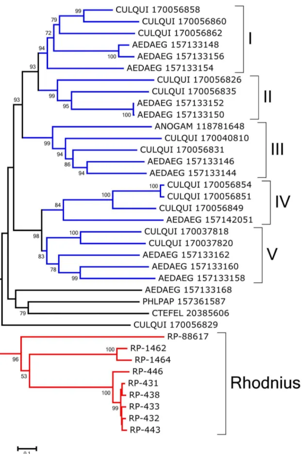
Group IV is a set of nine proteins that includes three which are significantly overexpressed in the gut tissues, such as RP-431, with a total of 782 reads on the gut libraries and only 57 on the WB. This peritrophin is evenly expressed in the three gut libraries, being a good marker of gut tissue, as are RP-433 and RP-438. None of these is expressed in the FB, MT, or OV libraries, but they are expressed in the TE library. These proteins have a CBD that is preceded and followed by a sequence with several conserved Cys residues. This framework is also observed among the best-matching proteins found in the non-redundant (NR) protein database.
The bootstrapped phylogram of Group IV peritrofins aligned with closely related sequences from other insects (Fig. 2) shows all R. prolixussequences fall within a single clade with strong bootstrap support, supporting the existence of at least three genes that differ more than 50% in sequence identity. The sequences 431, RP-434, RP-433, and RP-438 may be alleles. Notice also that the mosquitoesAedes aegyptiandCulex quinquefasciatus shown in Fig. 2 -have indications of at least five different genes with families that diverged before the separation of their genera as indicated by clades containing both genera and having strong bootstrap support (marked I–V in Fig. 2). Quite interestingly, all the proteins collected in this group are from bloodsucking insects that do not share a common bloodsucking ancestor withRhodnius, suggesting either convergent evolution or gene expansion of a common insect gene when associated with blood feeding. All the proteins of this group are predicted to be secreted except RP-88617 and RP-1462, which are predicted to lack a signal peptide or to be membrane-bound, respectively. Once in the midgut lumen, these proteins Table 2.Functional classification of AM-overexpressed transcripts (.106compared to posterior) fromRhodnius prolixus.
Class Number of contigs Number of reads Reads/contig Percent reads
Associated with digestive physiology
Digestive enzymes 6 965 160.8 8.6
Protease inhibitors 1 266 266.0 2.4
Transporters/storage 4 223 55.8 2.0
Other secreted 1 104 104.0 0.9
Mucins 1 47 47.0 0.4
Oxidant metabolism/detoxification 1 32 32.0 0.3
Associated with cellular function
Signal transduction 13 859 66.1 7.7
Transcription factor 3 722 240.7 6.5
Unknown, conserved 11 493 44.8 4.4
Cytoskeletal 3 466 155.3 4.2
Metabolism, amino acid 2 262 131.0 2.3
Protein export machinery 5 202 40.4 1.8
Transcription machinery 4 197 49.3 1.8
Metabolism, carbohydrate 2 90 45.0 0.8
Protein modification machinery 2 77 38.5 0.7
Metabolism, energy 1 56 56.0 0.5
Proteasome machinery 2 48 24.0 0.4
Unknown 68 5638 82.9 50.5
Transposable element 4 236 59.0 2.1
Viral 1 174 174.0 1.6
Total 135 11157
may bind heme, as AeIMUCI [41], possibly to catalyze the formation of hemozoin.
Group V corresponds to proteins that do not form a monophyletic clade in Fig. 1. They are probably cuticular proteins, as discussed for sequences from Groups I and III.
Supporting Information S2 (worksheet ‘‘Peritrophins’’) lists other proteins of this class, not necessarily with significant tissue differential expression.
Vertebrate-like mucins and other secreted proteins. The term mucin denotes two different molecules. Mucin may correspond to a highly glycosylated Ser+Thr-rich protein such as vertebrate mucin [42] or name a peritrophin with a very long mucin domain [43].R. prolixusmucins referred to here correspond to the first type. Thus, RP-5412 codes for a Ser+ Thr-rich protein with 70 putative N-acetyl-galactosamination sites. Its low complexity makes it difficult to assess close eukaryotic proteins, the best match by blastp to the NR database (with the filter of low complexity off) being with a bacterial protein. It is represented by 141 digestive transcripts and only 27 WB reads. 3746 and RP-3448 are overtranscribed somewhat equally in the three digestive tissues, while RP-15656 is overexpressed in the AM, where 43 of the 45 reads from the digestive tissues derive, none being found in
the WB, but two from the TE. The worksheet ‘‘Mucins’’ in Supporting Information S2 contains these and a few other mucins. The Smart ML domain predicts proteins involved with innate immunity and lipid metabolism. It is similar to the KOG domain for the major epididymal secretory protein HE1 and the PFAM E1_DerP2_DerF2 domain implicated in recognition of pathogen-related products. RP-5669 has such a domain and is 11.5-fold overexpressed in gut tissues. Five other transcripts are shown on the worksheet ‘‘Other’’ of Supporting Information S2, including homologs of accessory gland proteins and other proteins found in Triatomasialotranscriptomes and in the midgut transcriptome of sand flies, with unknown function.
Digestive enzymes. Carbohydrate digestion: It has been previously proposed that the digestive glycosidases of R. prolixus could help in digesting their endosymbiont cell walls [20]. Glycosidases could also have some importance in vector-parasite interactions, as several parasite surface molecules are heavily glycosylated. Glycosidases are classified in glycoside hydrolase families (GHFs) according to their amino acid sequence similarities (Carbohydrate Active Enzymes database, at http://www.cazy. org/; [44]). The worksheet ‘‘Carb digest’’ in Supporting Information S2 shows several of these enzymes, four of which Table 3.Functional classification of PM-overexpressed transcripts (.106compared to AM) fromRhodnius prolixus.
Class
Number of contigs
Number
of reads Reads/contig Percent reads
Associated with digestive physiology
Other secreted 1 132 132.0 0.4
Transporters/storage 8 428 53.5 1.3
Digestive enzymes 22 8549 388.6 26.9
Mucins 2 3609 1804.5 11.4
Odorant binding proteins 4 1020 255.0 3.2
Immunity 2 325 162.5 1.0
Oxidant metabolism/detoxification 2 137 68.5 0.4
Associated with cellular function
Nuclear regulation 2 148 74.0 0.5
Transcription factor 2 61 30.5 0.2
Transcription machinery 3 389 129.7 1.2
Protein synthesis machinery 5 129 25.8 0.4
Protein export machinery 2 72 36.0 0.2
Protein modification machinery 3 310 103.3 1.0
Proteasome machinery 1 64 64.0 0.2
Metabolism, carbohydrate 2 222 111.0 0.7
Metabolism, amino acid 2 45 22.5 0.1
Metabolism, lipid 2 266 133.0 0.8
Metabolism, intermediate 1 178 178.0 0.6
Signal transduction 7 191 27.3 0.6
Extracellular matrix/cell adhesion 6 4501 750.2 14.2
Cytoskeletal 5 187 37.4 0.6
Metabolism, energy 6 5158 859.7 16.2
Unknown, conserved 14 1109 79.2 3.5
Unknown 66 4527 68.6 14.2
Transposable element 1 29 29.0 0.1
Total 171 31786
are.10-fold overexpressed in digestive tissues. They comprise 13 enzymes belonging to nine different GHFs, namely families 1, 9, 13, 20, 29, 31, 35, 38, and 63.
The two hexosaminidases highly expressed in the R. prolixus midgut (RP-29656 and RP-25051) belong to family 20 of glycosyl hydrolases. Insect hexosaminidases from family 20 were already described as secreted or cytosolic enzymes [45], but in the case of R. prolixusenzymes, this information could not be assessed due to the lack of 59 sequence in both contigs. Interestingly, insect hexosaminidases are related to mammalian lysosomal hexosamin-idases, which raises the possibility that they were originally lysosomal enzymes recruited for digestion during the evolution of Hemiptera, as has been suggested already for proteolytic enzymes [7]. RP-25051 shares the catalytic residues Asp240 His294 Glu355 with human hexosaminidase but this information is lacking for RP-29656. These proteins can be involved in the digestion of N-linked oligosaccharides. RP-25051, however, does not seem to be exclusively digestive (141 reads in WB and 33 in gut libraries, 25 from RE). In contrast, RP-29656 has 19 reads, all from gut libraries, especially from AM. The distinct patterns of expression displayed by these two transcripts indicate distinct roles for these two proteins. These roles could correspond to the initial digestion of glycoproteins and intermediate or final digestion of chitin or bacterial cell wall polysaccharides, which would be consistent with the distinct compartmentalization of these two GHF20 proteins. In
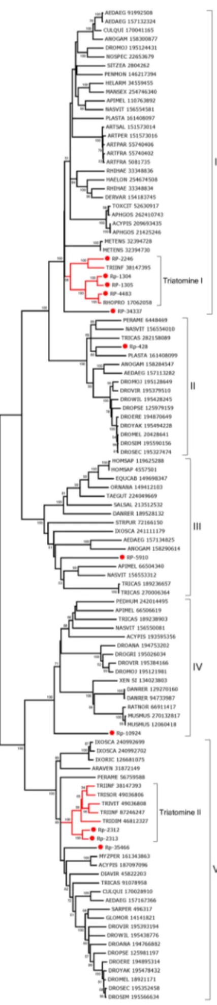
this respect, the expression of b-hexosaminidases should be concomitant with the production of chitinase, lysozymes, and proteinases. No chitinase is included in the set of highly transcribed midgut genes. In fact, from the four chitinases present in the whole-body screening (all from GHF18), only one showed significant expression in the gut (RP-13146), but this transcript belongs to insect chitinase family V, which is related to Imaginal Growth Factors (IGFs) and has no described catalytic role [46]. It is unlikely that thisR. prolixusIGF has catalytic activity, because its sequence lacks the glutamate identified as the catalytic proton donor in other family 18 chitinases, which in this case is substituted by a glutamine residue. Nevertheless, a highly active chitinase was recently purified and characterized fromR. prolixusmidgut (Genta, F.A., not published), but this activity seems to be secreted at later stages of blood digestion, which were not screened in this study. Perhaps the high lysozyme activity observed at later stages of digestion can account for the observed chitinase activity [20], since lysozyme have substantial chitinase activity in addition to hydrolyzing peptidoglycan [47]. It seems more likely that R. prolixushexosaminidases act on lysozyme products, as five of these proteins, belonging to GHF22, are highly expressed in the gut (RP-3602, RP-3604, RP-6482, RP-11146, and RP-24996, further discussed in the section on immune-related transcripts). Phyloge-netic analysis of insect proteins from GHF22 (Fig. 3) reveals that only threeR. prolixusGHF22 sequences (RP-24966, RP-3602, and Table 4.Functional classification of RE-overexpressed transcripts (.106compared to anterior+PMs) fromRhodnius prolixus.
Class
Number of contigs
Number
of reads Reads/contig Percent reads
Associated with digestive physiology
Transporters/storage 7 1292 184.6 3.6
Oxidant metabolism/detoxification 3 902 300.7 2.5
Other secreted 2 296 148.0 0.8
Digestive enzymes 3 244 81.3 0.7
Odorant binding proteins 3 193 64.3 0.5
Peritrophins 1 18 18.0 0.1
Associated with cellular function
Cytoskeletal 17 4562 268.4 12.8
Transcription machinery 7 3803 543.3 10.7
Unknown, conserved 21 3351 159.6 9.4
Protein synthesis machinery 5 2130 426.0 6.0
Metabolism, amino acid 3 1656 552.0 4.7
Extracellular matrix/cell adhesion 8 1367 170.9 3.8
Metabolism, lipid 3 824 274.7 2.3
Metabolism, energy 6 704 117.3 2.0
Protein modification machinery 2 656 328.0 1.8
Signal transduction 6 554 92.3 1.6
Nuclear regulation 5 534 106.8 1.5
Transcription factor 1 462 462.0 1.3
Protein export machinery 5 457 91.4 1.3
Metabolism, carbohydrate 1 423 423.0 1.2
Proteasome machinery 2 214 107.0 0.6
Unknown 69 8609 124.8 24.2
Transposable element 4 2355 588.8 6.6
Total 184 35606
RP-3604) group with other triatomine gut proteins (Triatomine clade I). In spite of that, they do not group with the other described insect digestive lysozymes from Diptera: Cyclorrapha, mainly from Musca domestica[48] and Drosophila melanogaster [49]. This suggests that some adaptive convergence could have occurred in these two insect groups, with the recruitment of lysozymes for digestion of bacteria. In the case of R. prolixus, digestion of the symbiont R. rhodnii seems to be a probable function of these enzymes.
The finding of a glycoside hydrolase from family 9 inR. prolixus (RP-10367; 4 reads from WB and 74 reads in gut, exclusively in PM) is quite unexpected, as GHF9 that were described in termites, beetles, and cockroaches are mainly cellulases (endo-b -1,4-glucanases) involved in plant cell-wall digestion [50]; however, GHF9 also contains several b-glycosidases, and it is difficult to ascertain a specificity or action pattern for these enzymes based only on a partial sequence. Twoa-mannosidases transcripts were identified: RP-3116 is markedly digestive with 65 reads in the gut, coming from PM and RE, and only 4 reads in WB and RP-2863, which showed 46 reads from WB and 37 reads coming from all three gut libraries. They belong to GHFs 38 and 63, respectively. Family 38 contains only mannosidases, mainly from lysosomal origin, which reinforces the use of lysosomal glycosidases in R. prolixusdigestion. Family 63, a poorly described glycoside family in eukaryotes, contains several a-glucosidases as well, making it difficult to construe the specificity or function to this member.
A complete sequence of a typical a-amylase (RP-10100) was found that is expressed mainly in AM. This amylase is predicted to
be activated by chloride ions and because of this, it should not be responsible for the amylase previously assayed inR. prolixusAM, which is secreted byR. rhodniiand is not activated by these ions [24]. From the four amylases highly expressed in the midgut (RP-10100, RP-8390, RP-5922, and RP-3792), three are from family 13 and only one (RP-5922) from family 31, which is related toa -glucosidases. RP-3792 has the same conserved catalytic residues of a-amylase but does not show complete calcium and chloride pockets, suggesting it is ana-glucosidase. As this sequence has a predicted signal peptide and GPI-anchor, it is a good candidate to correspond to thea-glucosidase activity that is a marker enzyme of the perimicrovillar membranes [51]. RP-10100 is a full-length transcript coding for an a-amylase overexpressed in gut tissues, mainly in AM (53 reads against only 9 reads from WB). While RP-10100 is more expressed in AM, RP-8390 and RP-3792 are more expressed in the PM. This could be related to different phases of polysaccharide digestion, corresponding to differences in the action pattern of these enzymes, e.g., liquefying or saccharifying amylases. AsR. prolixusis strictly hematophagous, the nature of the physiologic substrate of these enzymes remains unclear. An a -glucosidase from family 13 has been implicated in formation of hemozoin in theRhodniusmidgut [52], but no transcript coding for that enzyme (accession # FJ236283) was found here. The presence of several enzymes of this group raises the possibility that more than one protein may act in seeding formation of hemozoin crystals.
R. prolixusmidgutb-glycosidases are members of GHFs 1 (RP-12000 and RP-16121) and 35 (RP-4801). Family 35 members are
Figure 1. Cladogram ofRhodnius prolixusperitrophins.The dendrogram was generated with the neighbor-joining algorithm. Branches were statistically supported by bootstrap analysis (cut-off 45) based on ten thousand replicates. The Roman numerals indicate the perithrophin’s group classification.
Figure 2. Bootstrapped phylogram ofRhodnius prolixusand other insect peritrophin annotated as Group IV peritrophin in Fig. 1.
mainly b-galactosidases, and family 1 contains enzymes with differentb-glycosidase specificities. RP-12000 has a signal peptide and a GPI anchor and therefore can account for theb-glucosidase activity associated with the midgut cell microvillar membrane Insect ß-glycosidases can be divided into two classes. Class A includes the enzymes that hydrolyse substrates with hydrophilic aglycones, as disaccharides and oligosaccharides. Class B com-prises enzymes that have high activity only on substrates with hydrophobic aglycones, such as alkyl-, p-nitrophenyl-, and methylumbelliferyl-glycosides [47]. The physiological role of these b-glycosidases is thought to be the digestion of oligosaccharides and glycolipids, respectively [53]. It is possible thatR. prolixushas three active midgutb-glycosidases (twob-glucosidases and oneb -galactosidase) fulfilling these two roles, a situation already described in several insects [53].
One transcript coding for ana-fucosidase (RP-6619) pertains to GHF 29 and probably is involved in the release of L-fucose residues from oligosaccharide moieties attached to glycoproteins. The coding sequences for these and other carbohydrate-hydro-lyzing enzymes are shown on the worksheet ‘‘Carb digest’’ within Supporting Information S2.
Polypeptide digestion: Aspartyl and cysteinyl protease-coding transcripts dominate among those that are significantly over-transcribed in the gut tissues. Interestingly, despite no blood digestion being detected on the AM [54], several of those proteinases are highly expressed in the AM as well as in the RE, in addition of the PM. For example, the aspartyl protease coded by RP-2217 hits 2,857 reads from the digestive tract, and only 72 from the WB. From these 2,857 reads, 1,113 are from the AM, while 609 and 1,135 are from the PM and RE, respectively. A similar profile occurs with RP-2814. Also two different aspartyl proteases-encoding transcripts of Triatoma infestans—TiCatD and TiCatD2—were both expressed in AM and PM but active proteases were only isolated from PM [2] . Expression of aspartyl proteases in the AM can be interpreted as expression of pro-enzymes, such as pepsinogen, that might be activated in the PM. Alternatively, at least part of these enzymes, as well those expressed in RE (which epithelial cells are covered with a cuticle), may play intracellular roles.
The worksheet ‘‘Proteases’’ of Supporting Information S2 provides for 17 coding sequences from aspartyl proteases, most of them full length. All the aspartyl proteinases listed are actually cathepsin D-like enzymes. The motif [DxPxPx(G/A)P] - the proline loop - was suggested to be characteristic for lysosomal cathepsin D-like enzymes which were not secreted into the lumen of the digestive tract, because this motif is absent in digestive enzymes such as pepsin in vertebrates and digestive cathepsin D in cyclorrhaphan flies [55]. However, according to mass spectrom-etry of proteins from the lumen of the PM ofT. infestansand the sequencing of the respective genes, one cathepsin D without (TiCatD) and one with the entire proline loop (TiCatD2) are present in the lumen [2]. In contrast to the expression ofTiCatD, that of TiCatD2 changes only slightly after feeding, indicating different roles of both enzymes [2]. TiCatD is putatively a digestive enzyme, whereas the role of TiCatD2 remains unclear, although it branches with lysosomal enzymes in Fig. 4. RP-1760 is the onlyR. prolixussequence that has a proline loop and, although it may be a conserved lysosomal enzyme based on this evidence, also supported by its branching pattern in Fig. 4, it may be partially found in lumen as TiCatD2 [12]. It is worth mentioning
that enzymes like lysosomal acid phosphatase are partially discharged into midgut lumen [12]. RP-3415 and RP-2091 are probably non-digestive cathepsin Ds, the first because it misses most of the conserved residues that form the subsite binding pockets, and the second because it lacks the first catalytic residue in the sequence. RP-5007 has an incomplete (DxP) proline loop, which suggests a special function unknown until now. All the other sequences lack the proline loop and are, thus, candidates to be responsible for the midgut cathepsin D activity inR. prolixus.
Analysis of theR. prolixusaspartyl proteases aligned with their best-matching proteins from GenBank produces a phylogram (Fig. 4) showing most (13) of theR. prolixussequences forming a single clade, which includes aTriatoma infestanssequence. ThisT. infestanssequence - like those ofR. prolixus -lacks the proline loop. This triatomine gene expansion is indicative of divergence and gene conversion, suggesting this cluster of proteins originates from a chromosomal tandem array. This phenomenon probably occurred in the heteropteran ancestors. The aspartyl proteases RP-1760 and TiCatD2 exceptionally group with other vertebrate and invertebrate proteins, arguably lysosomal enzymes, despite RP-1760 being overexpressed in theR. prolixusmidgut.
Transcripts coding for three cysteinyl proteases are overex-pressed in the digestive tissues, RP-1305 being assembled from 97 transcripts from the WB and 761 from digestive tissues, 707 of which derive from the PM, allowing for the identification of its entire CDS. RP-2313 and RP-1304 are also overexpressed in the digestive tissues—especially in PM. Regarding these three cysteinyl proteases abundantly expressed in gut tissues, only 1 read is found for the TE library, suggesting that the reads from this organ that have a digestive expression (peritrophins, mucins, and aspartyl proteases) do not derive from tissue contamination. Several other transcripts coding for cysteinyl proteases are found with larger expression in the PM when compared to the AM, despite being also found in the WB. The worksheet ‘‘Proteases’’ (Supporting Information S2) presents the CDS of 11 cysteinyl proteases, mostly full length.
lysosomal rather than a secreted digestive function. Similarly, 5910, within clade III is not overexpressed in gut tissues. RP-34337—which belongs to clade I but not to the triatomine I subclade—is actually overexpressed in the WB library as compared with the digestive tract, which had only 1 read as opposed to 40 reads from the WB.
A CDS expressing a cathepsin F is presented in the form of RP-1287, overexpressed (12 fold) in gut tissues. Interestingly, this protein has four cystatin domains in its amino terminus followed by a typical papain-like domain, a structure that is conserved in human proteins as well [58,59], indicating it is an ancient gene structure.
Two CDS represent the carboxy region of trypsin-like serine proteases. RP-2259 showed only 64 reads from WB and 2,851 hits from gut tissues, 2,346 of these being from the RE, 504 from the PM, and only 1 read from the AM. RP-19173 is also overexpressed in the RE, where 154 of the 181 gut-derived reads originate. RP-19173 is also well expressed in the WB, with 141 reads. Trypsin-like serine proteases were found in the salivary glands ofT. infestansandPanstrongylus megistus[60,61] but no trypsin activity has been reported in the digestive tract of triatomine insects. These data—together with the predominance of cysteine and aspartic proteinases and the marked overexpression in RE— indicates that these enzymes will not have a digestive role, but act in the cells of the intestinal wall. Five carboxypeptidases containing the PFAM peptidase S10 domain are shown in the ‘‘Proteases’’ worksheet of Supporting Information S2, including RP-5638, which is overexpressed in the PM, and RP-3222, overexpressed in the AM. All these enzymes contain the catalytic triad of residues of a serine, aspartate, and histidine. Phylogenetic analysis of these carboxypeptidases aligned with their matches to the GenBank proteins shows distinct triatomine clades that do not group with any other sequences with significant bootstrap support except for RP-15295, which groups with 99% support in an animal clade (Fig. 6). The other triatomine sequences derive from T. infestans and from Triatoma brasiliensis. RP-15295, outside this triatomine clade, is underexpressed in the digestive tissues when compared to the WB, and may not have a specific digestive function.
Two additional terminal peptidases are conspicuously absent from the AM but present in PM and RE (RP-5555 and RP-2304, both full length). Both present the PFAM peptidase_S28 domain contained in the enzymes lysosomal Pro-X carboxypeptidase, dipeptidyl-peptidase II, and thymus-specific serine peptidase.
Three other peptidases that are significantly overexpressed in the digestive tract as compared to the WB, or between digestive organs are noted in the worksheet ‘‘Proteases’’ of Supporting Information S2.
Transporters. Following extracellular digestion of the meal, transporters are needed for nutrient intake as well as for maintaining pH and salt equilibrium in the gut. The worksheet ‘‘Transport’’ within Supporting Information S2 contains 76 coding sequences associated with this group and includes the subdivisions ‘‘amino acid and peptide transport’’, ‘‘nucleotide/ sugar transport’’, ‘‘ABC transporters’’, ‘‘permeases of the major facilitator superfamily’’, ‘‘sodium solute symporters,’’ ‘‘lipid transporters’’, ‘‘metal transporters’’, ‘‘ferritins’’, ‘‘aquaporins’’, ‘‘monovalent cation transport and homeostasis’’, ‘‘V-ATPase subunits’’ and ‘‘hemocyanin.’’
The following highlights are indicative of the digestive tract specialization of these families. RP-23175 codes for an amino acid transporter that is significantly overexpressed in the PM, where 15 of 15 digestive reads were found. The nucleotide/sugar transport-er coded by RP-2100 is ovtransport-erexpressed in the digestive tube, whtransport-ere all 757 reads were found, versus 70 in the WB. Similarly, RP-7749 is overexpressed in gut tissues. This sequence is similar to the major glucose uniporter (DpGLUT; GenBank accession number GU014570) that was functionally characterized in D. peruvianus [62]. A ubiquitous permease of the major facilitator superfamily (RP-28161) is overexpressed in the AM when compared to PM expression. RP-8563 is overexpressed in the digestive tissues and, principally, in the RE. This sequence is similar to that of the major midgut cation-glucose symporter (DpSGLT; GenBank accession number GU066262) functionally characterized in D. peruvianus [62]. Associated with water and monovalent cation transport, transcripts coding for theb-2 subunit of the Na+
+ K+ ATPase were overexpressed in the AM, where all 55 reads were found. The vacuolar ATPase is important for transepithelial acidification and water transport [63]. Several of its subunits are overexpressed in the digestive tissues.
Protease inhibitors. Twenty-six CDS coding for protease inhibitors from the Kazal and pacifastin family are shown in this section’s worksheet of Supporting Information S2. Proteins with multiple Kazal domains have been found in the AM of triatomine bugs where they act as inhibitors of blood coagulation enzymes and elastase. In some cases, their processing kinetics and crystal structure have been described [64–70].
The worksheet named ‘‘Prot. inhibitors’’ of Supporting Infor-mation S2 contains 22 CDS for proteins containing one or more Kazal domains, including previously described members of this family. RP-620, in particular, derives from an abundantly expressed transcript assembled from 4,447 digestive reads and 116 from the WB. It contains two Kazal domains and is 41% identical to the antithrombin named brasiliensin precursor ofT. brasiliensis [71] and 39% identical to infestin 1–7 precursor [69] from T. infestans. RP-620 presents inhibitory activity for bovine trypsin (data not published). The transcript RP-570 contains ten Kazal-type domains seeming to play the same role as infestin 1–7 precursor in T. infestans [67] and brasiliensin precursor in T. brasiliensis[71], providing anticoagulant molecules to theR. prolixus digestive tract. As it contains two copies of rhodniin, a potent thrombin inhibitor [65], we cannot discard the idea that other transcripts also supply the gut with rhodniin.
Several of the Kazal members shown in Supporting Information S2 were not found transcribed in the gut tissues but provide matches to sequences previously found in sialotranscriptomes of RhodniusandTriatoma, particularly the short single Kazal family— similar to vasotab, a potent vasodilator isolated from salivary glands of the horse flyHybomitra bimaculata[72].
The pacifastin family [73,74] is represented by four full-length and one truncated sequence, all providing matches to insect proteins annotated as pacifastin and having the Pacifastin_I PFAM domain. RP-8689 derives from an expressed transcript assembled from 75 digestive reads and 205 from the WB; it contains at least four pacifastin domains. Those pacifastin domains are not over transcribed in the gut tissues, which may suggest a physiologic role not related to digestion, possibly in the insect immune response [75].
Figure 3. Cladogram of insect Lysozymes from glycoside hydrolase Family 22.TheR. prolixussequences are shown by the notation RP-followed by a unique number. The remaining proteins were obtained from GenBank and they are annotated with accession number RP-followed by species name. The dendrogram was generated with the UPGMA algorithm. The branches were statistically supported by bootstrap analysis (cut-off 40) based on 1,000 replicates.
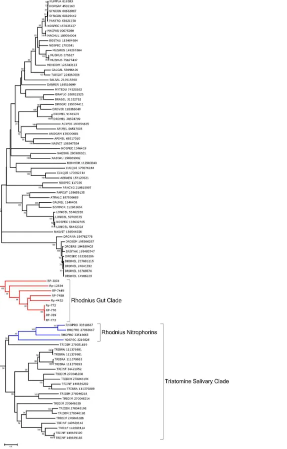
Lipocalins. The lipocalin family is ubiquitous and contains a typical barrel structure, or calyx, which is often used to carry hydrophobic compounds such as lipids in an aqueous environ-ment, thus the name lipocalin [76]. Many antihemostatic salivary proteins of triatomine bugs were found to belong to this family, including the nitric oxide (NO)-carrying heme proteins ofRhodnius, the biogenic amine- and adenosine-binding proteins of the same organism, and several clotting and platelet aggregation inhibitors ofRhodniusandTriatoma, which include the pallidipin and triabin proteins [76–83]. Contigs coding for these proteins are easily identified by the PFAM domains for nitrophorin, triabin, or lipocalin. Supporting Information S2 presents 88 CDS for this family, including an RE-specific transcript coding for RP-772, assembled from 4,242 reads from digestive tissues and only 94 from the WB. The deducted protein sequence provides many matches to salivary lipocalins of triatomines deposited in the NR database. RP-3004 matches Galleria gallerin, an insecticyanin homolog, and may function in lipid transport.
Phylogenetic analysis shows a strong clade (94% support) for a common origin of the salivary lipocalins of Rhodnius (including nitrophorins) and the salivary lipocalins of Triatoma (marked Triatomine salivary clade in Fig. 7). Six lipocalins overexpressed in the gut tissues can be aligned with their best matches to the NR database and form a robust clade by themselves, indicative of gene duplication and possible gene conversion independent of the salivary clade, marked as Rhodnius gut clade in Fig. 8. All six transcripts have predicted signal peptides, suggesting a role in binding and transport of dietary hydrophobic compounds such as lipids from the extracellular environment.
Odorant-binding, takeout, juvenile hormone-binding, and chemosensorial-binding proteins. Supporting Informa-tion S2 contains CDS informaInforma-tion for 46 contigs that contain domains from the takeout/juvenile hormone-binding protein (JHBP), odorant-binding protein (OBP), or chemosensorial protein (CSP) as identified by their sequence analysis and simple modular architecture research tool (SMART) or CDD matches. Four such CDS are noted in Supporting Information S2 as being overex-pressed in the gut tissues, including RP-828, RP-14075, RP-3723, and RP-1578.
The phylogenetic tree for these 46 contigs showed three clearly separate groups (Fig. 8). Group I corresponds totakeout/JHBP (24 contigs), Group II is classical OBPs (11 contigs), and Group III is CSPs (11 contigs). The four overexpressed contigs belong either to thetakeout/JHBP group (RP-14075, RP-1578, RP-828) or to the classical OBPs clade (RP-3723). RP-14075 and RP-7792 are members of the takeout/JHBP family with the two motifs characteristic of this protein family [84]. RP-828 did not show the motif 2 that characterizes atakeoutprotein and was grouped in the JHBP family.takeout/JHBP family proteins are carrier proteins of hydrophobic ligands and may have a role in binding or transport of signaling molecules or nutrients. JH synthesis is tightly coordinated with ingestion of a blood meal in hematophagous insects and was shown to control transcription in the midgut ofAe. aegyptiof both trypsin [85] and chymotrypsin [86]. Although RP-3723 has been grouped in classical OBPs—which are character-ized by the presence of six conserved cysteines—this transcript has only four cysteines, suggesting this is a member of CSP. In spite of its name, members of the OBP family have been ascribed roles that are not related to odor recognition, such as binding of heme
Figure 4. Bootstrapped phylogram of Rhodnius prolixus and other aspartyl proteinases.Bootstrap values above 50% are shown on the branches. The bottom line indicates 10% amino acid sequence divergence between the proteins.R. prolixussequences are shown by the notation RP followed by a unique number and have a red circle preceding their names. TheTriatoma infestanssequences from Balczun et. al. [2] have a green marker. The remaining sequences were obtained from GenBank and are annotated with the first three letters of the
genus name, followed by the first three letters of the species name, followed by their GenBank GI number. One thousand replicates were done for the bootstrap test using the neighbor joining test.
by theRhodniusheme-binding protein or the participation of a CSP in regeneration ofPeriplanetalegs [87]. The presence of this class of proteins overexpressed in the midgut ofR. prolixuscould suggest a role in the transport of nutrients or other molecules involved in the coordinating of physiological gut function.
Immunity related. Although lacking a classical adaptive immune response, insects have powerful innate immunity against several pathogens that have a cellular component involving hemocytes (leading to phagocytosis and encapsulation of patho-gens), as well as a humoral response carried out by several tissues such as the fat body, midgut, trachea, and salivary glands. Humoral immunity is based on production of antimicrobial peptides (AMPs), of reactive oxygen and nitrogen species, and melanization. In this way, synthesis and secretion of antimicrobial peptides and agents to the hemolymph is generally referred to as ‘‘systemic immunity,’’ while the same action at the level of the barrier epithelia (as observed in the gut, for example) is generally referred to as ‘‘epithelial immunity’’ [88].
Production of AMPs is regulated by three primary signaling pathways, namely, Toll, IMD, and Jak/STAT [89]. InDrosophila, Toll responds to gram-positive bacteria and fungi, while IMD response is elicited mainly by gram-negative bacteria. This separation does not seem to be so clear in mosquitoes, where both pathways seem highly interconnected and overlapping [90]. Activation of the Toll and IMD pathways occurs upon recognition of pathogen-associated molecular patterns (PAMPs), triggering a cascade that culminates with translocation of a NF-kB-like molecule to the nucleus and hence to the production of effector molecules. It is important to note that—although in several other immune tissues, such as the fat-body, both pathways can be potentially activated— in the presence of corresponding PAMP, it is believed that in epithelial gut and tracheal responses only IMD may be activated as a consequence of proliferation of gut commensal bacteria or the presence of pathogens [88,90,91].
Several immune-related transcripts were identified, ranging from PAMP recognition molecules to signal transducers and effector proteins, as described below.
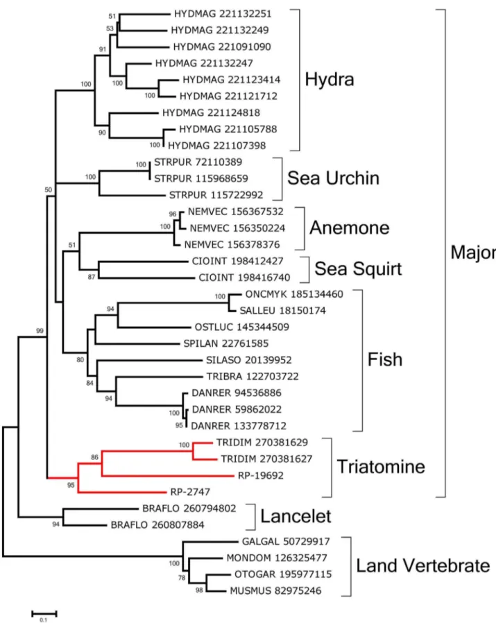
PAMP recognition molecules: Carbohydrate binding proteins, or lectins, could work as pathogen-recognition molecules that trigger insect defense responses [92,93] and/or could have a role in insect feeding [94]. Several b-galactoside-binding lectins (galectins) were overexpressed in Rhodnius digestive tissues. RP-2747 has the Gal_lectin PFAM domain and was assembled from 428 gut-derived reads and 47 from the WB. Also, RP-15084 derives from a different gene and is overexpressed in the gut tissues.
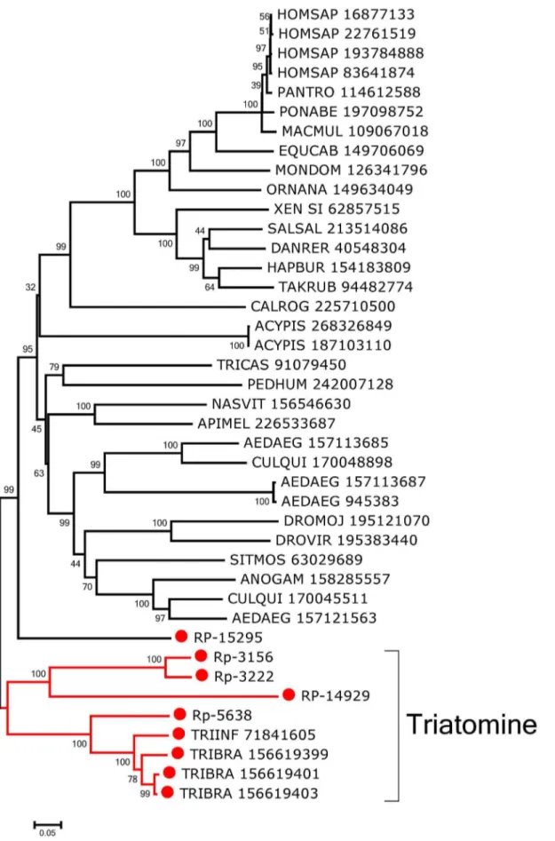
RP-2747 and RP-19692 are near full length in size and contain the CDD domain Gal_lectin. Alignment of these two sequences with their NR database matches produces the phylogram presented in Fig. 9 showing that a triatomine clade is formed with strong bootstrap support as part of a major clade formed with 99% support containing fish- and invertebrate-derived sequences. Lancelet and land vertebrate sequences lie on their own clades.
Figure 5. Bootstrapped phylogram of Rhodnius prolixus and other cysteinyl proteinases.Bootstrap values above 50% are shown on the branches. The bottom line indicates 10% amino acid sequence divergence between the proteins.R. prolixussequences are shown by the notation RP followed by a unique number and have a red circle preceding their names. The remaining sequences, obtained from GenBank, are annotated with the first three letters of the genus name, followed by the first three letters of the species name, followed by their GenBank GI number. One thousand replicates were done for the bootstrap test using the neighbor joining test.
The twoTriatoma dimidiatasequences that group with theRhodnius sequences have been described in a sialotranscriptome [95].
Sugar-inhibitable hemaglutinins have been described in the gut of triatomines [96,97]. Galectins are overexpressed in gut and salivary glands of Anopheles infected with bacteria or Plasmodium [98–100]. It is speculated that galectins are involved in insect immune response similarly to how they are in mammals—by opsonizing bacteria and other pathogens facilitating their recog-nition, agglutination, and/or phagocytosis for immune-competent cells. Also, a galectin (PpGalec) has been implicated inLeishmania majoradhesion to the midgut epithelia ofPhlebotomus papatasi. In this case, blockage of this protein with specific antibodies leads to an important decrease in vector parasite load after six days post infection [101]. It would be interesting to assess whether any of these proteins might be involved inT. cruzibinding to the midgut epithelium.
RP-16133 codes for a 59 truncated transcript producing matches against the NR database to proteins annotated as hemolectin. The best match (gi|193601326) has multiple domains, including von Willebrand, coagulation factor 5/8, TIL, and the C8 domains. Hemolectin is hemocyte-specific inDrosophilaand is involved in the fly’s clotting system [102–104].
Three contigs containing peptidoglycan recognition protein (PGRP) domains were also identified in the digestive tissues (Asb-69756, Asb-23314, Asb-48139). Asb-69756 and Asb-23314 do not present predicted trans-membrane regions and are likely to be soluble PGRPs. Interestingly, Asb-69756 probably presents amidase activity, as all five conserved catalytic amino acid residues are present in this protein. If that is the case, Asb-69756 could be involved in destruction of bacteria-released peptidoglycan, down-regulating the bug’s immune response. Asb-23314, on the other hand, is unlikely to present amidase activity, because one of the five conserved catalytic residues is missing. If that is the case, Asb-23314 could be involved in detecting peptidoglycan and activating an epithelial IMD response. The last PGRP domain containing transcript, Asb-23314, also does not present amidase activity but show a predicted transmembrane domain and is homologous to the Drosophila PGRP-LC (NP_729468.2). This transcript might constitute an actual PGRP-LC and may represent a receptor primarily responsible for activation of the IMD pathway in Rhodnius.
Immune signaling pathways: Transcripts coding for members of the immune signaling pathways were not overexpressed in gut compared to WB, but several of them showed a significant number of reads, indicating that they were operating in these tissues. Despite this, these transcripts were included in our analysis, because the midgut epithelia is the area of most intense contact between microorganisms and insects and is the only part of the triatomine body in contact withT. cruzi. Although it is generally accepted that the Toll pathway is not active in digestive tissues [88,105], several contigs putatively coding for proteins from this pathway were identified—namely, a Toll receptor (Asb-44175), its adaptor protein MyD88 (Asb-69782), the kinase pelle (Asb-15772) and the pelle-associated protein pellino (Asb-24337) [106]. The evolutionarily conserved intermediate in the Toll/IL-1 signal transduction pathway [107], ECSIT (Asb-9158) and a protein from the Spa¨tzle family (RP-45859) were identified in the transcriptome. Interestingly, contigs coding for two additional putative Toll-interacting proteins (Tollips; 22553 and Asb-45642), for an inhibitor of the Toll pathway transcription factor
rpDorsal Cactus (Asb-31044), the Cactus-binding protein cactin (Asb-33928), and a contig containing an NF-kB-repressing factor domain (Asb-17843) were also identified. Although these contigs were not overexpressed in the gut libraries when compared to WB, this is the first time that such a high number of Toll-related proteins were found consistently in a midgut transcriptome, suggesting that, in spite of the relative low abundance, this pathway may be of physiologic significance in gut immunity in Rhodnius.
In contrast to this high number of Toll-related transcripts, only one contig coding for a member of the IMD pathway was identified in the digestive tissues. It coded for the IMD negative regulator Caspar (Asb-145) [108]. This contig was highly expressed in the gut (80 reads) but also in WB (92 reads). Low expression levels also were found for the STAT pathway, where a transcript coding for a STAT (Asb-17321; 4 reads only in AM and none in WB) was identified. Together, these results suggest that all three main known immune pathways are active in the Rhodnius midgut.
A transcript resembling eiger was identified (Asb-21490; 21 reads from gut and 31 reads from WB). Eiger, the insect homolog of mammalian TNF, has been implicated in the immune response against extracellular pathogens [109] as well as against bacterial oral infection [110]. Eiger/TNF was suggested to be part of an ancient proof-reading pathway directed to suppress tumors in epithelial tissues based on alterations of polarity that are typical of malignant cells but that can also be found in cells that are either physically damaged or exposed to pathogens [111]. As mentioned below, there are several transcripts expressed in the gut that belong to signaling pathways related to cell polarity, indicating that sensing and control of cell polarity is a priority ofRhodnius intestinal cells. This could provide a link between tissue morphology and innate immunity related to intestinal pathogens. Interestingly, three contigs putatively coding for proteins with a double-strand (ds) RNA binding domain were identified in Rhodniusdigestive tissue libraries. One of these (Asb-16245) codes for a putative R2D2 protein. R2D2 is known to associate with Dicer-2 and is essential for channeling the siRNA generated by this protein to the RISC complex [112]. Tar RNA-binding proteins (TRBPs; Asb-26443) have a dsRNA binding domain and are structural components of the RISC complex [113]. Finally, also identified was a contig coding for a protein homologous to loquacious (Asb-21490), a protein that contains two dsRNA binding domains and was originally described as part of the miRNA generating machinery inDrosophila[114].
Effector proteins: Large amounts of lysozyme activity were described in the anterior and PM of R. prolixus [20]. Several lysozyme-coding transcripts were found to be overexpressed in gut tissues. RP-3602 was assembled from 7966 digestive reads and 619 WB-derived reads, hence being 23.1-fold overexpressed in digestive tissues. This lysozyme was previously reported as upregulated in the midgut following bacterial challenge as well as ingestion of T. cruzi [115]. In addition, RP-6482—although somewhat more mildly overexpressed in the digestive tissues than RP-3602—was reported to be upregulated in the FB after injection of bacteria into the hemocoel [115]. RP-24996 was the only lysozyme transcript not overexpressed in the digestive tissues. All lysozyme transcripts possess catalytic aspartate and glutamate residues except for lysozyme 2 fromT. infestans[116]. The function of this unusual lysozyme remains to be elucidated. InT. brasiliensis, annotated with the first three letters of the genus name, followed by the first three letters of the species name, followed by their GenBank GI number. One thousand replicates were done for the bootstrap test using the neighbor joining test.
Figure 7. Bootstrapped phylogram ofRhodnius prolixusmidgut lipocalins aligned with their best matches to the NR database.
Bootstrap values above 50% are shown on the branches. The bottom line indicates 20% amino acid sequence divergence between the proteins.R. prolixussequences are shown by the notation RP followed by a unique number. The remaining sequences, obtained from GenBank, are annotated with the first three letters of the genus name, followed by the first three letters of the species name, followed by their GenBank GI number. One thousand replicates were done for the bootstrap test using the neighbor joining test.