514 http://www.journal-imab-bg.org / J of IMAB. 2014, vol. 20, issue 1/
COMPARATIVE EVALUATION OF THE
EFFECTIVENESS OF THREE METHODS FOR
PROXIMAL CARIES DIAGNOSIS – a clinical study
Mirela Marinova-Takorova1, Radostina Anastasova2, Vladimir E. Panov2, Spartak Yanakiev3,
1) Department of Conservative Dentistry, Faculty of Dental medicine, Medical University - Sofia,
2) Department of Conservative dentistry and Oral pathology, Faculty of Dental medicine, Medical University - Varna,
3) Medical Colledge “Y. Filaretova”, Medical University - Sofia, Bulgaria
Journal of IMAB - Annual Proceeding (Scientific Papers) 2014, vol. 20, issue 1
Journal of IMAB
ISSN: 1312-773X (Online) http://www.journal-imab-bg.org
SUMMARY
Aim: The aim of the presented study is to compare the effectiveness of the diagnosis with a dental microscope, laser fluorescence (DIAGNOcam) and X-ray examination in proximal caries diagnosis.
Material and methods: Thirty-eight adult patients were examined. They were first examined with a dental mirror and a probe, under magnification 6.4 times. After that a diagnosis with DIAGNOcam was performed. Bitewing X-ray images were administered. The data from the three diagnostic methods was compared using SPSS 16 package of Windows. The lesions that were diagnosed as involving dentin were then excavated which served as a confirmation of the diagnosis.
Results: The results of the study showed that dentinal lesions were detected with a high degree of correlation with all three diagnostic methods. The visual examination seriously underestimated lesions involving only enamel. In these cases there was a good correlation between laser fluorescence and X-ray data.
Conclusions: Based on the conducted study we could conclude that the diagnosis of proximal caries with DIAGNOcam is equivalent to X-ray, both being more accurate in cases with early lesions, compared to visual diagnosis.
Key words: early caries detection, laser fluorescence,
INTRODUCTION
Dental caries is one of the most common chronic diseases worldwide. In the last decades there has been a change in the overall treatment strategies due to the increased understanding of the mechanism of the progress of the disease [1] as well as the development of the adhesive restorative materials. The philosophy of minimal intervention dentistry, involving prevention of the disease and preservation of sound tooth structure has emerged [2]. Early diagnosis and clinical staging of dental caries – its presence, activity and severity is of a significant importance for the minimally invasive treatment strategy, in order the dentist to have the opportunity to apply not only surgical but nonsurgical (fluorides, antimicrobial agents, sealants) procedures as well [3].
Detection of proximal lesions, especially in early stages has often been a diagnostic problem. The most commonly used method additional to visual inspection is radiography. The method has good sensitivity for dentin caries lesions involving proximal surfaces, but it has quite a limited diagnostic value in cases with enamel lesions [4].Due to the low sensitivity of this method in those cases the lesion size and depth is often underestimated or even in some cases it could remain undetected (5,6). As a result quite often there is no possibility to treat the lesion with conservative methods and reverse or arrest the process so it has to be treated operatively. Besides that there are present the hazardous effects of ionizing radiation.
A wide variety of new technologies has been developed and introduced to help clinicians to detect caries lesions and treat as many cases as possible conservatively [7]. A new device – DIAGNOcam has recently been introduced on the dental market. It uses laser diode, the wavelength being 780nm, to illuminate dental surface. Carious tooth tissue scatters and absorbs more light than surrounding healthy tissue. A camera digitally images the light emerging from the opposite surface. The images are displayed on a monitor and stored.
The aim of the presented study is to compare the effectiveness of diagnosis of non-cavitated proximal caries lesions with a dental microscope, digital imaging fiber-optic transillumination device - DIAGNOcam and X-ray examination.
MATERIAL AND METHODS
The survey was performed on 38 adult patients with a total number of 125 non-cavitated proximal lesions on permanent teeth. The consequence of application of the different diagnostic methods was the following:
1. Visual examination with dental microscope Leica M320 with magnification 6.4.
2. Examination of the proximal surfaces with DIAGNOcam.
3. Analysis of bitewing X-rays.
4. In cases where dentinal caries was diagnosed the lesions were excavated. This was used as a confirmation method for the validity of the diagnosis.
/ J of IMAB. 2014, vol. 20, issue 1/ http://www.journal-imab-bg.org 515 5. Enamel lesions were not treated operatively, so
validation could not be applied in these cases.
Statistical analysis was done using statistical software SPSS 16.0 (SPSS Inc.).
RESULTS
A total number of 125 non-cavitated proximal lesions on permanent teeth were diagnosed. Forty-seven of them (37.6%) were involving only enamel. For this type of lesions validation of the results with operative excavation of the lesion was not done, because it is accepted that they could eventually be treated conservatively (8). Visual diagnostic,
even under magnification showed to be insufficiently sensitive for lesions involving only enamel – only 11 (23.4%) caries lesions were diagnosed with this method. Using X-ray examination there were diagnosed 41 (87.2%) enamel lesions on the same patients, while with DIAGNOcam – 47. Three of the 6 more lesions diagnosed with DIAGNOcam were verified during the excavation of dentinal lesions situated on the neighboring surface of the teeth next to the one with the enamel lesion.
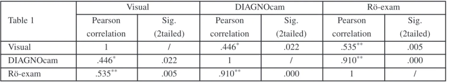
The correlations between the different diagnostic methods for lesions involving only enamel are presented on table 1.
Concerning the diagnosis of caries lesions involving both enamel and dentine the three methods showed very high level of correlation. In only 6.4% of the cases the visual examination was ineffective. There was a total concurrence of the results obtained with DIAGNOcam, Rö examination and the operative treatment in cases with lesions involving both enamel and dentine.
On figures one, two and three are presented some images of lesions, obtained with DIAGNOcam and their corresponding bitewings.
Fig. 2: Caries lesions involving enamel on teeth 24 and 25
Table 1: Correlation of different diagnostic methods for lesions involving only enamel.
Visual DIAGNOcam Rö-exam
Table 1 Pearson Sig. Pearson Sig. Pearson Sig.
correlation (2tailed) correlation (2tailed) correlation (2tailed)
Visual 1 / .446* .022 .535** .005
DIAGNOcam .446* .022 1 / .910** .000
Rö-exam .535** .005 .910** .000 1 /
* Correlation significant at 0.05 level, ** Correlation significant at 0.01 level
Fig.1: Caries lesions involving teeth 45 and 46 – the lesion on tooth 46 is seen only on DIAGNOcam image
Fig. 3: Caries lesions involving teeth 15 and 16
DISCUSSION
Early caries detection and quantification of lesions to establish their progression or arrest is crucial if dental approach is going to be changed from mainly operative to preventive. Early caries diagnosis is also important for clinical dental researches - the ability of accurate detection and determination of the size of early lesions may permit the use of shorter intervals and lesser number of patients to asses the effectiveness of caries preventive measures [7]. In this study DIFOTI method was compared with the traditional visual method for caries detection and radiographs done with bitewings. The efficacy of the second and third methods depends greatly on the skill and experience of the examiner [9]. The three methods proved to be equally effective in cases with caries lesions involving both enamel and dentin.
The visual method proved to be quite ineffective in cases with lesions involving only enamel. Dental radiographs also were less sensitive for early caries detection. This correlates with the data obtained from other studies [5, 9, 10, 11].
516 http://www.journal-imab-bg.org / J of IMAB. 2014, vol. 20, issue 1/ 1. Kudiyirickal MG, Ivancakova R.
Early enamel lesion. Part II. Histo-mor-phology and prevention. Acta Medica (Hradec Kralove). 2008 Mar;51(3): 151-6. [PubMed]
2. Dalli M, Colac H, Mustafa Hamidi M. Minimal intervention con-cept: a new paradigm for operative dentistry. J Investig Clin Dent. 2012 Aug;3(3):167-75. [PubMed] [CrossRef]
3. Frencken JE, Peters MS, Manton DJ, Leal SC, Gordan VV, Eden E. Minimal intervention dentistry (MID) for managing dental caries - a review: report of a FDI task group. Int Dent J.
2012 Oct;62(5):223-43. [PubMed] [CrossRef]
4. Bader JD, Shugars DA, Bonito AJ. A systematic review of the per-formance of methods for identifying carious lesions. J Public Health Dent.
(caries lesions), so prophylaxis should be done prior to exam in order to eliminate the possibility of getting false positive results.
CONCLUSIONS
Based on the data from the presented study we may conclude:
1. DIAGNOcam could be judged as a diagnostic
method equivalent to Ro-exam for non-cavitated proximal lesions
2. Visual examination proved to be an insufficient method for detection of proximal non-cavitated lesions involving only enamel.
3. We could recommend DIAGNOcam to be used in combination with the visual method as an alternative of Ro-exam
REFERENCES:
2002 Fall;62(4):201-213. [PubMed] 5. Stookey GK, Gonzalez-Cabezas C. Emerging methods of caries diagno-sis. J Dent Educ. 2001 Oct;65(10): 1001-6. [PubMed]
6. Maia AM, L Karlsson L, W Margulis W, Gomes AS. Evaluation of two imaging techniques: near-infrared transillumination and dental radio-graphs for the detection of early approximalenamel caries. Dento-maxillofac Radiol. 2011 Oct; 40(7):429-33. [PubMed] [CrossRef]
7. Amaechi BT. Emerging tech-nologies for diagnosis of dental caries: The road so far. J Appl Phys. 2009 May;105:102047. [CrossRef]
8. Pitts NB. Are we ready to move from operative to non-operative/pre-ventive treatment of dental caries in clinical practice? Caries Res. 2004 May-Jun;38(3):294-304. [PubMed]
[CrossRef]
9. Astvaldsdottir A, Ahlund K, Holbrook WP, de Verdier B, Tranæus S. Approximal Caries Detection by DIFOTI: In Vitro Comparison of Diag-nostic Accuracy/Efficacy with Film and DigitalRadiography. Int J Dent. 2012; 2012:326401 [PubMed] [CrossRef]
10. Young DA, Featherstone JD. Digital imaging fiber optic trans-illu-mination, F-speed radiographic film and depth of approximal lesions. J Am Dent Assoc. 2005 Dec;136(12): 1682-1687. [PubMed] [CrossRef]
11. Shinderman A, Elbaum M, Shultz T, Keem S, Greenebaum M, Driller J. Assessment of dental caries with Digital Imaging Fiber-Optic Tran-sillumination (DIFOTI): in vitro study. Caries Res. 1997 Feb;31(2):103-110. [PubMed]
Address for correspondence:
D-r. Mirela Marinova-Takorova
Department of Conservative dentistry, Faculty of Dental medicine, Medical University - Sofia